Introdução
O que você precisa saber de cara
Visão geral
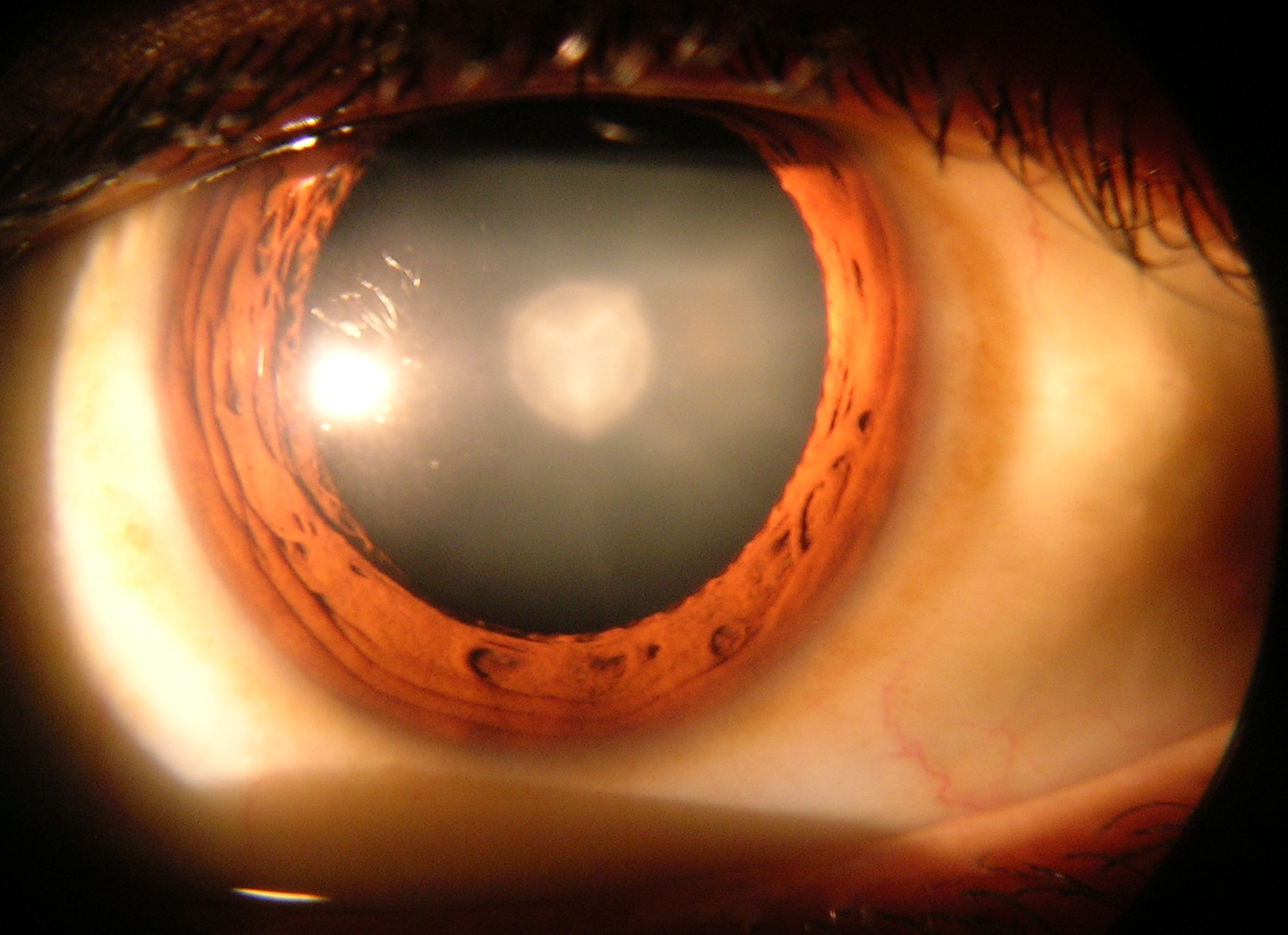
A catarata polar posterior de início precoce é uma condição ocular rara que se caracteriza pela presença de uma opacidade (nuvem) na parte central e posterior do cristalino, presente desde o nascimento ou nos primeiros meses de vida. Essa opacidade pode prejudicar a visão já na infância, pois bloqueia a passagem da luz para a retina. A doença é considerada rara e pode ocorrer de forma isolada ou associada a outras alterações oculares ou sistêmicas.[1]
Sinais e sintomas
Os principais sinais da catarata polar posterior de início precoce são a opacidade visível no centro do cristalino (geralmente detectada ao exame oftalmológico) e a dificuldade visual que pode se manifestar já nos primeiros meses de vida. Como a idade de início é neonatal ou na primeira infância, os bebês podem apresentar nistagmo (movimentos oculares involuntários) ou estrabismo, além de atraso no desenvolvimento visual. A gravidade da perda visual varia conforme o tamanho e a densidade da opacidade.[1]
Causas genéticas
A catarata polar posterior de início precoce tem causas genéticas. Mutações em pelo menos oito genes já foram associadas à condição: CRYAB, CHMP4B, MIP, PITX3, PANK4, CRYBA1, EPHA2 e GJA3. Esses genes estão envolvidos na produção de proteínas do cristalino, na comunicação entre as células do cristalino ou no desenvolvimento ocular. A herança pode ser autossômica dominante ou recessiva, dependendo do gene afetado, mas os dados específicos de herança para esta doença ainda não foram completamente estabelecidos.[1][3]
Diagnóstico
O diagnóstico é feito por um oftalmologista, que identifica a opacidade polar posterior ao exame de lâmpada de fenda. Para confirmar a causa genética, estão disponíveis no Brasil os seguintes exames pelo SUS: cariótipo com bandas G, Q ou R; pesquisa de microdeleções/microduplicações por FISH; sequenciamento completo do exoma (WES); e dosagem de alfa-fetoproteína. Atualmente, existem 336 testes genéticos registrados para essa condição e 136 variantes descritas no ClinVar, o que permite um diagnóstico molecular preciso na maioria dos casos.[1][3]
Tratamento e manejo
O tratamento da catarata polar posterior de início precoce é essencialmente cirúrgico, com a remoção do cristalino opaco (facoemulsificação) e implantação de lente intraocular, quando possível. O momento da cirurgia depende da densidade da catarata e do impacto na visão. Após a cirurgia, é necessário o uso de óculos ou lentes de contato para correção do foco. O acompanhamento com oftalmologista e reabilitação visual são fundamentais. No Brasil, o SUS oferece atendimento em reabilitação para doenças raras, que pode incluir terapia visual e suporte multidisciplinar.[1]
Prognóstico e qualidade de vida
O prognóstico visual depende do diagnóstico precoce e do tratamento adequado. Quando a cirurgia é realizada nos primeiros meses de vida, a maioria das crianças desenvolve visão funcional, embora possa ser necessário o uso de óculos por toda a vida. O atraso no tratamento pode levar a ambliopia (olho preguiçoso) irreversível. Com acompanhamento regular e reabilitação, a qualidade de vida pode ser boa, permitindo atividades escolares e sociais normais.[1]
Conteúdo informativo gerado e mantido automaticamente a partir de fontes oficiais (Orphanet, HPO, OMIM, SUS). Não substitui avaliação médica.
Catarata polar posterior de início precoce é uma opacidade congênita ou de início na infância, localizada na parte posterior do cristalino. Frequentemente associada a mutações nos genes CRYAB, CHMP4B, MIP, PITX3 ou PANK4, pode afetar a visão desde cedo.
Encontrou um erro ou informação desatualizada? Sugira uma correção →
Entender a doença
Do básico ao detalhe, leia no seu ritmo
Preparando trilha educativa...
Sinais e sintomas
O que aparece no corpo e com que frequência cada sintoma acontece
Visão geral
A catarata polar posterior de início precoce é uma condição ocular rara que se caracteriza pela presença de uma opacidade (nuvem) na parte central e posterior do cristalino, presente desde o nascimento ou nos primeiros meses de vida. Essa opacidade pode prejudicar a visão já na infância, pois bloqueia a passagem da luz para a retina. A doença é considerada rara e pode ocorrer de forma isolada ou associada a outras alterações oculares ou sistêmicas.[1]
Sinais e sintomas
Os principais sinais da catarata polar posterior de início precoce são a opacidade visível no centro do cristalino (geralmente detectada ao exame oftalmológico) e a dificuldade visual que pode se manifestar já nos primeiros meses de vida. Como a idade de início é neonatal ou na primeira infância, os bebês podem apresentar nistagmo (movimentos oculares involuntários) ou estrabismo, além de atraso no desenvolvimento visual. A gravidade da perda visual varia conforme o tamanho e a densidade da opacidade.[1]
Causas genéticas
A catarata polar posterior de início precoce tem causas genéticas. Mutações em pelo menos oito genes já foram associadas à condição: CRYAB, CHMP4B, MIP, PITX3, PANK4, CRYBA1, EPHA2 e GJA3. Esses genes estão envolvidos na produção de proteínas do cristalino, na comunicação entre as células do cristalino ou no desenvolvimento ocular. A herança pode ser autossômica dominante ou recessiva, dependendo do gene afetado, mas os dados específicos de herança para esta doença ainda não foram completamente estabelecidos.[1][3]
Diagnóstico
O diagnóstico é feito por um oftalmologista, que identifica a opacidade polar posterior ao exame de lâmpada de fenda. Para confirmar a causa genética, estão disponíveis no Brasil os seguintes exames pelo SUS: cariótipo com bandas G, Q ou R; pesquisa de microdeleções/microduplicações por FISH; sequenciamento completo do exoma (WES); e dosagem de alfa-fetoproteína. Atualmente, existem 336 testes genéticos registrados para essa condição e 136 variantes descritas no ClinVar, o que permite um diagnóstico molecular preciso na maioria dos casos.[1][3]
Tratamento e manejo
O tratamento da catarata polar posterior de início precoce é essencialmente cirúrgico, com a remoção do cristalino opaco (facoemulsificação) e implantação de lente intraocular, quando possível. O momento da cirurgia depende da densidade da catarata e do impacto na visão. Após a cirurgia, é necessário o uso de óculos ou lentes de contato para correção do foco. O acompanhamento com oftalmologista e reabilitação visual são fundamentais. No Brasil, o SUS oferece atendimento em reabilitação para doenças raras, que pode incluir terapia visual e suporte multidisciplinar.[1]
Prognóstico e qualidade de vida
O prognóstico visual depende do diagnóstico precoce e do tratamento adequado. Quando a cirurgia é realizada nos primeiros meses de vida, a maioria das crianças desenvolve visão funcional, embora possa ser necessário o uso de óculos por toda a vida. O atraso no tratamento pode levar a ambliopia (olho preguiçoso) irreversível. Com acompanhamento regular e reabilitação, a qualidade de vida pode ser boa, permitindo atividades escolares e sociais normais.[1]
Conteúdo informativo gerado e mantido automaticamente a partir de fontes oficiais (Orphanet, HPO, OMIM, SUS). Não substitui avaliação médica.
Linha do tempo da pesquisa
Encontrou um erro ou informação desatualizada? Sugira uma correção →
Genética e causas
O que está alterado no DNA e como passa nas famílias
Genes associados
8 genes identificados com associação a esta condição.
May contribute to the transparency and refractive index of the lens. Has chaperone-like activity, preventing aggregation of various proteins under a wide range of stress conditions. In lens epithelial cells, stabilizes the ATP6V1A protein, preventing its degradation by the proteasome (By similarity)
CytoplasmNucleusSecretedLysosome
Myopathy, myofibrillar, 2A, adult-onset
A form of myofibrillar myopathy, a group of chronic neuromuscular disorders characterized at ultrastructural level by disintegration of the sarcomeric Z disk and myofibrils, and replacement of the normal myofibrillar markings by small dense granules, or larger hyaline masses, or amorphous material. MFM2A is an autosomal dominant form characterized by weakness of the proximal and distal limb muscles, weakness of the neck, velopharynx and trunk muscles, respiratory insufficiency, hypertrophic cardiomyopathy, and cataract.
Probable core component of the endosomal sorting required for transport complex III (ESCRT-III) which is involved in multivesicular bodies (MVBs) formation and sorting of endosomal cargo proteins into MVBs. MVBs contain intraluminal vesicles (ILVs) that are generated by invagination and scission from the limiting membrane of the endosome and mostly are delivered to lysosomes enabling degradation of membrane proteins, such as stimulated growth factor receptors, lysosomal enzymes and lipids. The M
Cytoplasm, cytosolLate endosome membraneMidbodyNucleus envelope
Cataract 31, multiple types
An opacification of the crystalline lens of the eye that frequently results in visual impairment or blindness. Opacities vary in morphology, are often confined to a portion of the lens, and may be static or progressive. In general, the more posteriorly located and dense an opacity, the greater the impact on visual function. CTRCT31 includes posterior polar, progressive posterior subcapsular, nuclear, and anterior subcapsular cataracts.
Aquaporins form homotetrameric transmembrane channels, with each monomer independently mediating water transport across the plasma membrane along its osmotic gradient (PubMed:11001937, PubMed:24120416). Specifically expressed in lens fiber cells, this aquaporin is crucial for maintaining lens water homeostasis and transparency. Beyond water permeability, it also acts as a cell-to-cell adhesion molecule, forming thin junctions between lens fiber cells that are essential for maintaining the ordere
Cell membraneCell junction
Cataract 15, multiple types
An opacification of the crystalline lens of the eye that frequently results in visual impairment or blindness. Opacities vary in morphology, are often confined to a portion of the lens, and may be static or progressive. CTRCT15 includes polymorphic, progressive punctate lamellar, cortical, anterior and posterior polar, nonprogressive lamellar with sutural opacities, embryonic nuclear, and pulverulent cortical, among others.
Transcriptional regulator which is important for the differentiation and maintenance of meso-diencephalic dopaminergic (mdDA) neurons during development. In addition to its importance during development, it also has roles in the long-term survival and maintenance of the mdDA neurons. Activates NR4A2/NURR1-mediated transcription of genes such as SLC6A3, SLC18A2, TH and DRD2 which are essential for development of mdDA neurons. Acts by decreasing the interaction of NR4A2/NURR1 with the corepressor
Nucleus
Anterior segment dysgenesis 1
A form of anterior segment dysgenesis, a group of defects affecting anterior structures of the eye including cornea, iris, lens, trabecular meshwork, and Schlemm canal. Anterior segment dysgeneses result from abnormal migration or differentiation of the neural crest derived mesenchymal cells that give rise to components of the anterior chamber during eye development. Different anterior segment anomalies may exist alone or in combination, including iris hypoplasia, enlarged or reduced corneal diameter, corneal vascularization and opacity, posterior embryotoxon, corectopia, polycoria, abnormal iridocorneal angle, ectopia lentis, and anterior synechiae between the iris and posterior corneal surface. Clinical conditions falling within the phenotypic spectrum of anterior segment dysgeneses include aniridia, Axenfeld anomaly, Reiger anomaly/syndrome, Peters anomaly, and iridogoniodysgenesis.
Phosphatase which shows a preference for 4'-phosphopantetheine and its oxidatively damaged forms (sulfonate or S-sulfonate), providing strong indirect evidence that the phosphatase activity pre-empts damage in the coenzyme A (CoA) pathway (PubMed:27322068). Hydrolyzing excess 4'-phosphopantetheine could constitute a directed overflow mechanism to prevent its oxidation to the S-sulfonate, sulfonate, or other forms (PubMed:27322068). Hydrolyzing 4'-phosphopantetheine sulfonate or S-sulfonate would
Cytoplasm
Cataract 49
A form of cataract, an opacification of the crystalline lens of the eye that frequently results in visual impairment or blindness. Opacities vary in morphology, are often confined to a portion of the lens, and may be static or progressive. In general, the more posteriorly located and dense an opacity, the greater the impact on visual function. CTRCT49 is an autosomal dominant form characterized by congenital cataract located in the posterior region of the lens. Visual impairment has onset in early childhood.
Crystallins are the dominant structural components of the vertebrate eye lens
Cataract 10, multiple types
An opacification of the crystalline lens of the eye that frequently results in visual impairment or blindness. Opacities vary in morphology, are often confined to a portion of the lens, and may be static or progressive. CTRCT10 includes congenital zonular with sutural opacities, among others. This is a form of zonular cataract with an erect Y-shaped anterior and an inverted Y-shaped posterior sutural opacities. Zonular or lamellar cataracts are opacities, broad or narrow, usually consisting of powdery white dots affecting only certain layers or zones between the cortex and nucleus of an otherwise clear lens. The opacity may be so dense as to render the entire central region of the lens completely opaque, or so translucent that vision is hardly if at all impeded. Zonular cataracts generally do not involve the embryonic nucleus, though sometimes they involve the fetal nucleus. Usually sharply separated from a clear cortex outside them, they may have projections from their outer edges known as riders or spokes.
Receptor tyrosine kinase which binds promiscuously membrane-bound ephrin-A family ligands residing on adjacent cells, leading to contact-dependent bidirectional signaling into neighboring cells. The signaling pathway downstream of the receptor is referred to as forward signaling while the signaling pathway downstream of the ephrin ligand is referred to as reverse signaling. Activated by the ligand ephrin-A1/EFNA1 regulates migration, integrin-mediated adhesion, proliferation and differentiation
Cell membraneCell projection, ruffle membraneCell projection, lamellipodium membraneCell junction, focal adhesion
Cataract 6, multiple types
An opacification of the crystalline lens of the eye that frequently results in visual impairment or blindness. Opacities vary in morphology, are often confined to a portion of the lens, and may be static or progressive. CTRCT6 includes posterior polar and age-related cortical cataracts, among others. Posterior polar cataract is a subcapsular opacity, usually disk-shaped, located at the back of the lens. Age-related cortical cataract is a developmental punctate opacity restricted to the cortex. The cataract is white or cerulean, increases in number with age, but rarely affects vision.
Structural component of lens fiber gap junctions (PubMed:30044662). Gap junctions are dodecameric channels that connect the cytoplasm of adjoining cells (By similarity). They are formed by the docking of two hexameric hemichannels, one from each cell membrane. Small molecules and ions diffuse from one cell to a neighboring cell via the central pore (PubMed:30044662)
Cell membraneCell junction, gap junction
Cataract 14, multiple types
An opacification of the crystalline lens of the eye that frequently results in visual impairment or blindness. Opacities vary in morphology, are often confined to a portion of the lens, and may be static or progressive. CTRCT14 includes zonular pulverulent cataract, among others. Zonular or lamellar cataracts are opacities, broad or narrow, usually consisting of powdery white dots affecting only certain layers or zones between the cortex and nucleus of an otherwise clear lens. The opacity may be so dense as to render the entire central region of the lens completely opaque, or so translucent that vision is hardly if at all impeded. Usually sharply separated from a clear cortex outside them, they may have projections from their outer edges known as riders or spokes.
Variantes genéticas (ClinVar)
136 variantes patogênicas registradas no ClinVar.
Vias biológicas (Reactome)
25 vias biológicas associadas aos genes desta condição.
Diagnóstico
Os sinais que médicos procuram e os exames que confirmam
Tratamento e manejo
Remédios, cuidados de apoio e o que precisa acompanhar
Onde tratar no SUS
Hospitais de referência no Brasil e o protocolo oficial do SUS (PCDT)
🇧🇷 Atendimento SUS — Catarata polar posterior de início precoce
Selecione um estado ou use sua localização para ver resultados.
Dados de DATASUS/CNES, SBGM, ABNeuro e Ministério da Saúde. Sempre confirme a disponibilidade diretamente com o estabelecimento.
Pesquisa ativa
Ensaios clínicos abertos e novidades científicas recentes
Pesquisa e ensaios clínicos
Nenhum ensaio clínico registrado para esta condição.
Publicações mais relevantes
Publicações recentes
Prevalence and formation of primary cataracts and persistent hyperplastic tunica vasculosa lentis in the German Pinscher population in Germany.
A study to explore the risk factors for the early onset of cataract in India.
Two new cataract loci, Ccw and To3, and further mapping of the Npp and Opj cataracts in the mouse.
Ocular and systemic manifestations of cerebrotendinous xanthomatosis.
Associações
Organizações que acompanham esta doença — pra ter apoio e orientação
Ainda não temos associações cadastradas para Catarata polar posterior de início precoce.
É de uma associação que acompanha esta doença? Fale com a gente →
Comunidades
Grupos ativos de quem convive com esta doença aqui no Raras
Ainda não existe comunidade no Raras para Catarata polar posterior de início precoce
Pacientes, familiares e cuidadores se organizam em comunidades pra compartilhar experiências, fazer perguntas e se apoiar. Você pode ser o primeiro.
Tire suas dúvidas
Perguntas, dicas e experiências compartilhadas aqui na página
Participe da discussão
Faça login para postar dúvidas, compartilhar experiências e interagir com especialistas.
Fazer loginDoenças relacionadas
Doenças com sintomas parecidos — ajudam quem ainda está buscando diagnóstico
Ainda não achamos doenças com sintomas parecidos o suficiente.
Referências e fontes
Bases de dados externas citadas neste artigo
Publicações científicas
Artigos indexados no PubMed ligados a esta doença no grafo RarasNet — título, periódico e PMID direto da fonte, sem intermediação de IA.
- Prevalence and formation of primary cataracts and persistent hyperplastic tunica vasculosa lentis in the German Pinscher population in Germany.
- A study to explore the risk factors for the early onset of cataract in India.
- Two new cataract loci, Ccw and To3, and further mapping of the Npp and Opj cataracts in the mouse.
- Ocular and systemic manifestations of cerebrotendinous xanthomatosis.
Bases de dados e fontes oficiais
Identificadores e referências canônicas usadas para montar este verbete.
- ORPHA:98993(Orphanet)
- MONDO:0020378(MONDO)
- GARD:16889(GARD (NIH))
- Variantes catalogadas(ClinVar)
- Busca completa no PubMed(PubMed)
- Q56014426(Wikidata)
Dados compilados pelo RarasNet a partir de fontes abertas (Orphanet, OMIM, MONDO, PubMed/EuropePMC, ClinicalTrials.gov, DATASUS, PCDT/MS). Este conteúdo é informativo e não substitui avaliação médica.
Conteúdo mantido por Agente Raras · Médicos e pesquisadores podem colaborar
Catarata polar posterior de início precoce
📋 Origem dos dados
Esta página agrega dados de fontes públicas e oficiais. Dados sobre cobertura no SUS (PCDT, CEAF) são verificados ativamente por agente proativo (ver badge no infobox). Demais dados têm atribuição de fonte + data da última sincronização — clique para abrir o original.
- Doença rara (ontologia)
- fonte: Orphanet
- Identificador unificado
- fonte: MONDO
- Codificação WHO/SUS
- fonte: WHO ICD-10 / DATASUS
- CID-11 (futuro)
- fonte: WHO ICD-11
- NIH/GARD
- fonte: GARD (NIH)
- Dado público estruturado
- fonte: Wikidata