Anomalia congênita das cordas da válvula tricúspide, caracterizada por alterações estruturais nas cordas tendíneas que sustentam a válvula tricúspide. Pode levar a regurgitação tricúspide e, em casos graves, insuficiência cardíaca.
Introdução
O que você precisa saber de cara
Visão geral
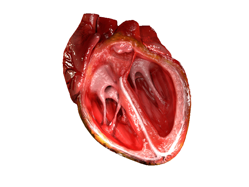
A anomalia das cordas da válvula tricúspide é uma condição cardíaca congênita rara, presente desde o nascimento. Ela afeta as estruturas que sustentam a válvula tricúspide do coração, chamadas de cordas tendíneas. Em vez de se prenderem normalmente na borda livre dos folhetos da válvula e nos músculos papilares, essas cordas se fixam em locais anormais, como na parte central do folheto (zona clara) ou diretamente no endocárdio (revestimento interno do coração). Isso pode prender um ou mais folhetos da válvula, prejudicando sua mobilidade e levando ao refluxo de sangue (regurgitação tricúspide). A condição pode estar associada a outras anomalias cardíacas congênitas.[1]
Sinais e sintomas
Os sinais e sintomas decorrem principalmente da regurgitação tricúspide, ou seja, do vazamento de sangue do ventrículo direito para o átrio direito a cada batimento cardíaco. Isso pode causar cansaço, falta de ar, inchaço nas pernas e abdômen (edema) e sensação de batimentos cardíacos irregulares ou acelerados (palpitações). Em muitos casos, a condição pode ser assintomática por anos, sendo descoberta em exames de rotina. A gravidade dos sintomas depende do grau de regurgitação e da presença de outras cardiopatias associadas.[1]
Causas genéticas
A anomalia das cordas da válvula tricúspide é uma condição congênita, mas sua causa genética específica ainda não foi identificada. Não há genes conhecidos associados a esta doença isoladamente. Acredita-se que fatores genéticos e ambientais durante o desenvolvimento fetal possam contribuir para sua ocorrência. A condição pode fazer parte de síndromes genéticas mais amplas que afetam o coração, mas, por si só, não tem um padrão de herança definido.[1][3]
Diagnóstico
O diagnóstico é geralmente feito por ecocardiograma, um exame de ultrassom do coração que permite visualizar a anatomia das válvulas e o fluxo sanguíneo. A ressonância magnética cardíaca pode ser usada para avaliar com mais detalhes a estrutura e a função do coração. Como a condição pode estar associada a outras anomalias, exames genéticos podem ser solicitados para investigar síndromes mais amplas. No Sistema Único de Saúde (SUS), estão disponíveis procedimentos como cariótipo, pesquisa de microdeleções/microduplicações por FISH, sequenciamento completo do exoma (WES) e dosagem de alfa-fetoproteína, além de atendimento em reabilitação para doenças raras.[1]
Tratamento e manejo
O tratamento depende da gravidade da regurgitação tricúspide e dos sintomas. Casos leves podem não necessitar de intervenção imediata, apenas acompanhamento regular com cardiologista. Casos moderados a graves podem exigir cirurgia para reparo ou substituição da válvula tricúspide. O manejo também inclui o tratamento de outras cardiopatias associadas, se houver. Não há medicamentos específicos aprovados para tratar a anomalia em si, mas medicamentos para insuficiência cardíaca (como diuréticos) podem ser usados para controlar os sintomas. O acompanhamento multidisciplinar é essencial.[1]
Prognóstico e qualidade de vida
O prognóstico varia amplamente. Pacientes com regurgitação leve e sem outras anomalias cardíacas podem ter uma expectativa de vida normal e boa qualidade de vida, com acompanhamento periódico. Já aqueles com regurgitação grave ou cardiopatias associadas podem necessitar de cirurgia e ter um risco maior de complicações como insuficiência cardíaca ou arritmias. O diagnóstico precoce e o manejo adequado são fundamentais para melhorar os resultados a longo prazo.[1]
Conteúdo informativo gerado e mantido automaticamente a partir de fontes oficiais (Orphanet, HPO, OMIM, SUS). Não substitui avaliação médica.
Anomalia congênita das cordas da válvula tricúspide, caracterizada por alterações estruturais nas cordas tendíneas que sustentam a válvula tricúspide. Pode levar a regurgitação tricúspide e, em casos graves, insuficiência cardíaca.
Encontrou um erro ou informação desatualizada? Sugira uma correção →
Entender a doença
Do básico ao detalhe, leia no seu ritmo
Preparando trilha educativa...
Sinais e sintomas
O que aparece no corpo e com que frequência cada sintoma acontece
Visão geral
A anomalia das cordas da válvula tricúspide é uma condição cardíaca congênita rara, presente desde o nascimento. Ela afeta as estruturas que sustentam a válvula tricúspide do coração, chamadas de cordas tendíneas. Em vez de se prenderem normalmente na borda livre dos folhetos da válvula e nos músculos papilares, essas cordas se fixam em locais anormais, como na parte central do folheto (zona clara) ou diretamente no endocárdio (revestimento interno do coração). Isso pode prender um ou mais folhetos da válvula, prejudicando sua mobilidade e levando ao refluxo de sangue (regurgitação tricúspide). A condição pode estar associada a outras anomalias cardíacas congênitas.[1]
Sinais e sintomas
Os sinais e sintomas decorrem principalmente da regurgitação tricúspide, ou seja, do vazamento de sangue do ventrículo direito para o átrio direito a cada batimento cardíaco. Isso pode causar cansaço, falta de ar, inchaço nas pernas e abdômen (edema) e sensação de batimentos cardíacos irregulares ou acelerados (palpitações). Em muitos casos, a condição pode ser assintomática por anos, sendo descoberta em exames de rotina. A gravidade dos sintomas depende do grau de regurgitação e da presença de outras cardiopatias associadas.[1]
Causas genéticas
A anomalia das cordas da válvula tricúspide é uma condição congênita, mas sua causa genética específica ainda não foi identificada. Não há genes conhecidos associados a esta doença isoladamente. Acredita-se que fatores genéticos e ambientais durante o desenvolvimento fetal possam contribuir para sua ocorrência. A condição pode fazer parte de síndromes genéticas mais amplas que afetam o coração, mas, por si só, não tem um padrão de herança definido.[1][3]
Diagnóstico
O diagnóstico é geralmente feito por ecocardiograma, um exame de ultrassom do coração que permite visualizar a anatomia das válvulas e o fluxo sanguíneo. A ressonância magnética cardíaca pode ser usada para avaliar com mais detalhes a estrutura e a função do coração. Como a condição pode estar associada a outras anomalias, exames genéticos podem ser solicitados para investigar síndromes mais amplas. No Sistema Único de Saúde (SUS), estão disponíveis procedimentos como cariótipo, pesquisa de microdeleções/microduplicações por FISH, sequenciamento completo do exoma (WES) e dosagem de alfa-fetoproteína, além de atendimento em reabilitação para doenças raras.[1]
Tratamento e manejo
O tratamento depende da gravidade da regurgitação tricúspide e dos sintomas. Casos leves podem não necessitar de intervenção imediata, apenas acompanhamento regular com cardiologista. Casos moderados a graves podem exigir cirurgia para reparo ou substituição da válvula tricúspide. O manejo também inclui o tratamento de outras cardiopatias associadas, se houver. Não há medicamentos específicos aprovados para tratar a anomalia em si, mas medicamentos para insuficiência cardíaca (como diuréticos) podem ser usados para controlar os sintomas. O acompanhamento multidisciplinar é essencial.[1]
Prognóstico e qualidade de vida
O prognóstico varia amplamente. Pacientes com regurgitação leve e sem outras anomalias cardíacas podem ter uma expectativa de vida normal e boa qualidade de vida, com acompanhamento periódico. Já aqueles com regurgitação grave ou cardiopatias associadas podem necessitar de cirurgia e ter um risco maior de complicações como insuficiência cardíaca ou arritmias. O diagnóstico precoce e o manejo adequado são fundamentais para melhorar os resultados a longo prazo.[1]
Conteúdo informativo gerado e mantido automaticamente a partir de fontes oficiais (Orphanet, HPO, OMIM, SUS). Não substitui avaliação médica.
Linha do tempo da pesquisa
Encontrou um erro ou informação desatualizada? Sugira uma correção →
Genética e causas
O que está alterado no DNA e como passa nas famílias
Nenhum gene associado encontrado
Os dados genéticos desta condição ainda estão sendo catalogados.
Diagnóstico
Os sinais que médicos procuram e os exames que confirmam
Tratamento e manejo
Remédios, cuidados de apoio e o que precisa acompanhar
Onde tratar no SUS
Hospitais de referência no Brasil e o protocolo oficial do SUS (PCDT)
🇧🇷 Atendimento SUS — Anomalia das cordas da válvula tricúspide
Centros de Referência SUS
24 centros habilitados pelo SUS para Anomalia das cordas da válvula tricúspide
Centros para Anomalia das cordas da válvula tricúspide
Detalhes dos centros
Hospital Universitário Prof. Edgard Santos (HUPES)
R. Dr. Augusto Viana, s/n - Canela, Salvador - BA, 40110-060 · CNES 0003808
Serviço de Referência
Hospital Infantil Albert Sabin
R. Tertuliano Sales, 544 - Vila União, Fortaleza - CE, 60410-794 · CNES 2407876
Serviço de Referência
Hospital de Apoio de Brasília (HAB)
AENW 3 Lote A Setor Noroeste - Plano Piloto, Brasília - DF, 70684-831 · CNES 0010456
Serviço de Referência
Hospital Estadual Infantil e Maternidade Alzir Bernardino Alves (HIABA)
Av. Min. Salgado Filho, 918 - Soteco, Vila Velha - ES, 29106-010 · CNES 6631207
Serviço de Referência
Hospital das Clínicas da UFG
Rua 235 QD. 68 Lote Área, Nº 285, s/nº - Setor Leste Universitário, Goiânia - GO, 74605-050 · CNES 2338424
Serviço de Referência
Hospital Universitário da UFJF
R. Catulo Breviglieri, Bairro - s/n - Santa Catarina, Juiz de Fora - MG, 36036-110 · CNES 2297442
Atenção Especializada
Hospital das Clínicas da UFMG
Av. Prof. Alfredo Balena, 110 - Santa Efigênia, Belo Horizonte - MG, 30130-100 · CNES 2280167
Serviço de Referência
Hospital Universitário Julio Müller (HUJM)
R. Luis Philippe Pereira Leite, s/n - Alvorada, Cuiabá - MT, 78048-902 · CNES 2726092
Atenção Especializada
Hospital Universitário João de Barros Barreto
R. dos Mundurucus, 4487 - Guamá, Belém - PA, 66073-000 · CNES 2337878
Serviço de Referência
Hospital Universitário Lauro Wanderley (HULW)
R. Tabeliao Estanislau Eloy, 585 - Castelo Branco, João Pessoa - PB, 58050-585 · CNES 0002470
Atenção Especializada
Instituto de Medicina Integral Prof. Fernando Figueira (IMIP)
R. dos Coelhos, 300 - Boa Vista, Recife - PE, 50070-902 · CNES 0000647
Serviço de Referência
Hospital Pequeno Príncipe
R. Des. Motta, 1070 - Água Verde, Curitiba - PR, 80250-060 · CNES 3143805
Serviço de Referência
Hospital Universitário Regional de Maringá (HUM)
Av. Mandacaru, 1590 - Parque das Laranjeiras, Maringá - PR, 87083-240 · CNES 2216108
Atenção Especializada
Hospital de Clínicas da UFPR
R. Gen. Carneiro, 181 - Alto da Glória, Curitiba - PR, 80060-900 · CNES 2364980
Serviço de Referência
Hospital Universitário Pedro Ernesto (HUPE-UERJ)
Blvd. 28 de Setembro, 77 - Vila Isabel, Rio de Janeiro - RJ, 20551-030 · CNES 2280221
Serviço de Referência
Instituto Nacional de Saúde da Mulher, da Criança e do Adolescente Fernandes Figueira (IFF/Fiocruz)
Av. Rui Barbosa, 716 - Flamengo, Rio de Janeiro - RJ, 22250-020 · CNES 2269988
Serviço de Referência
Hospital São Lucas da PUCRS
Av. Ipiranga, 6690 - Jardim Botânico, Porto Alegre - RS, 90610-000 · CNES 2232928
Serviço de Referência
Hospital de Clínicas de Porto Alegre (HCPA)
Rua Ramiro Barcelos, 2350 Bloco A - Av. Protásio Alves, 211 - Bloco B e C - Santa Cecília, Porto Alegre - RS, 90035-903 · CNES 2237601
Serviço de Referência
Hospital Universitário da UFSC (HU-UFSC)
R. Profa. Maria Flora Pausewang - Trindade, Florianópolis - SC, 88036-800 · CNES 2560356
Serviço de Referência
Hospital das Clínicas da FMUSP
R. Dr. Ovídio Pires de Campos, 225 - Cerqueira César, São Paulo - SP, 05403-010 · CNES 2077485
Serviço de Referência
Hospital de Base de São José do Rio Preto
Av. Brg. Faria Lima, 5544 - Vila Sao Jose, São José do Rio Preto - SP, 15090-000 · CNES 2079798
Atenção Especializada
Hospital de Clínicas da UNICAMP
R. Vital Brasil, 251 - Cidade Universitária, Campinas - SP, 13083-888 · CNES 2748223
Serviço de Referência
Hospital de Clínicas de Ribeirão Preto (HCRP-USP)
R. Ten. Catão Roxo, 3900 - Vila Monte Alegre, Ribeirão Preto - SP, 14015-010 · CNES 2082187
Serviço de Referência
UNIFESP / Hospital São Paulo
R. Napoleão de Barros, 715 - Vila Clementino, São Paulo - SP, 04024-002 · CNES 2688689
Serviço de Referência
Dados de DATASUS/CNES, SBGM, ABNeuro e Ministério da Saúde. Sempre confirme a disponibilidade diretamente com o estabelecimento.
Pesquisa ativa
Ensaios clínicos abertos e novidades científicas recentes
Pesquisa e ensaios clínicos
Nenhum ensaio clínico registrado para esta condição.
Publicações mais relevantes
Reconstruction of an unguarded tricuspid orifice using a simplified sliding plasty technique.
Unguarded tricuspid orifice is a rare anomaly of the tricuspid valve characterized by complete absence of tricuspid valve tissue and chordae on a proportion of the annulus. Management strategies vary widely. We report a case of successful repair of an unguarded tricuspid orifice with a simplified technique. A 35-year-old male presented with severe tricuspid regurgitation and right ventricular volume overload. Intraoperative inspection of the valve revealed an unguarded tricuspid orifice. For repair, sliding plasty of anterior and posterior leaflets were performed, followed by ring annuloplasty and commissuroplasty. Postoperative, echocardiogram showed minimal residual tricuspid regurgitation and significantly improved right ventricular dimensions. This case highlights the possibility of successful repair of an unguarded tricuspid orifice. If feasible, repair can be a good choice.
Dual-Patch Technique with Ventricular Septal Defect Closure for Straddling Chordae.
Surgical repair of ventricular septal defects (VSDs) with straddling atrioventricular (AV) valve chordae is challenging due to the risk of disrupting valve integrity. We report the successful use of a dual-patch technique in a 5-month-old girl (6.1 kg) with Down syndrome, presenting with a large inlet VSD, secundum atrial septal defect (ASD), and straddling chordae involving both AV valves. Ventricular septal defects closure was performed via right atriotomy using 2 glutaraldehyde-treated autologous pericardial patches placed on the superior and inferior septal margins, encasing the chordae without division. Mitral and tricuspid valve clefts were repaired, and the ASD was closed primarily. Postoperative echocardiography showed no residual VSD and only mild AV valve regurgitation. This approach preserved valvular geometry and avoided conduction disturbance. The dual-patch technique offers a physiologic and conservative solution when conventional VSD repair is precluded by straddling chordae. It avoids chordal translocation or reimplantation, maintaining the native architecture and function of the AV valves. Tricuspid regurgitation is a comparatively common anomaly. Although mild tricuspid regurgitation is commonly present, hemodynamically significant tricuspid regurgitation can lead to right ventricular dysfunction and cause substantial morbidity and mortality. Structural modifications of any or all of the tricuspid valve apparatus may cause the development of tricuspid regurgitation. Anatomy The right atrioventricular valve apparatus, or tricuspid valve complex (see Image. Valves of the Heart), consists of the following 4 components: Valve leaflets: Anterior, posterior, and septal: There may be as few as two and as many as 6 leaflets. The anterior leaflet is typically the largest, and the septal leaflet is the smallest. . Fibrous tricuspid valve annulus. Supporting chordae tendineae. Papillary muscles: from 2 to 9. : Just before the onset of the right ventricular systole, the papillary muscle contracts to increase tension in the chordae tendineae so that the 3-valve cusps coapt, preventing regurgitation across the tricuspid valve. The conduction system and the fibroelastic cardiac skeleton's supporting structure coordinate the tricuspid valve's actions. The tricuspid valve is located between the right atrium and the right ventricle and has a valve area of 4 to 6 cm2. The pathophysiological variants of the tricuspid valve include the following: Ebstein anomaly. Tricuspid atresia. Congenital tricuspid stenosis. Congenital cleft of the anterior leaflet.
[Intermediate and long-term outcomes of transcatheter closure of congenital coronary cameral fistulas in 66 children].
Objective: To evaluate the intermediate and long-term outcomes and technical aspects of transcatheter closure (TCC) of coronary cameral fistulas (CCF) in pediatric patients. Methods: This was a case-control study. All pediatric patients with CCF who underwent TCC between January 2005 and December 2019 were retrospectively reviewed. Data was collected from medical records, including demographic characteristics, procedural details, intraoperative and postoperative serious adverse events, follow-up results and prognosis. Patients with serious adverse events and without serious adverse events were compared regarding their clinical features and CCF characteristics. Comparisons between groups were performed with independent sample t test, chi-square test or Fisher exact test. Results: A total of 66 CCF patients (34 boys, 32 girls, 3.9 (1.9, 6.2) years old, 15 (11, 20) kg) underwent attempted TCC. All of the CCF were all medium or large fistulas including 55 proximal fistulas (83%) and 11 distal fistulas (17%). The CCF originated more frequently from the right coronary artery (38 cases (58%)), followed by the left coronary artery (28 cases (42%)). The incidence of coronary artery aneurysms (CAA) was 61% (40/66).Procedural treatment was achieved in 64 patients and procedural success was achieved in 59 patients (92%). Six (9%) serious adverse events occurred in 5 patients during the perioperative period. Acute complications included procedure-related death in one patient and acute myocardial infarction in one patient. Periprocedural complications occurred in 3 patients at one day postoperatively including acute myocardial infarction (2 cases), occluder detachment (1 case), and tricuspid chordae tendinae rupture (1 case). Clinical follow-up data were available in 58 of the 62 patients who underwent initial successful TCC with a follow-up period of 9.3 (6.5, 13.4) years. Ten adverse events occurred in 9 patients including 5 complications consisted of aortic valve perforation (1 case), coronary thrombosis (1 case), progressive aneurysmal dilation after reintervention (1 case), and new-onset tricuspid valve prolapse with significant regurgitation (2 cases) and large residual shunts due to fistula recanalization (5 cases). Therefore, the incidence of intermediate and long-term adverse events was 17% (10/58). During the periprocedural and follow-up period, 16 adverse events occurred in 13 patients, whereas no adverse events occurred in 51 patients. Patients with seriovs adverse events presented with larger proportion of large CCF (11/13 vs. 39% (20/51), P=0.005), giant CAA (10/13 vs.14% (7/51), P=0.030), and higher mean pulmonary artery pressure ((20±9) vs.(16±6) mmHg, 1 mmHg=0.133 kPa, t=2.02, P=0.048) compared to patients without serious adverse events. Conclusions: TCC in CCF children appears to be effective with favorable intermediate and long-term outcomes. Strict indication of TCC is mandatory. 目的: 总结经导管介入治疗儿童先天性冠状动脉-心腔瘘(CCF)的经验及中远期随访结果。 方法: 病例对照研究。选择2005年1月至2019年12月在广东省人民医院接受经导管介入治疗的66例先天性CCF患儿作为研究对象,收集并分析其临床基线资料、手术效果、术中及术后严重不良事件、随访结果及预后等资料。根据是否发生严重不良事件将患儿进行分组,比较严重不良事件组和无严重不良事件组一般情况及CCF特征的差异。组间比较采用独立样本t检验、χ2检验或Fisher确切概率法。 结果: 66例先天性CCF的患儿中男34例、女32例,年龄3.9(1.9,6.2)岁,体重15(11,20)kg。66例CCF均为中型或大型瘘管,其中近端型瘘管55例(83%),远端型瘘管11例(17%);瘘管起源于右冠状动脉38例(58%),起源于左冠状动脉28例(42%);合并冠状动脉瘤(CAA)40例(61%)。66例患儿中64例患儿完成封堵治疗,5例患儿发生6例次围手术期严重不良事件。围手术期手术成功率为92%(59/64),围手术期不良事件发生率为9%(6/64),其中2例患儿术中发生了2例次严重不良事件,包括手术相关死亡1例,急性心肌梗死1例;3例患儿术后随访1 d发生了4例次严重不良事件,包括急性心肌梗死2例、三尖瓣腱索断裂1例、封堵器脱落1例。共58例患儿完成中远期随访,随访时间9.3(6.5,13.4)年。9例患儿发生10例次严重不良事件,中远期严重不良事件发生率为17%(10/58),其中5例次并发症(1例次新发CAA、1例次主动脉瓣穿孔、1例次冠状动脉血栓形成、2例次新发三尖瓣脱垂伴重度反流)以及5例次大量残余分流(瘘管再通)。围手术期及随访期间,13例患儿发生16例次严重不良事件,51例患儿未发生严重不良事件。发生严重不良事件组患儿的大型瘘管[11/13比39%(20/51),P=0.005]、巨大CAA[10/13比14%(7/51),P=0.030]所占比例均明显高于无不良事件组患儿,肺动脉平均压明显高于无不良事件组患儿[(20±9)比(16±6)mmHg,1 mmHg=0.133 kPa,t=2.02,P=0.048]。 结论: 经导管介入治疗儿童先天性CCF即刻疗效确切,中远期有效性和安全性尚可,但需严格把握CCF介入治疗适应证。.
Word of caution: silent late device embolisation after perimembranous ventricular septal defect closure in a 6-Kg infant.
We report on a 6-month-old infant (6 Kg/ 64 cm) with perimembranous ventricular septal defect (absent sub-aortic rim, 10 mm left ventricular entry, and 4 and 6 mm right ventricular exists) and successful retrograde closure using an 8x6 mm KONAR-MF™ VSD occluder (Lifetech, China). Immediate and 48 hours post-procedure ultrasounds showed an accurately positioned device and two jets of mild-to-moderate residual shunts. At the 2-week follow-up, the device did not change position and the shunt was stable and intra-prosthetic. The scheduled 3-month follow-up was skipped for familial reasons. The patient came back without alarming symptoms for the regular 6-month follow-up, and the device was found embolised to the left pulmonary artery. The device was retrieved surgically, and the defect was patch-closed with excellent outcomes. There was a pseudoaneurysm involving the tricuspid valve chordae and the device was endothelialized partially on one edge suggesting that embolization occurred somewhere between 3 months and 6 months post-operative. Defects with compromised anatomies should be closed surgically to avoid suboptimal results, especially in small infants.
Tricuspid cleft or tetracuspid valve? Usefulness of three-dimensional echocardiogram in the assessment of isolated tricuspid regurgitation in pediatrics.
Tricuspid regurgitation (TR) in children may be secondary to congenital anomalies of the tricuspid valve complex which is composed by annulus, leaflets, commissures, chordae tendineae, and papillary muscles. The most common congenital cause is Ebstein's anomaly; however, there are less frequent causes such as abnormal number of tricuspid leaflets, tricuspid cleft, leaflet prolapse, double orifice tricuspid valve, and congenital tricuspid valve dysplasia. Identifying the precise cause is important to plan an appropriate repair surgery. In this article, the case of a 4-year-old patient with a tetracuspid valve with significant tricuspid regurgitation is presented and the morphological analysis was made by two-dimensional (2D) and three-dimensional (3D) transthoracic echocardiography. The morphological differences between a tetracuspid valve and a cleft of the anterior leaflet tricuspid valve are exposed. 3D echocardiographic evaluation of the tricuspid valve allowed a better understanding of the tricuspid valve anatomy, which includes evaluation of the tricuspid annulus, leaflets, commissures, and subvalvular apparatus. Recognizing the accurate cause of isolated tricuspid regurgitation allows better planning of the surgical technique.
Publicações recentes
Doppler signal analysis of the tricuspid valve in healthy fetuses during the first trimester: a cohort study.
The Combined Double-Orifice and Single-Patch Technique for Partial Atrioventricular Septal Defect in Adults: A Novel Strategy.
Regional mechanical and microstructural variations in ascending thoracic aortic aneurysms influenced by tricuspid and bicuspid aortic valves.
Use of right atrial wall to repair severely dysplastic tricuspid valve in an infant with Ebstein's anomaly.
Staged Single Ventricle Palliation with Pulmonary Artery Rehabilitation for Unguarded Tricuspid Orifice and Hypoplastic Left Pulmonary Artery.
📚 EuropePMCmostrando 23
Reconstruction of an unguarded tricuspid orifice using a simplified sliding plasty technique.
Interdisciplinary cardiovascular and thoracic surgeryDual-Patch Technique with Ventricular Septal Defect Closure for Straddling Chordae.
Interdisciplinary cardiovascular and thoracic surgery[Intermediate and long-term outcomes of transcatheter closure of congenital coronary cameral fistulas in 66 children].
Zhonghua er ke za zhi = Chinese journal of pediatricsWord of caution: silent late device embolisation after perimembranous ventricular septal defect closure in a 6-Kg infant.
Cardiology in the youngTricuspid cleft or tetracuspid valve? Usefulness of three-dimensional echocardiogram in the assessment of isolated tricuspid regurgitation in pediatrics.
Echocardiography (Mount Kisco, N.Y.)Ventricular Septal Defect Exposure by Tricuspid Valve Chordal Detachment-A Retrospective Matched Study.
World journal for pediatric & congenital heart surgeryA case of double-chambered right ventricle due to cap-like fibrous tissue and aberrant chordal insertion of tricuspid valve in adult.
Journal of cardiac surgery[Modified Carpentier Technique is Useful Method for Tricuspid Regurgitation in Hypoplastic Left Heart Syndrome].
Kyobu geka. The Japanese journal of thoracic surgeryBalloon Valvuloplasty via the Pulmonary Artery Trunk for Treating Neonates With Severe Pulmonary Valve Disease.
The heart surgery forumParachute tricuspid valve: a systematic review.
Orphanet journal of rare diseasesA rare case report: tricuspid valve prolapse due to spontaneous chordae rupture in a congenitally corrected transposition of the great arteries patient.
Journal of cardiothoracic surgeryTricuspid valvuloplasty for isolated tricuspid regurgitation in children.
Cardiology in the youngThree-Dimensional Echocardiography Reveals Extensive Congenital Anterior Tricuspid Valve Prolapse.
CASE (Philadelphia, Pa.)Late dislocation of an Amplatzer septal occluder in the chordae tendineae of the tricuspid valve after failed transcatheter closure of atrial septal defect.
Hellenic journal of cardiology : HJC = Hellenike kardiologike epitheoreseSeptal Leaflet Versus Chordal Detachment in Closure of Hard-to-Expose Ventricular Septal Defects.
The Annals of thoracic surgeryTotally Endoscopic Robotic Management of Failed Percutaneous Atrial Septal Defect Closure With Tricuspid Valve Injury.
Innovations (Philadelphia, Pa.)Tricuspid Valve Repair in Infancy Using Neochordae: Three-Dimensional Echocardiographic Imaging.
World journal for pediatric & congenital heart surgeryTricuspid Valve Repair With Artificial Chorda After Previous Ventricular Septal Defect Repair.
The Annals of thoracic surgeryCongenital absence of anterior papillary muscle of the tricuspid valve and surgical repair with artificial chordae.
Interactive cardiovascular and thoracic surgeryPalliative Mitral Valve Repair During Infancy for Neonatal Marfan Syndrome.
The Annals of thoracic surgeryLong-Term Follow-Up of the Conal Flap Method for Tricuspid Malinsertion in Transposition of the Great Arteries With Ventricular Septal Defect and Pulmonary Stenosis.
The Annals of thoracic surgerySurgery for Congenital Tricuspid Valve Cleft: Tricuspid Valve Repair with Neochordae and Annuloplasty.
The Journal of heart valve diseaseEbstein Anomaly With Right Atrial Clot.
Cardiology researchAssociações
Organizações que acompanham esta doença — pra ter apoio e orientação
Ainda não temos associações cadastradas para Anomalia das cordas da válvula tricúspide.
É de uma associação que acompanha esta doença? Fale com a gente →
Comunidades
Grupos ativos de quem convive com esta doença aqui no Raras
Ainda não existe comunidade no Raras para Anomalia das cordas da válvula tricúspide
Pacientes, familiares e cuidadores se organizam em comunidades pra compartilhar experiências, fazer perguntas e se apoiar. Você pode ser o primeiro.
Tire suas dúvidas
Perguntas, dicas e experiências compartilhadas aqui na página
Participe da discussão
Faça login para postar dúvidas, compartilhar experiências e interagir com especialistas.
Fazer loginDoenças relacionadas
Doenças com sintomas parecidos — ajudam quem ainda está buscando diagnóstico
Ainda não achamos doenças com sintomas parecidos o suficiente.
Referências e fontes
Bases de dados externas citadas neste artigo
Publicações científicas
Artigos indexados no PubMed ligados a esta doença no grafo RarasNet — título, periódico e PMID direto da fonte, sem intermediação de IA.
- Reconstruction of an unguarded tricuspid orifice using a simplified sliding plasty technique.
- Dual-Patch Technique with Ventricular Septal Defect Closure for Straddling Chordae.
- [Intermediate and long-term outcomes of transcatheter closure of congenital coronary cameral fistulas in 66 children].
- Word of caution: silent late device embolisation after perimembranous ventricular septal defect closure in a 6-Kg infant.
- Tricuspid cleft or tetracuspid valve? Usefulness of three-dimensional echocardiogram in the assessment of isolated tricuspid regurgitation in pediatrics.
- Doppler signal analysis of the tricuspid valve in healthy fetuses during the first trimester: a cohort study.
- The Combined Double-Orifice and Single-Patch Technique for Partial Atrioventricular Septal Defect in Adults: A Novel Strategy.
- Regional mechanical and microstructural variations in ascending thoracic aortic aneurysms influenced by tricuspid and bicuspid aortic valves.
- Use of right atrial wall to repair severely dysplastic tricuspid valve in an infant with Ebstein's anomaly.
- Staged Single Ventricle Palliation with Pulmonary Artery Rehabilitation for Unguarded Tricuspid Orifice and Hypoplastic Left Pulmonary Artery.
Bases de dados e fontes oficiais
Identificadores e referências canônicas usadas para montar este verbete.
- ORPHA:99055(Orphanet)
- MONDO:0020396(MONDO)
- GARD:19623(GARD (NIH))
- Busca completa no PubMed(PubMed)
- Q55789329(Wikidata)
Dados compilados pelo RarasNet a partir de fontes abertas (Orphanet, OMIM, MONDO, PubMed/EuropePMC, ClinicalTrials.gov, DATASUS, PCDT/MS). Este conteúdo é informativo e não substitui avaliação médica.
Conteúdo mantido por Agente Raras · Médicos e pesquisadores podem colaborar
Anomalia das cordas da válvula tricúspide
📋 Origem dos dados
Esta página agrega dados de fontes públicas e oficiais. Dados sobre cobertura no SUS (PCDT, CEAF) são verificados ativamente por agente proativo (ver badge no infobox). Demais dados têm atribuição de fonte + data da última sincronização — clique para abrir o original.
- Doença rara (ontologia)
- fonte: Orphanet
- Identificador unificado
- fonte: MONDO
- Codificação WHO/SUS
- fonte: WHO ICD-10 / DATASUS
- CID-11 (futuro)
- fonte: WHO ICD-11
- NIH/GARD
- fonte: GARD (NIH)
- Dado público estruturado
- fonte: Wikidata