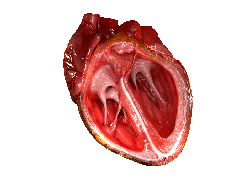
Uma válvula cardíaca é uma válvula biológica de sentido único que permite que o sangue flua em uma direção através das câmaras do coração. O coração de um mamífero geralmente possui quatro válvulas. Juntas, as válvulas determinam a direção do fluxo sanguíneo através do coração. As válvulas cardíacas são abertas ou fechadas por uma diferença na pressão sanguínea em cada lado.
Introdução
O que você precisa saber de cara
Visão geral
A Anomalia do aparelho mitral subvalvar é uma doença rara do desenvolvimento que afeta as estruturas responsáveis por sustentar e controlar o movimento da válvula mitral do coração. Essa condição está classificada no banco de dados de doenças raras MONDO sob o código MONDO:0015109 e na Classificação Internacional de Doenças (CID-10) como Q23.8. Por ser uma condição pouco frequente, ainda não há dados precisos sobre sua prevalência na população, padrão de herança ou idade habitual de aparecimento dos sintomas.[1][2]
Sinais e sintomas
Até o momento, não há uma lista padronizada de fenótipos (sinais e sintomas) registrada nas bases de dados oficiais para esta doença. Isso significa que as manifestações clínicas podem variar amplamente entre os pacientes e dependem do grau de comprometimento das estruturas subvalvares da válvula mitral. É importante que qualquer suspeita seja avaliada por um cardiologista especializado em doenças cardíacas congênitas.[1]
Causas genéticas
A causa genética específica da Anomalia do aparelho mitral subvalvar ainda não foi identificada. As bases de dados científicas não listam genes associados confirmados, e não há variantes patogênicas registradas no ClinVar para esta condição. A pesquisa genética continua em andamento para esclarecer os mecanismos hereditários envolvidos.[1][3]
Diagnóstico
O diagnóstico é baseado na avaliação clínica e em exames de imagem do coração, como ecocardiograma, que permite visualizar as alterações nas estruturas subvalvares da válvula mitral. Não há um teste genético específico para confirmar a doença, mas exames genéticos podem ser solicitados para excluir outras condições que cursam com anomalias cardíacas. Entre os procedimentos disponíveis no Sistema Único de Saúde (SUS) para investigação estão: cariótipo com bandas G, Q ou R; pesquisa de microdeleções/microduplicações por FISH; sequenciamento completo do exoma (WES); e dosagem de alfa-fetoproteína. O nível de cobertura pelo SUS é classificado como 'Cobertura mínima'.[1][3]
Tratamento e manejo
O tratamento da Anomalia do aparelho mitral subvalvar depende da gravidade do comprometimento da válvula mitral e dos sintomas apresentados. As opções podem incluir acompanhamento clínico regular, uso de medicamentos para controlar sintomas de insuficiência cardíaca (quando presente) e, em casos mais graves, cirurgia cardíaca para reparo ou substituição da válvula mitral. Não existem medicamentos específicos aprovados exclusivamente para esta condição. O SUS oferece atendimento em reabilitação para doenças raras como parte do suporte multidisciplinar.[1]
Prognóstico e qualidade de vida
O prognóstico é variável e depende da gravidade da anomalia, da presença de outras malformações cardíacas associadas e da resposta ao tratamento. Com acompanhamento médico adequado e, quando necessário, intervenção cirúrgica, muitos pacientes podem manter boa qualidade de vida. É fundamental o seguimento regular com cardiologista especializado em cardiopatias congênitas.[1]
Conteúdo informativo gerado e mantido automaticamente a partir de fontes oficiais (Orphanet, HPO, OMIM, SUS). Não substitui avaliação médica.
Uma válvula cardíaca é uma válvula biológica de sentido único que permite que o sangue flua em uma direção através das câmaras do coração. O coração de um mamífero geralmente possui quatro válvulas. Juntas, as válvulas determinam a direção do fluxo sanguíneo através do coração. As válvulas cardíacas são abertas ou fechadas por uma diferença na pressão sanguínea em cada lado.
Encontrou um erro ou informação desatualizada? Sugira uma correção →
Entender a doença
Do básico ao detalhe, leia no seu ritmo
Preparando trilha educativa...
Sinais e sintomas
O que aparece no corpo e com que frequência cada sintoma acontece
Visão geral
A Anomalia do aparelho mitral subvalvar é uma doença rara do desenvolvimento que afeta as estruturas responsáveis por sustentar e controlar o movimento da válvula mitral do coração. Essa condição está classificada no banco de dados de doenças raras MONDO sob o código MONDO:0015109 e na Classificação Internacional de Doenças (CID-10) como Q23.8. Por ser uma condição pouco frequente, ainda não há dados precisos sobre sua prevalência na população, padrão de herança ou idade habitual de aparecimento dos sintomas.[1][2]
Sinais e sintomas
Até o momento, não há uma lista padronizada de fenótipos (sinais e sintomas) registrada nas bases de dados oficiais para esta doença. Isso significa que as manifestações clínicas podem variar amplamente entre os pacientes e dependem do grau de comprometimento das estruturas subvalvares da válvula mitral. É importante que qualquer suspeita seja avaliada por um cardiologista especializado em doenças cardíacas congênitas.[1]
Causas genéticas
A causa genética específica da Anomalia do aparelho mitral subvalvar ainda não foi identificada. As bases de dados científicas não listam genes associados confirmados, e não há variantes patogênicas registradas no ClinVar para esta condição. A pesquisa genética continua em andamento para esclarecer os mecanismos hereditários envolvidos.[1][3]
Diagnóstico
O diagnóstico é baseado na avaliação clínica e em exames de imagem do coração, como ecocardiograma, que permite visualizar as alterações nas estruturas subvalvares da válvula mitral. Não há um teste genético específico para confirmar a doença, mas exames genéticos podem ser solicitados para excluir outras condições que cursam com anomalias cardíacas. Entre os procedimentos disponíveis no Sistema Único de Saúde (SUS) para investigação estão: cariótipo com bandas G, Q ou R; pesquisa de microdeleções/microduplicações por FISH; sequenciamento completo do exoma (WES); e dosagem de alfa-fetoproteína. O nível de cobertura pelo SUS é classificado como 'Cobertura mínima'.[1][3]
Tratamento e manejo
O tratamento da Anomalia do aparelho mitral subvalvar depende da gravidade do comprometimento da válvula mitral e dos sintomas apresentados. As opções podem incluir acompanhamento clínico regular, uso de medicamentos para controlar sintomas de insuficiência cardíaca (quando presente) e, em casos mais graves, cirurgia cardíaca para reparo ou substituição da válvula mitral. Não existem medicamentos específicos aprovados exclusivamente para esta condição. O SUS oferece atendimento em reabilitação para doenças raras como parte do suporte multidisciplinar.[1]
Prognóstico e qualidade de vida
O prognóstico é variável e depende da gravidade da anomalia, da presença de outras malformações cardíacas associadas e da resposta ao tratamento. Com acompanhamento médico adequado e, quando necessário, intervenção cirúrgica, muitos pacientes podem manter boa qualidade de vida. É fundamental o seguimento regular com cardiologista especializado em cardiopatias congênitas.[1]
Conteúdo informativo gerado e mantido automaticamente a partir de fontes oficiais (Orphanet, HPO, OMIM, SUS). Não substitui avaliação médica.
Linha do tempo da pesquisa
Encontrou um erro ou informação desatualizada? Sugira uma correção →
Genética e causas
O que está alterado no DNA e como passa nas famílias
Nenhum gene associado encontrado
Os dados genéticos desta condição ainda estão sendo catalogados.
Diagnóstico
Os sinais que médicos procuram e os exames que confirmam
Tratamento e manejo
Remédios, cuidados de apoio e o que precisa acompanhar
Onde tratar no SUS
Hospitais de referência no Brasil e o protocolo oficial do SUS (PCDT)
🇧🇷 Atendimento SUS — Anomalia do aparelho mitral subvalvar
Centros de Referência SUS
24 centros habilitados pelo SUS para Anomalia do aparelho mitral subvalvar
Centros para Anomalia do aparelho mitral subvalvar
Detalhes dos centros
Hospital Universitário Prof. Edgard Santos (HUPES)
R. Dr. Augusto Viana, s/n - Canela, Salvador - BA, 40110-060 · CNES 0003808
Serviço de Referência
Hospital Infantil Albert Sabin
R. Tertuliano Sales, 544 - Vila União, Fortaleza - CE, 60410-794 · CNES 2407876
Serviço de Referência
Hospital de Apoio de Brasília (HAB)
AENW 3 Lote A Setor Noroeste - Plano Piloto, Brasília - DF, 70684-831 · CNES 0010456
Serviço de Referência
Hospital Estadual Infantil e Maternidade Alzir Bernardino Alves (HIABA)
Av. Min. Salgado Filho, 918 - Soteco, Vila Velha - ES, 29106-010 · CNES 6631207
Serviço de Referência
Hospital das Clínicas da UFG
Rua 235 QD. 68 Lote Área, Nº 285, s/nº - Setor Leste Universitário, Goiânia - GO, 74605-050 · CNES 2338424
Serviço de Referência
Hospital Universitário da UFJF
R. Catulo Breviglieri, Bairro - s/n - Santa Catarina, Juiz de Fora - MG, 36036-110 · CNES 2297442
Atenção Especializada
Hospital das Clínicas da UFMG
Av. Prof. Alfredo Balena, 110 - Santa Efigênia, Belo Horizonte - MG, 30130-100 · CNES 2280167
Serviço de Referência
Hospital Universitário Julio Müller (HUJM)
R. Luis Philippe Pereira Leite, s/n - Alvorada, Cuiabá - MT, 78048-902 · CNES 2726092
Atenção Especializada
Hospital Universitário João de Barros Barreto
R. dos Mundurucus, 4487 - Guamá, Belém - PA, 66073-000 · CNES 2337878
Serviço de Referência
Hospital Universitário Lauro Wanderley (HULW)
R. Tabeliao Estanislau Eloy, 585 - Castelo Branco, João Pessoa - PB, 58050-585 · CNES 0002470
Atenção Especializada
Instituto de Medicina Integral Prof. Fernando Figueira (IMIP)
R. dos Coelhos, 300 - Boa Vista, Recife - PE, 50070-902 · CNES 0000647
Serviço de Referência
Hospital Pequeno Príncipe
R. Des. Motta, 1070 - Água Verde, Curitiba - PR, 80250-060 · CNES 3143805
Serviço de Referência
Hospital Universitário Regional de Maringá (HUM)
Av. Mandacaru, 1590 - Parque das Laranjeiras, Maringá - PR, 87083-240 · CNES 2216108
Atenção Especializada
Hospital de Clínicas da UFPR
R. Gen. Carneiro, 181 - Alto da Glória, Curitiba - PR, 80060-900 · CNES 2364980
Serviço de Referência
Hospital Universitário Pedro Ernesto (HUPE-UERJ)
Blvd. 28 de Setembro, 77 - Vila Isabel, Rio de Janeiro - RJ, 20551-030 · CNES 2280221
Serviço de Referência
Instituto Nacional de Saúde da Mulher, da Criança e do Adolescente Fernandes Figueira (IFF/Fiocruz)
Av. Rui Barbosa, 716 - Flamengo, Rio de Janeiro - RJ, 22250-020 · CNES 2269988
Serviço de Referência
Hospital São Lucas da PUCRS
Av. Ipiranga, 6690 - Jardim Botânico, Porto Alegre - RS, 90610-000 · CNES 2232928
Serviço de Referência
Hospital de Clínicas de Porto Alegre (HCPA)
Rua Ramiro Barcelos, 2350 Bloco A - Av. Protásio Alves, 211 - Bloco B e C - Santa Cecília, Porto Alegre - RS, 90035-903 · CNES 2237601
Serviço de Referência
Hospital Universitário da UFSC (HU-UFSC)
R. Profa. Maria Flora Pausewang - Trindade, Florianópolis - SC, 88036-800 · CNES 2560356
Serviço de Referência
Hospital das Clínicas da FMUSP
R. Dr. Ovídio Pires de Campos, 225 - Cerqueira César, São Paulo - SP, 05403-010 · CNES 2077485
Serviço de Referência
Hospital de Base de São José do Rio Preto
Av. Brg. Faria Lima, 5544 - Vila Sao Jose, São José do Rio Preto - SP, 15090-000 · CNES 2079798
Atenção Especializada
Hospital de Clínicas da UNICAMP
R. Vital Brasil, 251 - Cidade Universitária, Campinas - SP, 13083-888 · CNES 2748223
Serviço de Referência
Hospital de Clínicas de Ribeirão Preto (HCRP-USP)
R. Ten. Catão Roxo, 3900 - Vila Monte Alegre, Ribeirão Preto - SP, 14015-010 · CNES 2082187
Serviço de Referência
UNIFESP / Hospital São Paulo
R. Napoleão de Barros, 715 - Vila Clementino, São Paulo - SP, 04024-002 · CNES 2688689
Serviço de Referência
Dados de DATASUS/CNES, SBGM, ABNeuro e Ministério da Saúde. Sempre confirme a disponibilidade diretamente com o estabelecimento.
Pesquisa ativa
Ensaios clínicos abertos e novidades científicas recentes
Pesquisa e ensaios clínicos
Nenhum ensaio clínico registrado para esta condição.
Publicações mais relevantes
Congenital Mitral Rings: Insights From a Single-Institution Study and Review of Current Literature.
BackgroundCongenital mitral rings represent a rare subset of congenital mitral stenosis often associated with complex cardiac anomalies. Comprehensive literature on congenital mitral rings, particularly from large single-institution experiences, remains sparse. Methods: This retrospective study, conducted at All India Institute of Medical Sciences, New Delhi, analyzed data from 52 patients who underwent surgical correction for congenital mitral rings between 2014 and 2024. Data on demographics, clinical presentation, preoperative echocardiographic and cardiac catheterization parameters, associated cardiac anomalies, intraoperative findings, and postoperative outcomes were collected. Patients had a mean follow-up of 72 months.ResultsThe cohort had a mean age of 6 years. An isolated vestibular mitral ring was noted in only 1 patient, with 51 cases showing associated cardiac anomalies. The mean preoperative diastolic gradient was 13 mm Hg, significantly reduced to 2.69 mm Hg in the immediate postoperative period and 3.35 mm Hg at follow-up. Two distinct morphological types were identified: "distinct vestibular supramitral ring" (43 patients) and "adherent intramitral ring" (9 patients), with the latter demonstrating a higher association with subvalvular apparatus abnormalities. Preoperative transesophageal echocardiography proved crucial in detecting these rings in 4 cases where the diagnosis was doubtful on transthoracic echocardiography. Surgical outcomes were favorable, with 1 early postoperative mortality and 3 late mortalities. Four patients required reinterventions for residual lesions, achieving successful resolution.ConclusionThis large single institution series provides valuable insights into associated anomalies, morphological variants, and long-term surgical outcomes of congenital mitral rings. Accurate diagnosis and timely surgical intervention led to favorable outcomes, reinforcing the need for thorough preoperative evaluation.
Obstructive hypertrophic cardiomyopathy: pathophysiology and diagnosis.
Hypertrophic cardiomyopathy (HCM) is a genetic cardiac disorder characterized predominantly by left ventricular (LV) hypertrophy, frequently leading to dynamic obstruction of the left ventricular outflow tract (LVOT). Obstructive HCM is driven by structural abnormalities including asymmetric septal hypertrophy, systolic anterior motion of often elongated mitral valve leaflets, and alterations in the mitral sub-valvular apparatus such as displaced or hypertrophied papillary muscles. Pathophysiological mechanisms underlying HCM include hypercontractility due to increased actin-myosin cross bridges, myocyte hypertrophy and disarray, interstitial fibrosis, and coronary microvascular dysfunction-which contribute variably to disease expression, impaired myocardial relaxation, ischemia, fibrosis, and arrhythmogenesis. Diagnosis relies on integrating clinical presentation, physical examination, electrocardiographic features, genetic testing, and advanced imaging techniques. Transthoracic echocardiography remains the primary diagnostic and monitoring tool, accurately assessing patterns of hypertrophy, dynamic LVOT gradients, mitral valve abnormalities, and ventricular function, including strain imaging for early functional impairment. Cardiac magnetic resonance imaging complements echocardiography, providing superior anatomical delineation, precise quantification of LV mass, detection of apical and distal-dominant forms, identification of fibrosis via late gadolinium enhancement, and detailed tissue characterization. This review emphasizes the complex interplay of genetic, structural, and functional elements in obstructive HCM, underscoring the importance of comprehensive evaluation to facilitate individualized and effective therapeutic decisions.
Anterior mitral valve leaflet length and response to mavacamten in obstructive hypertrophic cardiomyopathy.
This study examines whether anterior mitral valve leaflet (AMVL) length is associated with response to mavacamten in patients with obstructive hypertrophic cardiomyopathy (HCM). Obstruction of the left-ventricular outflow tract (LVOT) in HCM has been associated with asymmetric septal hypertrophy and abnormalities of the mitral valve and sub-valvular apparatus. Mavacamten is a myosin-inhibitor shown to decrease LVOT gradient and improve functional status in patients with obstructive HCM. Measurements of cardiac structural elements were obtained from magnetic resonance imaging and echocardiography data among patients with obstructive HCM treated with mavacamten. Endpoints were effective mavacamten dose, defined as the dose required to achieve a Valsalva LVOT gradient <30 mmHg, and rapid response to mavacamten therapy, defined as achieved Valsalva LVOT gradient <20 mmHg within 8 weeks of initiation. Among 33 patients, patients with an effective dose of 5 mg (n = 13) had a shorter AMVL length [20.00 (18.50, 20.80) mm] compared with patients with a dose of 10 mg (n = 12) [23.30 (22.45, 26.10) mm] and 15 mg (n = 8) [25.45 (24.20, 26.85) mm] (P < 0.001). After adjusting for age and sex, the 5 mg dose was associated with a shorter AMVL length (P = 0.003). AMVL length was shorter in rapid responders [20.9 (19.9, 22.5) mm] compared with patients without a rapid response [24.9 (23.3, 26.5) mm] (P = 0.006). Shorter AMVL length is associated with a lower effective dose and a rapid response to mavacamten. If confirmed in larger studies, AMVL length may inform optimal dosing of myosin inhibitors in obstructive HCM.
Managing The Mitral Valve In HCM Surgery: Tips And Tricks.
Mitral regurgitation (MR) in hypertrophic cardiomyopathy (HCM) patients is mainly due to systolic anterior motion (SAM) of the mitral valve (MV). However, other mechanisms contributing to mitral regurgitation may coexist as a result of further structural abnormalities. SAM might occur because of the increased septal thickness alone or due to simultaneous MV or subvalvular apparatus anomalies, such as mitral leaflet elongation, papillary muscle body anomalies, accessory papillary muscles or additional papillary muscle heads. Additionally, anomalous mitral chordae or the recently described mitral-aortic discontinuity (leading to a longer anterior mitral leaflet (AML)) can contribute to abnormal physiology. A closed aortomitral angle may also contribute. During intraoperative echocardiographic assessment, it is important to thoroughly evaluate the MV and the regurgitant jet to understand the mechanism(s) that cause MR in HCM patients. Although myectomy alone is frequently enough to correct SAM, concomitant MV procedures may be needed, especially when the septum is thin (<16-18 mm) and/or there is intrinsic MV disease. Detection of concomitant regurgitation mechanisms beyond SAM can eventually be identified preoperatively, either by direct structural detection (valve prolapse), by pharmacological palliation of SAM with vasopressors and negative inotropic agents or suspected by identification of anteriorly and centrally directed regurgitant mitral jets. Surgical techniques that can be employed to contribute to SAM elimination include plication/extension/retention plasty of the AML, resection/release/reorientation of papillary muscles, division of anomalous chordae, edge-to-edge repair, or, at times, prosthetic MV replacement. If there is structural MV disease concomitant to HCM, appropriately tailored techniques to address the MV may be used. Transoesophageal echocardiography at the end of the procedure should demonstrate elimination of SAM, resolution of LVOT obstruction, and appropriate coaptation of the MV leaflets and nearly resolution of MR. Provocation with inotropes can be used to ensure no latent obstruction persists.
Transcatheter edge-to-edge repair for arcade-like mitral apparatus: expanding treatment options and assessing clinical outcome.
The arcade-like mitral apparatus is a rare congenital anomaly characterized by complex subvalvular pathology, often resulting in mitral dysfunction. These anatomical complexities make conventional surgical interventions particularly challenging, especially for elderly high-risk patients. For these patients, less invasive options like Transcatheter Edge-to-Edge Repair (TEER) present a promising alternative, addressing both anatomical challenges and procedural risks. To assess the feasibility, procedural success, safety, and clinical outcomes of TEER in patients with isolated severe MR due to arcade-like mitral apparatus. This case series involved four high-risk patients with isolated severe mitral regurgitation (MR) secondary to arcade-like mitral apparatus treated with TEER between August 2022 and August 2023. Each patient was evaluated by a multidisciplinary Heart Team to ensure optimal selection and procedural planning. Detailed anatomical assessment using advanced imaging techniques, was performed to customize the approach and ensure procedural success. The MitraClip XTR device was employed in all cases, with careful attention to patient-specific anatomical challenges. TEER was successfully performed in all patients, with immediate and sustained reductions in MR severity. At the one-year follow-up, all patients demonstrated improved cardiac function, an increase in New York Heart Association (NYHA) functional class, and a reduction in Borg dyspnea scores. Significant improvements in myocardial mechanics and work parameters were observed. Global Longitudinal Strain (GLS) improved significantly compared to baseline. The Global Work Index (GWI), Global Constructive Work (GCW), Global Pressure-Volume Work (GPW), Global Systolic Constructive Work (GSCW) and Global Work Efficiency (GWE) also showed a marked increase. TEER represents a promising, minimally invasive option for managing severe MR due to arcade-like mitral apparatus in high-risk patients. This case series underscores TEER's potential to offer significant symptom relief and improved hemodynamics, presenting a new therapeutic perspective for treating isolated MR in this anatomically challenging condition. Further large-scale studies are warranted to validate these findings and establish TEER's role in broader clinical practice.
📚 EuropePMCmostrando 42
Congenital Mitral Rings: Insights From a Single-Institution Study and Review of Current Literature.
World journal for pediatric & congenital heart surgeryObstructive hypertrophic cardiomyopathy: pathophysiology and diagnosis.
Indian journal of thoracic and cardiovascular surgeryManaging The Mitral Valve In HCM Surgery: Tips And Tricks.
Portuguese journal of cardiac thoracic and vascular surgeryTranscatheter edge-to-edge repair for arcade-like mitral apparatus: expanding treatment options and assessing clinical outcome.
Frontiers in cardiovascular medicineAnterior mitral valve leaflet length and response to mavacamten in obstructive hypertrophic cardiomyopathy.
European heart journal. Imaging methods and practiceA surgeon's toolkit for mitral valve-induced left ventricular outflow tract obstruction with minimal septal hypertrophy.
The Journal of thoracic and cardiovascular surgeryThree-dimensional, totally endoscopic mitral valve repair of anomalous mitral arcade.
Multimedia manual of cardiothoracic surgery : MMCTSDouble Orifice Mitral Valve: Two Patients with Contrasting Presentations.
Journal of cardiovascular echographyA rare presentation of an accessory mitral valve chordae.
The international journal of cardiovascular imagingMidterm Outcomes: A Comprehensive Approach to Surgery for Hypertrophic Obstructive Cardiomyopathy.
The Annals of thoracic surgeryIn vivo mitral valve repair for the transplanted donor heart in orthotopic heart transplantation.
Journal of cardiothoracic surgeryInvasive therapies for symptomatic obstructive hypertrophic cardiomyopathy.
Progress in cardiovascular diseasesPercutaneous transluminal septal myocardial ablation was effective in hypertrophic obstructive cardiomyopathy with anomalous mitral papillary muscles: a case report.
European heart journal. Case reportsSurgical Management for Systolic Anterior Motion (SAM) of the Mitral Valve in Obstructive Hypertrophic Myopathy.
Annals of thoracic and cardiovascular surgery : official journal of the Association of Thoracic and Cardiovascular Surgeons of AsiaSeptal Thickness Does Not Impact Outcome After Hypertrophic Obstructive Cardiomyopathy Surgery (Septal Myectomy and Subvalvular Mitral Apparatus Remodeling): A 15-Years of Experience.
Frontiers in cardiovascular medicineA Case of Transient Mitral Regurgitation: Not Everything Is Always What It Seems.
Journal of cardiothoracic and vascular anesthesiaSurgery for Hypertrophic Obstructive Cardiomyopathy: Comprehensive LVOT Management beyond Septal Myectomy.
Journal of clinical medicineComprehensive left ventricular outflow tract management beyond septal reduction to relieve obstruction.
Asian cardiovascular & thoracic annalsCommentary: "Postoperative Changes in Left Ventricular Systolic Function after Combined Mitral and Aortic Valve Replacement in Patients with Rheumatic Heart Disease" Sang-Mee An, MD, Jae-Sik Nam, MD, Ho Jin Kim, MD, PhD, Hyeun Joon Bae, MD, Ji-Hyun Chin, MD, PhD, Eun-Ho Lee, MD, PhD, In-Cheol Choi, MD, PhD.
Journal of cardiac surgeryTransaortic extended left ventricular septal myectomy in an adult with hypertrophic obstructive cardiomyopathy.
Multimedia manual of cardiothoracic surgery : MMCTSTHE PLACE OF CARDIAC COMPUTED TOMOGRAPHY IN PREOPERATIVE PLANNING OF EXTENDED SEPTAL MYECTOMY IN PATIENTS WITH OBSTRUCTIVE FORM OF HYPERTROPHIC CARDIOMYOMATHY.
Problemy radiatsiinoi medytsyny ta radiobiolohiiSurgery for Anomalous Papillary Muscle Directly Into the Anterior Mitral Leaflet.
The Annals of thoracic surgeryCongenital double mitral orifice with severe mitral regurgitation-associated rheumatoid arthritis: a case report.
European heart journal. Case reportsAberrant right coronary artery in a grown up congenital cardiac patient, successfully treated 46 years earlier with a double Starr-Edwards silastic ball valve replacement: a case report.
BMC cardiovascular disordersIsolated anterior mitral cleft.
Echocardiography (Mount Kisco, N.Y.)Muscular Mitral Chord Contribution to Left Ventricular Outflow Tract Obstruction in HOCM.
The Thoracic and cardiovascular surgeon reportsStructural abnormalities in hypertrophic cardiomyopathy beyond left ventricular hypertrophy by multimodality imaging evaluation.
Echocardiography (Mount Kisco, N.Y.)Double orifice and atrioventricular septal defect: dealing with the zone of apposition†.
European journal of cardio-thoracic surgery : official journal of the European Association for Cardio-thoracic SurgerySeptal Myectomy With Vs Without Subvalvular Apparatus Intervention in Patients With Hypertrophic Obstructive Cardiomyopathy: A Prospective Randomized Study.
Seminars in thoracic and cardiovascular surgeryStarfish in the heart: Congenital anomaly of the papillary muscles.
Echocardiography (Mount Kisco, N.Y.)Intraoperative Two- and Three-Dimensional Transesophageal Echocardiography in Combined Myectomy-Mitral Operations for Hypertrophic Cardiomyopathy.
Journal of the American Society of Echocardiography : official publication of the American Society of EchocardiographyTransmitral Septal Myectomy for Hypertrophic Obstructive Cardiomyopathy.
The Annals of thoracic surgeryAnatomy of the mitral subvalvular apparatus.
The Journal of thoracic and cardiovascular surgeryNew insights into mitral valve dystrophy: a Filamin-A genotype-phenotype and outcome study.
European heart journalPapillary muscle approximation in mitral valve repair for secondary MR.
Journal of thoracic diseaseBiomechanics raises solution to avoid geometric mitral valve configuration abnormalities in ischemic mitral regurgitation.
Journal of thoracic diseaseAssessment of mitral apparatus in patients with acute inferoposterior myocardial infarction and ischaemic mitral regurgitation with two-dimensional echocardiography from anatomically correct imaging planes.
Kardiologia polskaExercise Testing and Stress Imaging in Mitral Valve Disease.
Current treatment options in cardiovascular medicineAbnormalities of Mitral Subvalvular Apparatus in Hypertrophic Cardiomyopathy: Role of Intraoperative 3D Transesophageal Echocardiography.
Anesthesia and analgesiaComputed tomography imaging to quantify the area of the endocardial subvalvular apparatus in hypertrophic cardiomyopathy - Relationship to outflow tract obstruction and symptoms.
Journal of cardiovascular computed tomographySurgery for Congenital Tricuspid Valve Cleft: Tricuspid Valve Repair with Neochordae and Annuloplasty.
The Journal of heart valve diseaseThree-Dimensional Transesophageal Echocardiography in the Anatomical Assessment of Isolated Parachute Mitral Valve in an Adult Patient.
Echocardiography (Mount Kisco, N.Y.)Associações
Organizações que acompanham esta doença — pra ter apoio e orientação
Ainda não temos associações cadastradas para Anomalia do aparelho mitral subvalvar.
É de uma associação que acompanha esta doença? Fale com a gente →
Comunidades
Grupos ativos de quem convive com esta doença aqui no Raras
Ainda não existe comunidade no Raras para Anomalia do aparelho mitral subvalvar
Pacientes, familiares e cuidadores se organizam em comunidades pra compartilhar experiências, fazer perguntas e se apoiar. Você pode ser o primeiro.
Tire suas dúvidas
Perguntas, dicas e experiências compartilhadas aqui na página
Participe da discussão
Faça login para postar dúvidas, compartilhar experiências e interagir com especialistas.
Fazer loginDoenças relacionadas
Doenças com sintomas parecidos — ajudam quem ainda está buscando diagnóstico
Ainda não achamos doenças com sintomas parecidos o suficiente.
Referências e fontes
Bases de dados externas citadas neste artigo
Publicações científicas
Artigos indexados no PubMed ligados a esta doença no grafo RarasNet — título, periódico e PMID direto da fonte, sem intermediação de IA.
- Congenital Mitral Rings: Insights From a Single-Institution Study and Review of Current Literature.
- Obstructive hypertrophic cardiomyopathy: pathophysiology and diagnosis.
- Anterior mitral valve leaflet length and response to mavacamten in obstructive hypertrophic cardiomyopathy.
- Managing The Mitral Valve In HCM Surgery: Tips And Tricks.
- Transcatheter edge-to-edge repair for arcade-like mitral apparatus: expanding treatment options and assessing clinical outcome.
Bases de dados e fontes oficiais
Identificadores e referências canônicas usadas para montar este verbete.
- ORPHA:101932(Orphanet)
- MONDO:0015109(MONDO)
- GARD:19784(GARD (NIH))
- Q55785257(Wikidata)
Dados compilados pelo RarasNet a partir de fontes abertas (Orphanet, OMIM, MONDO, PubMed/EuropePMC, ClinicalTrials.gov, DATASUS, PCDT/MS). Este conteúdo é informativo e não substitui avaliação médica.
Conteúdo mantido por Agente Raras · Médicos e pesquisadores podem colaborar
Anomalia do aparelho mitral subvalvar
📋 Origem dos dados
Esta página agrega dados de fontes públicas e oficiais. Dados sobre cobertura no SUS (PCDT, CEAF) são verificados ativamente por agente proativo (ver badge no infobox). Demais dados têm atribuição de fonte + data da última sincronização — clique para abrir o original.
- Doença rara (ontologia)
- fonte: Orphanet
- Identificador unificado
- fonte: MONDO
- Codificação WHO/SUS
- fonte: WHO ICD-10 / DATASUS
- CID-11 (futuro)
- fonte: WHO ICD-11
- NIH/GARD
- fonte: GARD (NIH)
- Dado público estruturado
- fonte: Wikidata