A distrofia muscular (DM) refere-se a um grupo de mais de 30 doenças genéticas caracterizadas por fraqueza progressiva e degeneração dos músculos esqueléticos que controlam o movimento. Algumas formas de DM são observadas em recém-nascidos, bebés ou crianças, enquanto outras têm início tardio e podem não aparecer até à meia-idade ou mais tarde. Os distúrbios diferem em termos de distribuição e extensão da fraqueza muscular (algumas formas de DM também afetam o músculo cardíaco), idade de início, taxa de progressão e padrão de herança. O prognóstico para pessoas com DM varia de acordo com o tipo e progressão do transtorno. Não existe tratamento específico para interromper ou reverter qualquer forma de DM. O tratamento é de suporte e pode incluir fisioterapia, terapia respiratória, fonoaudiologia, aparelhos ortopédicos usados para suporte, cirurgia ortopédica corretiva e medicamentos, incluindo corticosteróides, anticonvulsivantes (medicamentos para convulsões), imunossupressores e antibióticos. Alguns indivíduos podem precisar de ventilação assistida para tratar fraqueza muscular respiratória ou de marcapasso para anormalidades cardíacas.
Introdução
O que você precisa saber de cara
A distrofia muscular (DM) refere-se a um grupo de mais de 30 doenças genéticas caracterizadas por fraqueza progressiva e degeneração dos músculos esqueléticos que controlam o movimento. Algumas formas de DM são observadas em recém-nascidos, bebés ou crianças, enquanto outras têm início tardio e podem não aparecer até à meia-idade ou mais tarde. Os distúrbios diferem em termos de distribuição e extensão da fraqueza muscular (algumas formas de DM também afetam o músculo cardíaco), idade de início, taxa de progressão e padrão de herança. O prognóstico para pessoas com DM varia de acordo com o tipo e progressão do transtorno. Não existe tratamento específico para interromper ou reverter qualquer forma de DM. O tratamento é de suporte e pode incluir fisioterapia, terapia respiratória, fonoaudiologia, aparelhos ortopédicos usados para suporte, cirurgia ortopédica corretiva e medicamentos, incluindo corticosteróides, anticonvulsivantes (medicamentos para convulsões), imunossupressores e antibióticos. Alguns indivíduos podem precisar de ventilação assistida para tratar fraqueza muscular respiratória ou de marcapasso para anormalidades cardíacas.
Tem tratamento?
Encontrou um erro ou informação desatualizada? Sugira uma correção →
Entender a doença
Do básico ao detalhe, leia no seu ritmo
Preparando trilha educativa...
Sinais e sintomas
O que aparece no corpo e com que frequência cada sintoma acontece
Partes do corpo afetadas
+ 363 sintomas em outras categorias
Características mais comuns
Os sintomas variam de pessoa para pessoa. Abaixo estão as 962 características clínicas mais associadas, ordenadas por frequência.
Linha do tempo da pesquisa
Encontrou um erro ou informação desatualizada? Sugira uma correção →
Genética e causas
O que está alterado no DNA e como passa nas famílias
Genes associados
62 genes identificados com associação a esta condição.
Curadoria gene-doença
fontes oficiaisAnchors the extracellular matrix to the cytoskeleton via F-actin. Ligand for dystroglycan. Component of the dystrophin-associated glycoprotein complex which accumulates at the neuromuscular junction (NMJ) and at a variety of synapses in the peripheral and central nervous systems and has a structural function in stabilizing the sarcolemma. Also implicated in signaling events and synaptic transmission
Cell membrane, sarcolemmaCytoplasm, cytoskeletonPostsynaptic cell membrane
Duchenne muscular dystrophy
Most common form of muscular dystrophy; a sex-linked recessive disorder. It typically presents in boys aged 3 to 7 year as proximal muscle weakness causing waddling gait, toe-walking, lordosis, frequent falls, and difficulty in standing up and climbing up stairs. The pelvic girdle is affected first, then the shoulder girdle. Progression is steady and most patients are confined to a wheelchair by age of 10 or 12. Flexion contractures and scoliosis ultimately occur. About 50% of patients have a lower IQ than their genetic expectations would suggest. There is no treatment.
Type XII collagen interacts with type I collagen-containing fibrils, the COL1 domain could be associated with the surface of the fibrils, and the COL2 and NC3 domains may be localized in the perifibrillar matrix
Secreted, extracellular space, extracellular matrix
Ullrich congenital muscular dystrophy 2
A form of Ullrich congenital muscular dystrophy, a disease characterized by generalized muscle weakness and striking hypermobility of distal joints in conjunction with variable contractures of more proximal joints and normal intelligence. Additional findings may include kyphoscoliosis, protruded calcanei, and follicular hyperkeratosis (rough skin). More severely affected patients manifest at birth and never achieve independent ambulation, while patients with milder phenotypes might maintain ambulation into adulthood. UCMD2 is a severe, autosomal recessive form with onset at birth.
Stabilizer subunit of the dolichol-phosphate mannose (DPM) synthase complex; tethers catalytic subunit DPM1 to the endoplasmic reticulum
Endoplasmic reticulum membrane
Muscular dystrophy-dystroglycanopathy congenital with impaired intellectual development B15
An autosomal recessive, congenital muscular disorder characterized by hyperCKemia, myopathic features observed on muscle biopsy, developmental delay, mildly impaired intellectual development with learning difficulties, epilepsy, and mild white matter abnormalities.
Participates in O-mannosyl glycosylation by catalyzing the addition of N-acetylglucosamine to O-linked mannose on glycoproteins (PubMed:11709191, PubMed:27493216, PubMed:28512129). Catalyzes the synthesis of the GlcNAc(beta1-2)Man(alpha1-)O-Ser/Thr moiety on alpha-dystroglycan and other O-mannosylated proteins, providing the necessary basis for the addition of further carbohydrate moieties (PubMed:11709191, PubMed:27493216). Is specific for alpha linked terminal mannose and does not have MGAT3,
Golgi apparatus membrane
Muscular dystrophy-dystroglycanopathy congenital with brain and eye anomalies A3
An autosomal recessive disorder characterized by congenital muscular dystrophy, ocular abnormalities, cobblestone lissencephaly, and cerebellar and pontine hypoplasia. Patients present severe congenital myopia, congenital glaucoma, pallor of the optic disks, retinal hypoplasia, intellectual disability, hydrocephalus, abnormal electroencephalograms, generalized muscle weakness and myoclonic jerks. Included diseases are the more severe Walker-Warburg syndrome and the slightly less severe muscle-eye-brain disease.
Interlinks intermediate filaments with microtubules and microfilaments and anchors intermediate filaments to desmosomes or hemidesmosomes. Could also bind muscle proteins such as actin to membrane complexes in muscle. May be involved not only in the filaments network, but also in the regulation of their dynamics. Structural component of muscle. Isoform 9 plays a major role in the maintenance of myofiber integrity
Cytoplasm, cytoskeletonCell junction, hemidesmosomeCell projection, podosome
Epidermolysis bullosa simplex 5C, with pyloric atresia
A form of epidermolysis bullosa, a genodermatosis characterized by recurrent blistering, fragility of the skin and mucosal epithelia, and erosions caused by minor mechanical trauma. EBS5C is an autosomal recessive disorder characterized by severe skin blistering at birth and congenital pyloric atresia. Death usually occurs in infancy.
Broad substrate specificity ATP-dependent transporter of the ATP-binding cassette (ABC) family that catalyzes the transport of long-chain fatty acids (LCFA)-CoA, dicarboxylic acids-CoA, long-branched-chain fatty acids-CoA and bile acids from the cytosol to the peroxisome lumen for beta-oxydation (PubMed:11248239, PubMed:24333844, PubMed:25168382, PubMed:29397936). Has fatty acyl-CoA thioesterase and ATPase activities (PubMed:29397936). Probably hydrolyzes fatty acyl-CoAs into free fatty acids pr
Peroxisome membrane
Congenital bile acid synthesis defect 5
An autosomal recessive disorder characterized by hepatosplenomegaly, hepatic fibrosis, progressive liver failure, and accumulation of peroxisomal C27-bile acid intermediates in plasma.
Has a key role in phospholipid metabolism, and catalyzes the first step of phosphatidylethanolamine and phosphatidylcholine biosynthesis
Muscular dystrophy, congenital, megaconial type
An autosomal recessive, congenital muscular dystrophy characterized by early-onset muscle wasting, intellectual disability, and dilated cardiomyopathy in half of affected individuals. Some patients may die from cardiomyopathy in the first or second decade of life. Muscle biopsy shows peculiar enlarged mitochondria that are prevalent toward the periphery of the fibers but are sparse in the center.
Bifunctional glycosyltransferase with both alpha-1,3-xylosyltransferase and beta-1,3-glucuronyltransferase activities involved in the maturation of alpha-dystroglycan (DAG1) by glycosylation leading to DAG1 binding to laminin G-like domain-containing extracellular proteins with high affinity (PubMed:15661757, PubMed:15752776, PubMed:21987822, PubMed:22223806, PubMed:23125099, PubMed:25279697, PubMed:25279699). Elongates the glucuronyl-beta-1,4-xylose-beta disaccharide primer structure initiated
Golgi apparatus membrane
Muscular dystrophy-dystroglycanopathy congenital with impaired intellectual development B6
A congenital muscular dystrophy associated with profound intellectual disability, white matter changes and structural brain abnormalities. Skeletal muscle biopsies show reduced immunolabeling of alpha-dystroglycan.
Key regulator of transforming growth factor beta (TGFB1, TGFB2 and TGFB3) that controls TGF-beta activation by maintaining it in a latent state during storage in extracellular space. Associates specifically via disulfide bonds with the Latency-associated peptide (LAP), which is the regulatory chain of TGF-beta, and regulates integrin-dependent activation of TGF-beta
Secreted, extracellular space, extracellular matrix
Urban-Rifkin-Davis syndrome
A syndrome characterized by disrupted pulmonary, gastrointestinal, urinary, musculoskeletal, craniofacial and dermal development. Clinical features include cutis laxa, mild cardiovascular lesions, respiratory distress with cystic and atelectatic changes in the lungs, and diverticulosis, tortuosity and stenosis at various levels of the intestinal tract. Craniofacial features include microretrognathia, flat midface, receding forehead and wide fontanelles.
Human-specific protein that promotes neural progenitor proliferation and evolutionary expansion of the brain neocortex by regulating the Notch signaling pathway (PubMed:29561261, PubMed:29856954, PubMed:29856955). Able to promote neural progenitor self-renewal, possibly by down-regulating neuronal differentiation genes, thereby delaying the differentiation of neuronal progenitors and leading to an overall final increase in neuronal production (PubMed:29856954). Acts by enhancing the Notch signal
Secreted
Neuronal intranuclear inclusion disease
An autosomal dominant, slowly progressive, neurodegenerative disease characterized by eosinophilic hyaline intranuclear inclusions in the central and peripheral nervous system, and also in the visceral organs. Clinical manifestations are variable and include pyramidal and extrapyramidal symptoms, cerebellar ataxia, cognitive decline and dementia, peripheral neuropathy, and autonomic dysfunction.
Processive microtubule plus-end directed motor protein involved in neuronal axon guidance. Is recruited by KANK1 to cortical microtubule stabilizing complexes (CMSCs) at focal adhesions (FAs) rims where it promotes microtubule capture and stability. Controls microtubule polymerization rate at axonal growth cones and suppresses microtubule growth without inducing microtubule disassembly once it reaches the cell cortex
Cytoplasm, cytoskeletonCytoplasm, cell cortexCell projection, axonCell projection, dendriteCell projection, growth cone
Fibrosis of extraocular muscles, congenital, 1
A congenital ocular motility disorder marked by restrictive ophthalmoplegia affecting extraocular muscles innervated by the oculomotor and/or trochlear nerves. It is clinically characterized by anchoring of the eyes in downward gaze, ptosis, and backward tilt of the head. Patients affected by congenital fibrosis of extraocular muscles type 1 show an absence of the superior division of the oculomotor nerve (cranial nerve III) and corresponding oculomotor subnuclei.
May be involved in G protein-linked signaling
CytoplasmMembrane
Oculopharyngodistal myopathy 2
A form of oculopharyngodistal myopathy, a muscle disorder characterized by progressive ptosis, external ophthalmoplegia, and weakness of the masseter, facial, pharyngeal, and distal limb muscles. The myopathological features are presence of rimmed vacuoles in the muscle fibers and myopathic changes of differing severity. OPDM2 inheritance pattern is autosomal dominant.
Involved in endoplasmic reticulum to Golgi apparatus trafficking at a very early stage
Golgi apparatusGolgi apparatus, cis-Golgi network
Muscular dystrophy, limb-girdle, autosomal recessive 18
A form of limb-girdle muscular dystrophy characterized by proximal muscle weakness with childhood onset, resulting in gait abnormalities and scapular winging. Serum creatine kinase is increased. A subset of patients may show a hyperkinetic movement disorder with chorea, ataxia, or dystonia and global developmental delay.
Calcium-regulated non-lysosomal thiol-protease. Proteolytically cleaves CTBP1 at 'His-409'. Mediates, with UTP25, the proteasome-independent degradation of p53/TP53 (PubMed:23357851, PubMed:27657329)
CytoplasmNucleus, nucleolus
Muscular dystrophy, limb-girdle, autosomal recessive 1
An autosomal recessive degenerative myopathy characterized by progressive symmetrical atrophy and weakness of the proximal limb muscles and elevated serum creatine kinase. The symptoms usually begin during the first two decades of life, and the disease gradually worsens, often resulting in loss of walking ability 10 or 20 years after onset.
Collagen VI acts as a cell-binding protein
Secreted, extracellular space, extracellular matrixMembrane
Bethlem myopathy 1B
A form of Bethlem myopathy, a slowly progressive muscular dystrophy characterized by joint contractures, most frequently affecting the elbows and ankles, and muscle weakness and wasting involving the proximal and extensor muscles more than the distal and flexor ones. The clinical onset more often occurs in childhood or adulthood, but it can be prenatal with decreased fetal movements or neonatal with hypotonia. The hallmark of Bethlem myopathy is long finger flexion contractures. Inheritance can be autosomal dominant or autosomal recessive.
RNA-binding protein involved in the regulation of alternative pre-RNA splicing and mRNA translation by binding to uridine-rich (U-rich) RNA sequences (PubMed:11106748, PubMed:12486009, PubMed:17488725, PubMed:8576255). Binds to U-rich sequences immediately downstream from a 5' splice sites in a uridine-rich small nuclear ribonucleoprotein (U snRNP)-dependent fashion, thereby modulating alternative pre-RNA splicing (PubMed:11106748, PubMed:8576255). Preferably binds to the U-rich IAS1 sequence in
NucleusCytoplasmCytoplasm, Stress granule
Welander distal myopathy
An autosomal dominant disorder characterized by adult onset of distal muscle weakness predominantly affecting the distal long extensors of the hands, with slow progression to involve all small hand muscles and the lower legs. Skeletal muscle biopsy shows myopathic changes and prominent rimmed vacuoles. Rare homozygous patients showed earlier onset, faster progression, and proximal muscle involvement.
May have an important role in maintaining nuclear envelope structure by organizing protein complexes at the inner nuclear membrane. Required for retaining emerin at the inner nuclear membrane (By similarity). Plays a role in the modulation of innate immune signaling through the cGAS-STING pathway by interacting with RNF26 (PubMed:32614325). In addition, functions as a critical signaling component in mediating NF-kappa-B activation by acting downstream of EGFR and upstream of CARD10 (PubMed:27991
Endoplasmic reticulum membraneNucleus inner membraneCell membrane
Arrhythmogenic right ventricular dysplasia, familial, 5
A congenital heart disease characterized by infiltration of adipose and fibrous tissue into the right ventricle and loss of myocardial cells, resulting in ventricular and supraventricular arrhythmias.
Molecular adapter required for selective macroautophagy (aggrephagy) by acting as a bridge between polyubiquitinated proteins and autophagosomes (PubMed:15340068, PubMed:15953362, PubMed:16286508, PubMed:17580304, PubMed:20168092, PubMed:22017874, PubMed:22622177, PubMed:24128730, PubMed:28404643, PubMed:29343546, PubMed:29507397, PubMed:31857589, PubMed:33509017, PubMed:34471133, PubMed:34893540, PubMed:35831301, PubMed:37306101, PubMed:37802024). Promotes the recruitment of ubiquitinated cargo
Cytoplasmic vesicle, autophagosomePreautophagosomal structureCytoplasm, cytosolNucleus, PML bodyLate endosomeLysosomeNucleusEndoplasmic reticulumCytoplasm, myofibril, sarcomere
Paget disease of bone 3
A disorder of bone remodeling characterized by increased bone turnover affecting one or more sites throughout the skeleton, primarily the axial skeleton. Osteoclastic overactivity followed by compensatory osteoblastic activity leads to a structurally disorganized mosaic of bone (woven bone), which is mechanically weaker, larger, less compact, more vascular, and more susceptible to fracture than normal adult lamellar bone.
Component of a complex of multiple actin cross-linking proteins. Involved in the control of myofibril assembly and stability at the Z lines in muscle cells
Cell membrane, sarcolemmaCytoplasm, cytoskeletonCytoplasm, myofibril, sarcomere, Z line
Myopathy, myofibrillar, 3
A form of myofibrillar myopathy, a group of chronic neuromuscular disorders characterized at ultrastructural level by disintegration of the sarcomeric Z disk and myofibrils, and replacement of the normal myofibrillar markings by small dense granules, or larger hyaline masses, or amorphous material. MFM3 is characterized by progressive skeletal muscle weakness greater distally than proximally, tight heel cords, hyporeflexia, cardiomyopathy and peripheral neuropathy in some patients. Affected muscle exhibits disorganization and streaming of the Z-line, presence of large hyaline structures, excessive accumulation of myotilin and other ectopically expressed proteins and prominent congophilic deposits.
Cytidylyltransferase required for protein O-linked mannosylation (PubMed:22522420, PubMed:22522421, PubMed:26687144, PubMed:26923585, PubMed:27130732, PubMed:27601598). Catalyzes the formation of CDP-ribitol nucleotide sugar from D-ribitol 5-phosphate (PubMed:26687144, PubMed:26923585, PubMed:27130732). CDP-ribitol is a substrate of FKTN during the biosynthesis of the phosphorylated O-mannosyl trisaccharide (N-acetylgalactosamine-beta-3-N-acetylglucosamine-beta-4-(phosphate-6-)mannose), a carboh
Cytoplasm, cytosol
Muscular dystrophy-dystroglycanopathy congenital with brain and eye anomalies A7
An autosomal recessive disorder characterized by congenital muscular dystrophy associated with cobblestone lissencephaly and other brain anomalies, eye malformations, profound intellectual disability, and death usually in the first years of life. Included diseases are the more severe Walker-Warburg syndrome and the slightly less severe muscle-eye-brain disease.
Transfers mannosyl residues to the hydroxyl group of serine or threonine residues. Coexpression of both POMT1 and POMT2 is necessary for enzyme activity, expression of either POMT1 or POMT2 alone is insufficient (PubMed:14699049, PubMed:28512129). Essentially dedicated to O-mannosylation of alpha-DAG1 and few other proteins but not of cadherins and protocaherins (PubMed:28512129)
Endoplasmic reticulum membrane
Muscular dystrophy-dystroglycanopathy congenital with brain and eye anomalies A2
An autosomal recessive disorder characterized by congenital muscular dystrophy associated with cobblestone lissencephaly and other brain anomalies, eye malformations, profound intellectual disability, and death usually in the first years of life. Included diseases are the more severe Walker-Warburg syndrome and the slightly less severe muscle-eye-brain disease.
Component of the sarcoglycan complex, a subcomplex of the dystrophin-glycoprotein complex which forms a link between the F-actin cytoskeleton and the extracellular matrix
Cell membrane, sarcolemmaCytoplasm, cytoskeleton
Muscular dystrophy, limb-girdle, autosomal recessive 4
An autosomal recessive degenerative myopathy characterized by pelvic and shoulder muscle wasting, onset usually in childhood and variable progression rate.
May contribute to the transparency and refractive index of the lens. Has chaperone-like activity, preventing aggregation of various proteins under a wide range of stress conditions. In lens epithelial cells, stabilizes the ATP6V1A protein, preventing its degradation by the proteasome (By similarity)
CytoplasmNucleusSecretedLysosome
Myopathy, myofibrillar, 2A, adult-onset
A form of myofibrillar myopathy, a group of chronic neuromuscular disorders characterized at ultrastructural level by disintegration of the sarcomeric Z disk and myofibrils, and replacement of the normal myofibrillar markings by small dense granules, or larger hyaline masses, or amorphous material. MFM2A is an autosomal dominant form characterized by weakness of the proximal and distal limb muscles, weakness of the neck, velopharynx and trunk muscles, respiratory insufficiency, hypertrophic cardiomyopathy, and cataract.
Inositol 5-phosphatase which acts on inositol 1,4,5-trisphosphate, inositol 1,3,4,5-tetrakisphosphate, phosphatidylinositol 4,5-bisphosphate and phosphatidylinositol 3,4,5-trisphosphate (PubMed:10753883, PubMed:16824732). Has 6-fold higher affinity for phosphatidylinositol 4,5-bisphosphate than for inositol 1,4,5-trisphosphate (PubMed:10753883). Negatively regulates assembly of the actin cytoskeleton. Controls insulin-dependent glucose uptake among inositol 3,4,5-trisphosphate phosphatases; ther
Endoplasmic reticulumCytoplasm
Muscular dystrophy, congenital, with cataracts and impaired intellectual development
An autosomal recessive form of muscular dystrophy with onset in early childhood and characterized by progressive muscle weakness. Almost all patients also have early-onset cataracts and intellectual disability of varying severity. Some patients have seizures.
Protein O-mannose kinase that specifically mediates phosphorylation at the 6-position of an O-mannose of the trisaccharide (N-acetylgalactosamine (GalNAc)-beta-1,3-N-acetylglucosamine (GlcNAc)-beta-1,4-mannose) to generate phosphorylated O-mannosyl trisaccharide (N-acetylgalactosamine-beta-1,3-N-acetylglucosamine-beta-1,4-(phosphate-6-)mannose). Phosphorylated O-mannosyl trisaccharide is a carbohydrate structure present in alpha-dystroglycan (DAG1), which is required for binding laminin G-like d
Endoplasmic reticulum membrane
Muscular dystrophy-dystroglycanopathy congenital with brain and eye anomalies A12
An autosomal recessive disorder characterized by congenital muscular dystrophy associated with cobblestone lissencephaly and other brain anomalies, eye malformations, profound intellectual disability, and death usually in the first years of life. Included diseases are the more severe Walker-Warburg syndrome and the slightly less severe muscle-eye-brain disease.
May act as a scaffolding protein within caveolar membranes. Interacts directly with G-protein alpha subunits and can functionally regulate their activity. May also regulate voltage-gated potassium channels. Plays a role in the sarcolemma repair mechanism of both skeletal muscle and cardiomyocytes that permits rapid resealing of membranes disrupted by mechanical stress (By similarity). Mediates the recruitment of CAVIN2 and CAVIN3 proteins to the caveolae (PubMed:19262564)
Golgi apparatus membraneCell membraneMembrane, caveolaCell membrane, sarcolemma
HyperCKmia
Characterized by persistent elevated levels of serum creatine kinase without muscle weakness.
Binding to cells via a high affinity receptor, laminin is thought to mediate the attachment, migration and organization of cells into tissues during embryonic development by interacting with other extracellular matrix components
Secreted, extracellular space, extracellular matrix, basement membrane
Merosin-deficient congenital muscular dystrophy 1A
Characterized by difficulty walking, hypotonia, proximal weakness, hyporeflexia, and white matter hypodensity on MRI.
Collagen VI acts as a cell-binding protein
Secreted, extracellular space, extracellular matrix
Bethlem myopathy 1C
A form of Bethlem myopathy, a slowly progressive muscular dystrophy characterized by joint contractures, most frequently affecting the elbows and ankles, and muscle weakness and wasting involving the proximal and extensor muscles more than the distal and flexor ones. The clinical onset more often occurs in childhood or adulthood, but it can be prenatal with decreased fetal movements or neonatal with hypotonia. The hallmark of Bethlem myopathy is long finger flexion contractures. BTHLM1C inheritance is autosomal dominant.
May have an involvement in muscle development or hypertrophy
CytoplasmNucleusCytoplasm, cytosol
Emery-Dreifuss muscular dystrophy 6, X-linked
A form of Emery-Dreifuss muscular dystrophy, a degenerative myopathy characterized by weakness and atrophy of muscle without involvement of the nervous system, early contractures of the elbows, Achilles tendons and spine, and cardiomyopathy associated with cardiac conduction defects.
Single-stranded DNA-binding protein that preferentially binds to the sterol regulatory element (SRE) sequence 5'-GTGCGGTG-3', and thereby mediates transcriptional repression (PubMed:2562787). Has a role as transactivator of the Myc promoter (By similarity). Binds single-stranded RNA in a sequence-specific manner (By similarity) Binds G-rich elements in target mRNA coding sequences (PubMed:28329689). Prevents G-quadruplex structure formation in vitro, suggesting a role in supporting translation b
NucleusCytoplasmEndoplasmic reticulum
Dystrophia myotonica 2
A multisystem disease characterized by the association of proximal muscle weakness with myotonia, cardiac manifestations and cataract. Additional features can include hyperhidrosis, testicular atrophy, insulin resistance and diabetes and central nervous system anomalies in rare cases.
O-linked mannose beta-1,4-N-acetylglucosaminyltransferase that transfers UDP-N-acetyl-D-glucosamine to the 4-position of the mannose to generate N-acetyl-D-glucosamine-beta-1,4-O-D-mannosylprotein. Involved in the biosynthesis of the phosphorylated O-mannosyl trisaccharide (N-acetylgalactosamine-beta-3-N-acetylglucosamine-beta-4-(phosphate-6-)mannose), a carbohydrate structure present in alpha-dystroglycan (DAG1), which is required for binding laminin G-like domain-containing extracellular prote
Endoplasmic reticulum membrane
Muscular dystrophy-dystroglycanopathy congenital with brain and eye anomalies A8
An autosomal recessive disorder characterized by congenital muscular dystrophy associated with cobblestone lissencephaly and other brain anomalies, eye malformations, profound intellectual disability, and death usually in the first years of life. Included diseases are the more severe Walker-Warburg syndrome and the slightly less severe muscle-eye-brain disease.
The dystroglycan complex is involved in a number of signaling events and processes including laminin deposition and extracellular matrix assembly, acetylcholine receptor clustering, sarcolemmal stability, cell survival, peripheral nerve myelination, nodal structure, cell migration, epithelial polarization, and epithelium branching morphogenesis (By similarity). Required for the formation of photoreceptor ribbon synapses, and long-term maintenance of inhibitory synapses in cerebellar Purkinje cel
Secreted, extracellular spaceSecreted, extracellular space, extracellular matrix, basement membraneSynapseCell membraneCytoplasm, cytoskeletonNucleus, nucleoplasmCell membrane, sarcolemmaPostsynaptic cell membrane
Muscular dystrophy-dystroglycanopathy limb-girdle C9
An autosomal recessive muscular dystrophy showing onset in early childhood, and associated with intellectual disability without structural brain anomalies.
May be involved in regulating the specificity of expression of the catecholamine biosynthetic genes. Acts as a transcription activator/factor. Could maintain the noradrenergic phenotype
Nucleus
Fibrosis of extraocular muscles, congenital, 2
A congenital ocular motility disorder marked by restrictive ophthalmoplegia affecting extraocular muscles innervated by the oculomotor and/or trochlear nerves. It is clinically characterized by anchoring of the eyes in downward gaze, ptosis, and backward tilt of the head. Congenital fibrosis of extraocular muscles type 2 may result from the defective development of the oculomotor (nIII), trochlear (nIV) and abducens (nVI) cranial nerve nuclei.
Probable receptor, which may be involved in the internalization of lipophilic molecules and/or signal transduction. May act as a tumor suppressor
MembraneMembrane, coated pit
Oculopharyngodistal myopathy 1
A form of oculopharyngodistal myopathy, a muscle disorder characterized by progressive ptosis, external ophthalmoplegia, and weakness of the masseter, facial, pharyngeal, and distal limb muscles. The myopathological features are presence of rimmed vacuoles in the muscle fibers and myopathic changes of differing severity. OPDM1 inheritance pattern is autosomal dominant.
Myosins are actin-based motor molecules with ATPase activity essential for muscle contraction. Forms regular bipolar thick filaments that, together with actin thin filaments, constitute the fundamental contractile unit of skeletal and cardiac muscle
Cytoplasm, myofibrilCytoplasm, myofibril, sarcomere
Cardiomyopathy, familial hypertrophic, 1
A hereditary heart disorder characterized by ventricular hypertrophy, which is usually asymmetric and often involves the interventricular septum. The symptoms include dyspnea, syncope, collapse, palpitations, and chest pain. They can be readily provoked by exercise. The disorder has inter- and intrafamilial variability ranging from benign to malignant forms with high risk of cardiac failure and sudden cardiac death.
Heterogeneous nuclear ribonucleoprotein (hnRNP) that associates with nascent pre-mRNAs, packaging them into hnRNP particles. The hnRNP particle arrangement on nascent hnRNA is non-random and sequence-dependent and serves to condense and stabilize the transcripts and minimize tangling and knotting. Packaging plays a role in various processes such as transcription, pre-mRNA processing, RNA nuclear export, subcellular location, mRNA translation and stability of mature mRNAs (PubMed:19099192). Forms
NucleusNucleus, nucleoplasmCytoplasmCytoplasmic granuleSecreted, extracellular exosome
Inclusion body myopathy with early-onset Paget disease with or without frontotemporal dementia 2
An autosomal dominant disease characterized by disabling muscle weakness clinically resembling to limb girdle muscular dystrophy, osteolytic bone lesions consistent with Paget disease, and premature frontotemporal dementia. Clinical features show incomplete penetrance.
Catalytic subunit of the GMPPA-GMPPB mannose-1-phosphate guanylyltransferase complex (PubMed:33986552). Catalyzes the formation of GDP-mannose, an essential precursor of glycan moieties of glycoproteins and glycolipids (PubMed:33986552). Can catalyze the reverse reaction in vitro (PubMed:33986552). Together with GMPPA regulates GDP-alpha-D-mannose levels (PubMed:33986552)
Cytoplasm
Muscular dystrophy-dystroglycanopathy congenital with brain and eye anomalies A14
An autosomal recessive disorder characterized by congenital muscular dystrophy associated with brain anomalies, eye malformations, and profound intellectual disability. The disorder includes a severe form designated as Walker-Warburg syndrome and a less severe phenotype known as muscle-eye-brain disease. MDDGA14 features include increased muscle tone, microcephaly, cleft palate, feeding difficulties, severe muscle weakness, sensorineural hearing loss, cerebellar hypoplasia, ataxia, and retinal dysfunction.
Collagen VI acts as a cell-binding protein
Secreted, extracellular space, extracellular matrix
Bethlem myopathy 1A
A form of Bethlem myopathy, a slowly progressive muscular dystrophy characterized by joint contractures, most frequently affecting the elbows and ankles, and muscle weakness and wasting involving the proximal and extensor muscles more than the distal and flexor ones. The clinical onset more often occurs in childhood or adulthood, but it can be prenatal with decreased fetal movements or neonatal with hypotonia. The hallmark of Bethlem myopathy is long finger flexion contractures. Inheritance can be autosomal dominant or autosomal recessive.
Component of the sarcoglycan complex, a subcomplex of the dystrophin-glycoprotein complex which forms a link between the F-actin cytoskeleton and the extracellular matrix
Cell membrane, sarcolemmaCytoplasm, cytoskeleton
Muscular dystrophy, limb-girdle, autosomal recessive 6
An autosomal recessive degenerative myopathy initially affecting the proximal limb girdle musculature. Muscle from patients shows a complete loss of delta-sarcoglycan as well as of the others components of the sarcoglycan complex.
Plays an important role in cell protection against oxidative stress and in the regulation of redox-related calcium homeostasis. Regulates the calcium level of the ER by protecting the calcium pump ATP2A2 against the oxidoreductase ERO1A-mediated oxidative damage. Within the ER, ERO1A activity increases the concentration of H(2)O(2), which attacks the luminal thiols in ATP2A2 and thus leads to cysteinyl sulfenic acid formation (-SOH) and SEPN1 reduces the SOH back to free thiol (-SH), thus restor
Endoplasmic reticulum membrane
Congenital myopathy 3 with rigid spine
An autosomal recessive, slowly progressive muscular disorder apparent from birth or early childhood and characterized by hypotonia, proximal muscle weakness, poor axial muscle strength, scoliosis and neck weakness, and a variable degree of spinal rigidity. Most patients remain ambulatory. Early ventilatory insufficiency may lead to death by respiratory failure. Additional features may include facial muscle weakness, amyotrophy, joint contractures, distal hyperlaxity, pulmonary hypertension with secondary cardiac dysfunction, and insulin resistance in patients with a low BMI. Skeletal muscle biopsy typically shows multiminicores and other abnormal non-specific myopathic findings.
Component of the sarcoglycan complex, a subcomplex of the dystrophin-glycoprotein complex which forms a link between the F-actin cytoskeleton and the extracellular matrix
Cell membrane, sarcolemmaCytoplasm, cytoskeleton
Muscular dystrophy, limb-girdle, autosomal recessive 3
An autosomal recessive degenerative myopathy characterized by progressive muscle wasting from early childhood with loss of independent ambulation by teenage years. Muscle biopsy shows necrosis, decreased immunostaining for alpha sarcoglycan, and adhalin deficiency.
Importin, which transports target proteins into the nucleus (PubMed:10366588, PubMed:10713112, PubMed:11517331, PubMed:12628928, PubMed:24449914). Specifically mediates the nuclear import of splicing factor serine/arginine (SR) proteins, such as RBM4, SFRS1 and SFRS2, by recognizing phosphorylated SR domains (PubMed:10366588, PubMed:10713112, PubMed:11517331, PubMed:12628928, PubMed:24449914). Also mediates the nuclear import of serine/arginine (SR) protein CPSF6, independently of CPSF6 phosphor
Nucleus envelopeCytoplasm
Muscular dystrophy, limb-girdle, autosomal dominant 2
An autosomal dominant myopathy characterized by proximal muscle weakness primarily affecting the lower limbs, but also affecting the upper limbs in most patients. Affected individuals also have distal muscle weakness of the hands and lower leg muscles. The disease has generally a benign clinical course but some individuals with childhood or juvenile onset manifest severe widespread myopathy, leading to wheelchair dependency and respiratory insufficiency. Muscle biopsy shows dystrophic changes with abnormal nuclei, rimmed vacuoles, and filamentous inclusions.
Lamins are intermediate filament proteins that assemble into a filamentous meshwork, and which constitute the major components of the nuclear lamina, a fibrous layer on the nucleoplasmic side of the inner nuclear membrane (PubMed:10080180, PubMed:10580070, PubMed:10587585, PubMed:10814726, PubMed:11799477, PubMed:12075506, PubMed:12927431, PubMed:15317753, PubMed:18551513, PubMed:18611980, PubMed:2188730, PubMed:22431096, PubMed:2344612, PubMed:23666920, PubMed:24741066, PubMed:31434876, PubMed:
Nucleus laminaNucleus envelopeNucleus, nucleoplasmNucleus matrixNucleus speckle
Emery-Dreifuss muscular dystrophy 2, autosomal dominant
A form of Emery-Dreifuss muscular dystrophy, a degenerative myopathy characterized by weakness and atrophy of muscle without involvement of the nervous system, early contractures of the elbows, Achilles tendons and spine, and cardiomyopathy associated with cardiac conduction defects.
Catalyzes the conversion of (3S)-hydroxy-3-methylglutaryl-CoA (HMG-CoA) to mevalonic acid, the rate-limiting step in the synthesis of cholesterol and other isoprenoids, thus plays a critical role in cellular cholesterol homeostasis (PubMed:21357570, PubMed:2991281, PubMed:36745799, PubMed:6995544). HMGCR is the main target of statins, a class of cholesterol-lowering drugs (PubMed:11349148, PubMed:18540668, PubMed:36745799)
Endoplasmic reticulum membranePeroxisome membrane
Muscular dystrophy, limb-girdle, autosomal recessive 28
An autosomal recessive form of limb girdle muscular dystrophy, a group of genetically heterogeneous muscular disorders that share proximal muscle weakness as the major attribute. Most limb girdle muscular dystrophies present with elevated creatinine kinase and myopathic electromyographic features. Disease is usually progressive to a variable degree, ranging from minor disability to complete inability to ambulate, and can involve the large proximal muscles, as well as axial and facial muscles. Different disease forms may exhibit skeletal muscle hypertrophy, kyphoscoliosis, and contractures or involve other muscle groups and manifest with distal weakness, cardiomyopathy, dysphagia, and respiratory difficulties. LGMDR28 is characterized by progressive muscle weakness affecting the proximal and axial muscles of the upper and lower limbs, and highly variable age at onset. Most patients have limited ambulation or become wheelchair-bound within a few decades, and respiratory insufficiency commonly occurs.
May play a role in transcription or may interact with other nuclear matrix proteins to form the internal fibrogranular network. In association with the SFPQ-NONO heteromer may play a role in nuclear retention of defective RNAs. Plays a role in the regulation of DNA virus-mediated innate immune response by assembling into the HDP-RNP complex, a complex that serves as a platform for IRF3 phosphorylation and subsequent innate immune response activation through the cGAS-STING pathway (PubMed:2871272
Nucleus matrix
Amyotrophic lateral sclerosis 21
A neurodegenerative disorder affecting upper and lower motor neurons, resulting in muscle weakness and respiratory failure. Some patients may develop myopathic features or dementia.
Plays a role in plasma membrane repair in a process involving annexins (PubMed:33496727). Does not exhibit calcium-activated chloride channel (CaCC) activity
Endoplasmic reticulum membraneCell membrane
Gnathodiaphyseal dysplasia
Rare skeletal syndrome characterized by bone fragility, sclerosis of tubular bones, and cemento-osseous lesions of the jawbone. Patients experience frequent bone fractures caused by trivial accidents in childhood; however the fractures heal normally without bone deformity. The jaw lesions replace the tooth-bearing segments of the maxilla and mandible with fibrous connective tissues, including various amounts of cementum-like calcified mass, sometimes causing facial deformities. Patients also have a propensity for jaw infection and often suffer from purulent osteomyelitis-like symptoms, such as swelling of and pus discharge from the gums, mobility of the teeth, insufficient healing after tooth extraction and exposure of the lesions into the oral cavity.
Required for genome-wide de novo methylation and is essential for the establishment of DNA methylation patterns during development. DNA methylation is coordinated with methylation of histones. May preferentially methylates nucleosomal DNA within the nucleosome core region. May function as transcriptional co-repressor by associating with CBX4 and independently of DNA methylation. Seems to be involved in gene silencing (By similarity). In association with DNMT1 and via the recruitment of CTCFL/BOR
Nucleus
Immunodeficiency-centromeric instability-facial anomalies syndrome 1
A rare disorder characterized by a variable immunodeficiency resulting in recurrent infections, facial anomalies, and branching of chromosomes 1, 9, and 16. Other variable symptoms include growth retardation, failure to thrive, and psychomotor retardation. Laboratory studies show limited hypomethylation of DNA in a small fraction of the genome in some, but not all, patients.
Beta-1,4-glucuronyltransferase involved in O-mannosylation of alpha-dystroglycan (DAG1) (PubMed:19587235, PubMed:23359570, PubMed:25279697, PubMed:25279699). Transfers a glucuronic acid (GlcA) residue onto a xylose (Xyl) acceptor to produce the glucuronyl-beta-1,4-xylose-beta disaccharide primer, which is further elongated by LARGE1, during synthesis of phosphorylated O-mannosyl glycan (PubMed:25279697, PubMed:25279699). Phosphorylated O-mannosyl glycan is a carbohydrate structure present in alp
Golgi apparatus membrane
Muscular dystrophy-dystroglycanopathy congenital with brain and eye anomalies A13
An autosomal recessive disorder characterized by congenital muscular dystrophy associated with cobblestone lissencephaly and other brain anomalies, eye malformations, profound intellectual disability, and death usually in the first years of life. Included diseases are the more severe Walker-Warburg syndrome and the slightly less severe muscle-eye-brain disease.
Transfers mannosyl residues to the hydroxyl group of serine or threonine residues. Coexpression of both POMT1 and POMT2 is necessary for enzyme activity, expression of either POMT1 or POMT2 alone is insufficient (PubMed:12369018, PubMed:14699049, PubMed:28512129). Essentially dedicated to O-mannosylation of alpha-DAG1 and few other proteins but not of cadherins and protocaherins (PubMed:28512129)
Endoplasmic reticulum membrane
Muscular dystrophy-dystroglycanopathy congenital with impaired intellectual development B1
An autosomal recessive disorder characterized by congenital muscular dystrophy associated with intellectual disability and mild structural brain abnormalities.
Acts as a UDP-D-xylose:ribitol-5-phosphate beta1,4-xylosyltransferase, which catalyzes the transfer of UDP-D-xylose to ribitol 5-phosphate (Rbo5P) to form the Xylbeta1-4Rbo5P linkage on O-mannosyl glycan (Probable) (PubMed:27733679, PubMed:29477842). Participates in the biosynthesis of the phosphorylated O-mannosyl trisaccharide (N-acetylgalactosamine-beta-3-N-acetylglucosamine-beta-4-(phosphate-6-)mannose), a carbohydrate structure present in alpha-dystroglycan (DAG1), which is required for bin
Golgi apparatus membrane
Muscular dystrophy-dystroglycanopathy congenital with brain and eye anomalies A10
An autosomal recessive disorder characterized by congenital muscular dystrophy associated with cobblestone lissencephaly and other brain anomalies, eye malformations, profound intellectual disability, and death usually in the first years of life. Included diseases are the more severe Walker-Warburg syndrome and the slightly less severe muscle-eye-brain disease.
Catalyzes the transfer of a ribitol 5-phosphate from CDP-L-ribitol to the ribitol 5-phosphate previously attached by FKTN/fukutin to the phosphorylated O-mannosyl trisaccharide (N-acetylgalactosamine-beta-3-N-acetylglucosamine-beta-4-(phosphate-6-)mannose), a carbohydrate structure present in alpha-dystroglycan (DAG1) (PubMed:26923585, PubMed:27194101, PubMed:29477842, PubMed:31949166). This constitutes the second step in the formation of the ribose 5-phosphate tandem repeat which links the phos
Golgi apparatus membraneSecretedCell membrane, sarcolemmaRough endoplasmic reticulumCytoplasm
Muscular dystrophy-dystroglycanopathy congenital with brain and eye anomalies A5
An autosomal recessive disorder characterized by congenital muscular dystrophy associated with cobblestone lissencephaly and other brain anomalies, eye malformations, profound intellectual disability, and death usually in the first years of life. Included diseases are the more severe Walker-Warburg syndrome and the slightly less severe muscle-eye-brain disease.
Non-receptor serine/threonine protein kinase which is necessary for the maintenance of skeletal muscle structure and function. May play a role in myocyte differentiation and survival by regulating the integrity of the nuclear envelope and the expression of muscle-specific genes. May also phosphorylate PPP1R12A and inhibit the myosin phosphatase activity to regulate myosin phosphorylation. Also critical to the modulation of cardiac contractility and to the maintenance of proper cardiac conduction
Endoplasmic reticulum membraneNucleus outer membraneMitochondrion outer membraneSarcoplasmic reticulum membraneCell membraneCytoplasm, cytosolMitochondrion membrane
Dystrophia myotonica 1
A muscular disorder characterized by myotonia, muscle wasting in the distal extremities, cataract, hypogonadism, defective endocrine functions, male baldness and cardiac arrhythmias.
Tubulin is the major constituent of microtubules, protein filaments consisting of alpha- and beta-tubulin heterodimers (PubMed:34996871, PubMed:38305685, PubMed:38609661). Microtubules grow by the addition of GTP-tubulin dimers to the microtubule end, where a stabilizing cap forms (PubMed:34996871, PubMed:38305685, PubMed:38609661). Below the cap, alpha-beta tubulin heterodimers are in GDP-bound state, owing to GTPase activity of alpha-tubulin (PubMed:34996871, PubMed:38609661). TUBB3 plays a cr
Cytoplasm, cytoskeletonCell projection, growth coneCell projection, lamellipodiumCell projection, filopodium
Fibrosis of extraocular muscles, congenital, 3A
A congenital ocular motility disorder marked by restrictive ophthalmoplegia affecting extraocular muscles innervated by the oculomotor and/or trochlear nerves. It is clinically characterized by anchoring of the eyes in downward gaze, ptosis, and backward tilt of the head. Congenital fibrosis of extraocular muscles type 3 presents as a non-progressive, autosomal dominant disorder with variable expression. Patients may be bilaterally or unilaterally affected, and their oculo-motility defects range from complete ophthalmoplegia (with the eyes fixed in a hypo- and exotropic position), to mild asymptomatic restrictions of ocular movement. Ptosis, refractive error, amblyopia, and compensatory head positions are associated with the more severe forms of the disorder. In some cases, the ocular phenotype is accompanied by additional features including developmental delay, corpus callosum agenesis, basal ganglia dysmorphism, facial weakness, polyneuropathy.
Beta-1,3-N-acetylgalactosaminyltransferase that synthesizes a unique carbohydrate structure, GalNAc-beta-1-3GlcNAc, on N- and O-glycans. Has no galactose nor galactosaminyl transferase activity toward any acceptor substrate. Involved in alpha-dystroglycan (DAG1) glycosylation: acts coordinately with GTDC2/POMGnT2 to synthesize a GalNAc-beta3-GlcNAc-beta-terminus at the 4-position of protein O-mannose in the biosynthesis of the phosphorylated O-mannosyl trisaccharide (N-acetylgalactosamine-beta-3
Golgi apparatus membraneEndoplasmic reticulum
Muscular dystrophy-dystroglycanopathy congenital with brain and eye anomalies A11
An autosomal recessive disorder characterized by congenital muscular dystrophy associated with cobblestone lissencephaly and other brain anomalies, eye malformations, profound intellectual disability, and death usually in the first years of life. Included diseases are the more severe Walker-Warburg syndrome and the slightly less severe muscle-eye-brain disease.
Stabilizes and promotes the formation of a nuclear actin cortical network. Stimulates actin polymerization in vitro by binding and stabilizing the pointed end of growing filaments. Inhibits beta-catenin activity by preventing its accumulation in the nucleus. Acts by influencing the nuclear accumulation of beta-catenin through a CRM1-dependent export pathway. Links centrosomes to the nuclear envelope via a microtubule association. Required for proper localization of non-farnesylated prelamin-A/C.
Nucleus inner membraneNucleus outer membrane
Emery-Dreifuss muscular dystrophy 1, X-linked
A form of Emery-Dreifuss muscular dystrophy, a degenerative myopathy characterized by weakness and atrophy of muscle without involvement of the nervous system, early contractures of the elbows, Achilles tendons and spine, and cardiomyopathy associated with cardiac conduction defects.
Probable muscle-intrinsic activator of MUSK that plays an essential role in neuromuscular synaptogenesis. Acts in aneural activation of MUSK and subsequent acetylcholine receptor (AchR) clustering in myotubes. Induces autophosphorylation of MUSK
Cell membraneSynapse
Myasthenic syndrome, congenital, 10
A form of congenital myasthenic syndrome, a group of disorders characterized by failure of neuromuscular transmission, including pre-synaptic, synaptic, and post-synaptic disorders that are not of autoimmune origin. Clinical features are easy fatigability and muscle weakness affecting the axial and limb muscles (with hypotonia in early-onset forms), the ocular muscles (leading to ptosis and ophthalmoplegia), and the facial and bulbar musculature (affecting sucking and swallowing, and leading to dysphonia). The symptoms fluctuate and worsen with physical effort. CMS10 is an autosomal recessive, post-synaptic form characterized by a typical 'limb girdle' pattern of muscle weakness with small, simplified neuromuscular junctions but normal acetylcholine receptor and acetylcholinesterase function.
Key calcium ion sensor involved in the Ca(2+)-triggered synaptic vesicle-plasma membrane fusion. Plays a role in the sarcolemma repair mechanism of both skeletal muscle and cardiomyocytes that permits rapid resealing of membranes disrupted by mechanical stress (By similarity)
Cell membrane, sarcolemmaCytoplasmic vesicle membraneCell membraneLate endosome membrane
Muscular dystrophy, limb-girdle, autosomal recessive 2
An autosomal recessive degenerative myopathy characterized by weakness and atrophy starting in the proximal pelvifemoral muscles, with onset in the late teens or later, massive elevation of serum creatine kinase levels and slow progression. Scapular muscle involvement is minor and not present at onset. Upper limb girdle involvement follows some years after the onset in lower limbs.
May function as an adapter in striated muscle to couple protein kinase C-mediated signaling via its LIM domains to the cytoskeleton
Cytoplasm, perinuclear regionCell projection, pseudopodiumCytoplasm, cytoskeletonCytoplasm, myofibril, sarcomere, Z line
Cardiomyopathy, dilated, 1C, with or without left ventricular non-compaction
A disorder characterized by ventricular dilation and impaired systolic function, resulting in congestive heart failure and arrhythmia. Patients are at risk of premature death. Cardiomyopathy dilated type 1C is associated with left ventricular non-compaction in some patients. Left ventricular non-compaction is characterized by numerous prominent trabeculations and deep intertrabecular recesses in hypertrophied and hypokinetic segments of the left ventricle.
Catalyzes the transfer of a ribitol-phosphate from CDP-ribitol to the distal N-acetylgalactosamine of the phosphorylated O-mannosyl trisaccharide (N-acetylgalactosamine-beta-3-N-acetylglucosamine-beta-4-(phosphate-6-)mannose), a carbohydrate structure present in alpha-dystroglycan (DAG1) (PubMed:26923585, PubMed:27194101, PubMed:29477842). This constitutes the first step in the formation of the ribitol 5-phosphate tandem repeat which links the phosphorylated O-mannosyl trisaccharide to the ligan
Golgi apparatus membraneCytoplasmNucleus
Muscular dystrophy-dystroglycanopathy congenital with brain and eye anomalies A4
An autosomal recessive disorder characterized by congenital muscular dystrophy associated with cobblestone lissencephaly and other brain anomalies, eye malformations, profound intellectual disability, and death usually in the first years of life. Included diseases are the more severe Walker-Warburg syndrome and the slightly less severe muscle-eye-brain disease.
Muscle assembly regulating factor. Mediates the antiparallel assembly of titin (TTN) molecules at the sarcomeric Z-disk
Cytoplasm, myofibril, sarcomere
Cardiomyopathy, familial hypertrophic, 25
A hereditary heart disorder characterized by ventricular hypertrophy, which is usually asymmetric and often involves the interventricular septum. The symptoms include dyspnea, syncope, collapse, palpitations, and chest pain. They can be readily provoked by exercise. The disorder has inter- and intrafamilial variability ranging from benign to malignant forms with high risk of cardiac failure and sudden cardiac death.
Dual specificity glycosyltransferase that catalyzes the transfer of glucose and xylose from UDP-glucose and UDP-xylose, respectively, to a serine residue found in the consensus sequence of C-X-S-X-P-C (PubMed:21081508, PubMed:21490058, PubMed:21949356, PubMed:27807076, PubMed:28775322). Specifically targets extracellular EGF repeats of protein such as CRB2, F7, F9 and NOTCH2 (PubMed:21081508, PubMed:21490058, PubMed:21949356, PubMed:27807076, PubMed:28775322). Acts as a positive regulator of Not
Endoplasmic reticulum lumen
Dowling-Degos disease 4
A form of Dowling-Degos disease, a genodermatosis manifesting with postpubertal reticulate hyperpigmentation that is progressive and disfiguring, and small hyperkeratotic dark brown papules that affect mainly the flexures and great skin folds. Patients usually show no abnormalities of the hair or nails. DDD4 is characterized by prominent involvement of non-flexural skin areas.
Regulates the biosynthesis of dolichol phosphate-mannose (PubMed:10835346). Regulatory subunit of the dolichol-phosphate mannose (DPM) synthase complex; essential for the ER localization and stable expression of DPM1 (PubMed:10835346). Part of the glycosylphosphatidylinositol-N-acetylglucosaminyltransferase (GPI-GnT) complex that catalyzes the transfer of N-acetylglucosamine from UDP-N-acetylglucosamine to phosphatidylinositol and participates in the first step of GPI biosynthesis (PubMed:161628
Endoplasmic reticulum membrane
Congenital disorder of glycosylation 1U
A form of congenital disorder of glycosylation, a multisystem disorder caused by a defect in glycoprotein biosynthesis and characterized by under-glycosylated serum glycoproteins. Congenital disorders of glycosylation result in a wide variety of clinical features, such as defects in the nervous system development, psychomotor retardation, dysmorphic features, hypotonia, coagulation disorders, and immunodeficiency. The broad spectrum of features reflects the critical role of N-glycoproteins during embryonic development, differentiation, and maintenance of cell functions. Some CDG1U patients have dystrophic changes seen on muscle biopsy and reduced O-mannosyl glycans on alpha-dystroglycan.
Medicamentos e terapias
Mecanismo: Dystrophin pre-mRNA positive modulator
Mecanismo: 80S Ribosome modulator
Variantes genéticas (ClinVar)
10.571 variantes patogênicas registradas no ClinVar.
Classificação de variantes (ClinVar)
Distribuição de 83.709 variantes classificadas pelo ClinVar.
Vias biológicas (Reactome)
130 vias biológicas associadas aos genes desta condição.
Diagnóstico
Os sinais que médicos procuram e os exames que confirmam
Tratamento e manejo
Remédios, cuidados de apoio e o que precisa acompanhar
Onde tratar no SUS
Hospitais de referência no Brasil e o protocolo oficial do SUS (PCDT)
🇧🇷 Atendimento SUS — Distrofia muscular
Selecione um estado ou use sua localização para ver resultados.
Dados de DATASUS/CNES, SBGM, ABNeuro e Ministério da Saúde. Sempre confirme a disponibilidade diretamente com o estabelecimento.
Pesquisa ativa
Ensaios clínicos abertos e novidades científicas recentes
Ensaios em destaque
🟢 Recrutando agora
18 pesquisas recrutando participantes. Converse com seu médico sobre a possibilidade de participar.
Outros ensaios clínicos
856 ensaios clínicos encontrados, 29 ativos.
Publicações mais relevantes
Mostrando amostra de 200 publicações de um total de 11.548
Wearable technologies in clinical trials for drug development: trends and emerging opportunities.
As tecnologias vestíveis (wearables) estão transformando os ensaios clínicos, permitindo coletar dados contínuos e em tempo real sobre a saúde de pacientes em seu ambiente natural. Isso oferece a médicos e pacientes uma compreensão mais precisa de como os medicamentos atuam e podem ser otimizados, além de reduzir a necessidade de visitas frequentes ao hospital. Na Distrofia Muscular de Duchenne, por exemplo, uma medida específica (SV95C) obtida via wearables já tem qualificação regulatória, demonstrando o potencial dessas ferramentas no avanço do desenvolvimento de tratamentos.
🇧🇷 traduzidoPrediction of relapse in myelin oligodendrocyte glycoprotein antibody-associated disease: external validation of the MOG-AR score.
Este estudo avaliou a pontuação MOG-AR, desenvolvida para prever recaídas na doença associada a anticorpos anti-MOG (MOGAD) e guiar decisões de tratamento. Contudo, numa coorte de pacientes do Reino Unido, a pontuação teve um desempenho abaixo do ideal, mostrando-se limitada na previsão precisa de recaídas em três anos e superestimando as probabilidades. Isso destaca a necessidade de ferramentas preditivas mais eficazes para ajudar médicos e pacientes a planejar as estratégias de tratamento desde o início da MOGAD.
🇧🇷 traduzidoInvestigation of Walking Ability in 161 Patients With Duchenne Muscular Dystrophy.
Este estudo com 161 pacientes com Distrofia Muscular de Duchenne (DMD) revelou que a terapia hormonal precoce, a frequência escolar e um bom suporte social são fatores cruciais para preservar a capacidade de caminhar. Níveis mais baixos de vitamina D também foram associados à perda de ambulação. Para otimizar e prolongar a capacidade de andar, é fortemente recomendado para pacientes e médicos combinar terapia hormonal com reabilitação consistente e engajamento escolar.
🇧🇷 traduzidoCTLA4-Ig reduces proliferation and inflammatory gene expression in muscle fibroblasts, corresponding to less fibrosis and inflammation in mdx muscular dystrophy.
Um estudo pré-clínico em camundongos com distrofia muscular (modelo mdx) demonstrou que o tratamento com CTLA4-Ig reduziu significativamente o dano muscular, a inflamação e a fibrose, aspectos cruciais da progressão da doença. Curiosamente, essa melhora ocorreu por uma ação direta do CTLA4-Ig nos fibroblastos musculares – células que contribuem para a fibrose – diminuindo sua proliferação e a expressão de genes pró-inflamatórios, e não primariamente pelo bloqueio da ativação de células T. Isso sugere uma nova e promissora via terapêutica para pacientes, focada na modulação direta da fibrose.
🇧🇷 traduzidoCracking the Code: Genotype-Phenotype Correlation Models in Sarcoglycanopathies.
Este estudo estabeleceu correlações genótipo-fenótipo robustas para as sarcoglicanopatias, um grupo de distrofias musculares graves com apresentações variáveis, cuja progressão era anteriormente difícil de prever. Ao analisar uma grande coorte e identificar variantes genéticas que afetam criticamente o transporte de proteínas, os pesquisadores desenvolveram um modelo preditivo de alta precisão, agora disponível como uma ferramenta de acesso aberto. Para pacientes e médicos, isso significa uma melhoria significativa no prognóstico, na gestão do tratamento e na seleção de indivíduos para ensaios clínicos.
🇧🇷 traduzidoPublicações recentes
Development of a DUX4-targeting antibody oligonucleotide conjugate as a therapy for FSHD.
Predictors of quality of life in parents of children with rare diseases: a tertiary care center cross-sectional study in Saudi Arabia.
Single nuclei/cell transcriptomics reveal DMD driven cell dynamics and mechanisms of fibroblast inflammatory tissue priming in human dystrophic muscle.
WNT5a-Mediated Aberrant Actin Filament Dynamics Drive Cardiac Pathogenic Phenotypes in LMNA-Related Emery-Dreifuss Muscular Dystrophy.
Vertebral fractures and muscle function in glucocorticoid-treated individuals with Duchenne muscular dystrophy: a cohort study.
📚 EuropePMC17.095 artigos no totalmostrando 198
Wearable technologies in clinical trials for drug development: trends and emerging opportunities.
Nature reviews. Drug discoveryCorrelative multimodal imaging for microscale spatial mapping of collagen-gene activity interactions in human tissues.
Npj imagingComplete genetic and epigenetic architecture of D4Z4 macrosatellites in FSHD, BAMS, and reference cohorts with D4Z4End2End.
Genome researchProgress on cell therapy for skeletal muscle disorders.
Advanced drug delivery reviewsUnmet Needs in the Care of Patients with Duchenne Muscular Dystrophy in Brazil.
Arquivos de neuro-psiquiatriaPrediction of relapse in myelin oligodendrocyte glycoprotein antibody-associated disease: external validation of the MOG-AR score.
Journal of neurologyOptical Genome Mapping and Long-Read Sequencing Identifies a Novel Dystrophin Gene Inversion in a Patient With Duchenne Muscular Dystrophy.
American journal of medical genetics. Part ADAB2 in LGMD R2: a molecular link between disease progression and lipid dysregulation.
JCI insightGlobal MyoG research 2004-2024: a bibliometric analysis of trends and translational implications.
Experimental biology and medicine (Maywood, N.J.)Investigation of Walking Ability in 161 Patients With Duchenne Muscular Dystrophy.
Clinical pediatricsThorough QT Study on the Effect of Therapeutic and Supratherapeutic Dosing of Givinostat in Healthy Volunteers.
Clinical pharmacology in drug developmentEvaluation of the methodology of independent Community Advisory Boards in health products research and development: a mixed-methods cross-sectional survey study.
Research involvement and engagementLate-onset facioscapulohumeral muscular dystrophy defines a distinct clinical subgroup.
Neuromuscular disorders : NMDMotor Function and Growth Outcomes With Early Corticosteroid Initiation in Duchenne Muscular Dystrophy: An Adjusted Cross-Trial Comparison.
Muscle & nerveViscoelastic properties of dystrophin-deficient mouse skeletal muscles are resilient to isometric fatiguing exercise.
Physiological reportsEuglycemic ketoacidosis in a nondiabetic patient with Duchenne muscular dystrophy on dapagliflozin: comment.
Internal and emergency medicineLaminin-α2 is required for the maintenance of the myotendinous junction in vivo.
Matrix biology : journal of the International Society for Matrix BiologyCTLA4-Ig reduces proliferation and inflammatory gene expression in muscle fibroblasts, corresponding to less fibrosis and inflammation in mdx muscular dystrophy.
American journal of physiology. Cell physiologyClinico-genetic heterogeneity in Pakistani families affected with muscular dystrophies.
Molecular biology reportsSirolimus for the treatment of steroid-refractory hepatotoxicity following AAV gene therapy in patients with Duchenne muscular dystrophy.
Journal of neuromuscular diseasesCracking the Code: Genotype-Phenotype Correlation Models in Sarcoglycanopathies.
Annals of clinical and translational neurologyCo-occurring DMD, GJA1, and novel FYCO1 variants in a proband from a consanguineous oculodentodigital dysplasia family: a rare multi-locus case report.
Frontiers in geneticsMapping neurogenic dysphagia diagnostics in Germany: accessibility, implementation practices, and barriers to swallowing endoscopy.
Neurological research and practiceLysosomal phosphoinositide turnover acts upstream of RagGTPase-mTORC1 and controls muscle growth.
Nature metabolismImpact of C4BPA on Muscle progenitor cell differentiation: insights for Duchenne muscular dystrophy treatment.
Cell death & diseaseMyofibre Density Reveals a Critical Threshold Around Age 6 in Steroid-Naïve Duchenne Muscular Dystrophy: A Retrospective Observational Study.
Neuropathology and applied neurobiologyMolecular and Cellular Mechanisms of Sarcopenia: Integrating Fiber-Type Remodeling, Contractile Protein Dynamics, and Systemic Regulatory Pathways.
Cell biochemistry and functionWernicke Encephalopathy Complicating a Distinctive POLG Phenotype With MNGIE-Like Features.
European journal of neurologyCRISPR Gene Editing for Nucleotide Repeat Expansion Disorders: A Systematic Review of Preclinical and Clinical Evidence.
Genetic testing and molecular biomarkersX-linked Emery-Dreifuss muscular dystrophy caused by a novel FHL1 mutation: A case report.
The Journal of international medical researchPearls & Oy-sters: Use of Short-Acting B-Agonist in DOK7-Related Congenital Myasthenic Syndrome Treatment.
NeurologyApproaching Serious Illness Conversations in Amyotrophic Lateral Sclerosis Using Telehealth: A Practical Guide.
Neurology. Clinical practiceRethinking Corticosteroid Therapy in Pediatric Neurology.
Journal of inflammation researchEconomic burden of Duchenne muscular dystrophy from a societal perspective in Mumbai, India.
Journal of postgraduate medicinePhase separation of DUX family proteins drives totipotent-like state via 3D genome reorganization and retrotransposon activation.
Protein & cellHypertransaminasemia as an initial manifestation of muscular dystrophy in childhood.
Medicina clinicaExosome-Mediated Benefits of Cell Therapy in Mouse and Human Models of Duchenne Muscular Dystrophy.
Stem cell reportsBurden of illness of Duchenne muscular dystrophy in Belgium: A retrospective, descriptive, cross-sectional study.
Journal of neuromuscular diseasesAdult SMA REACH: A UK clinical network and real-world data collection study for adults living with spinal muscular atrophy.
Journal of neuromuscular diseasesSkipping the Biopsy: Real-World Experience of Whole-Exome Sequencing as First-Tier Testing in Pediatric Muscular Disorders.
International journal of molecular sciencesBest reference genes for unbiased normalized transcript expression in normal and dystrophic human cell models of myogenesis.
PloS oneA Novel Dysferlin-Binding Kinase CK2α Promotes Plasma Membrane Repair in Dysferlinopathy.
FASEB journal : official publication of the Federation of American Societies for Experimental BiologyMutation of leucine 170 alters the subcellular distribution, neurite outgrowth and three-dimensional structure of dystrophins Dp71 and Dp40.
Molecular biology reportsHearing, Voice and Speech Disorders in 10-Year-Old-Boy with Facio-Scapulo-Humeral Dystrophy (FSHD) - Case Study.
The application of clinical geneticsEvaluation of Dysphagia in Myositis and Muscular Dystrophy Using Real-Time MRI and Quantitative Muscle Ultrasound.
Journal of cachexia, sarcopenia and muscleDelpacibart etedesiran improves the molecular pathology of myotonic dystrophy type 1 in the phase 1/2 MARINA study.
Molecular therapy : the journal of the American Society of Gene TherapyPTBP1 inhibition reprograms myogenesis to rescue impaired muscle regeneration in mdx mice through correcting E2A splicing.
Nature communicationsDystrophin-Deficient Cardiomyopathy Due to a Novel Hemizygous DMD Indel Variant.
JACC. Case reportsA Retrospective Analysis of Human Angiotensin II Receptor Blockers (ARB) Pharmacokinetics, Dosages and Cmax Values for the Experimental Modelling of Pleiotropic and Supratherapeutic Activities.
Basic & clinical pharmacology & toxicologySORT LNPs encapsulating Cas9 mRNA achieve efficient editing in skeletal muscle in a dystrophic mouse model.
Molecular therapy : the journal of the American Society of Gene TherapyConjugated Antisense Oligonucleotides for Skipping of Duchenne Muscular Dystrophy Exon 53: A Cautionary Study.
Nucleic acid therapeuticsEarly postsynaptic instability and acetylcholine receptor compartmentalization precede neuromuscular synapse dismantling.
Communications biologyAn Assessment of Paediatricians' Knowledge and Perspectives of Duchenne Muscular Dystrophy in Oman.
Sultan Qaboos University medical journalLiver health in myotubular and centronuclear myopathies: a patient-driven data collection study to better understand liver health and improve standards of care.
Neuromuscular disorders : NMDGait analysis reveals new outcome measures for monitoring disease progression in individuals with late-onset Pompe disease.
Journal of neuroengineering and rehabilitationPharyngeal electrical stimulation for postextubation dysphagia after stroke: a randomized trial on hospitalization costs from a health insurance perspective.
Scientific reportsDuchenne Muscular Dystrophy Presenting as Acute Flaccid Paralysis: Authors' Reply.
Indian journal of pediatricsLAMA2 Triple Variant in a Mexican Child with Congenital Muscular Dystrophy.
Annals of Indian Academy of NeurologyGenetic insights into syndromic anophthalmia/microphthalmia: novel molecular findings in a prenatal context.
Ophthalmic geneticsIdentifying Neurodevelopmental Domain Subgroups in Autism and ADHD.
Journal of autism and developmental disordersBioengineered AAV9 and Optimised Microdystrophin Vectors Augment Phenotypic Rescue in a Murine Model of Duchenne Muscular Dystrophy.
Journal of cellular and molecular medicineEvaluation and management of DMD gene copy number variations detected by prenatal SNP-array testing.
BMC medical genomicsKey Principles and Disease-Specific Considerations to Guide Management of Bone Health and Osteoporosis Among Individuals With Neuromuscular Disorders: The Path Forward.
Muscle & nerveFraming childhood-onset facioscapulohumeral dystrophy: from first symptoms to future trials.
Neuromuscular disorders : NMDReal-world walking speed as a digital biomarker and outcome measure for clinical trials-a systematic review, regulatory status and future directions.
Frontiers in digital healthCosegregation of congenital dysferlinopathy phenotype and marinesco-sjögren syndrome: a case report with literature review.
BMC pediatricsOverview of facioscapulohumeral dystrophy clinical features and diagnostic pathway.
Neuromuscular disorders : NMDIf Becker Muscular Dystrophy Initially Manifests with Heart Disease and Rhabdomyolysis, Neurological Work-up Is Imperative.
Internal medicine (Tokyo, Japan)Status of Clinical Care of Duchenne Muscular Dystrophy: Global Perspective and Situation in India.
Indian journal of pediatricsA muscular dystrophy associated with bi-allelic LEMD2 variants: Expanding the genotype of nuclear envelopathies.
Brain pathology (Zurich, Switzerland)Longitudinal ankle range of motion and functional decline in Duchenne muscular dystrophy.
Neuromuscular disorders : NMDResults of a phase II open-label, multiple-dose study of vamorolone (VBP15-006) in 7- to < 18-year-old boys with duchenne muscular dystrophy.
Journal of neurologyFat embolism syndrome in Duchenne muscular dystrophy: an underdiagnosed complication.
Acta myologica : myopathies and cardiomyopathies : official journal of the Mediterranean Society of MyologyPyroxd1 is essential for murine viability with the homozygous N155S recurrent variant linked to myopathy, muscle hypotrophy and osteopenia.
Acta neuropathologica communicationsProspective Validation of the New PLS Diagnostic Criteria From PLS Natural History Study: EMG and Neurofilament Analyses.
Muscle & nerveDecoding genetic complexity in glycogen storage diseases: three novel variants in SLC37A4, GAA, and PHKG2 identified in an Iranian cohort.
Neuromuscular disorders : NMDCargo Recognition of Nesprin-2 by the Dynein Adapter Bicaudal D2 for a Nuclear Positioning Pathway That Is Important for Brain Development.
BiochemistryThe Amyotrophic Lateral Sclerosis House Call Program: A Single-Center Experience in the United States.
Neurology research internationalMiglustat: a first-in-class enzyme stabilizer for cipaglucosidase alfa for the treatment of late-onset Pompe disease.
Therapeutic advances in rare diseaseRandomised trial of Aureobasidium pullulans-produced beta 1,3-1,6-glucans in patients with Duchenne muscular dystrophy: favourable changes in gut microbiota and clinical outcomes indicating their potential in epigenetic manipulation.
BMJ nutrition, prevention & healthWhat Is in the Myopathy Literature?
Journal of clinical neuromuscular diseaseExpanding the phenotypic spectrum of LAMA2-related disorders: Axonal neuropathy in the absence of muscular dystrophy.
Journal of human geneticsPersonalized antioxidant supplementation improves muscle strength, physical activity, and quality of life in patients with FSHD1: A real-world longitudinal study.
Free radical biology & medicineRespiratory function in Becker muscular dystrophy: a comprehensive longitudinal study.
Journal of neurology, neurosurgery, and psychiatrySMCHD1 loss re-wires MYOD1 enhancer nexuses and chromatin accessibility landscapes in muscle cells.
bioRxiv : the preprint server for biologyMolecular Insights and Orthopedic Management in Muscular Dystrophies: A Comprehensive Review.
International journal of molecular sciencesPotential Involvement of Ferroptosis in Duchenne Muscular Dystrophy-Associated Cardiomyopathy.
BiomedicinesEffects of Bisphosphonates on Bone Micro-Architecture of Children With Duchenne Muscular Dystrophy: A Prospective Comparative Study.
Journal of cachexia, sarcopenia and muscleA comprehensive framework for the interpretation of TTN missense variants.
Genome medicinePan-immune-inflammation value as a predictor of loss of ambulation in duchenne muscular dystrophy: a retrospective cohort study.
BMC pediatricsManagement of Duchenne Muscular Dystrophy in Clinical Practice: A Survey-Based Study in Spain.
Neurology and therapySenolytics and exercise: Dual modalities for rejuvenating muscle.
The Journal of physiologyAnalysis of adverse event reporting with casimersen: a pharmacovigilance study based on the United States food and drug administration adverse event reporting system database.
International journal of clinical pharmacyInflammation at the core: rethinking pathology in limb-girdle muscular dystrophy R3.
Brain : a journal of neurologyElevated Risk of Endometrial Cancer and Precursor Lesions in Patients With Myotonic Dystrophy: A Retrospective Study at a Single Institution in Japan.
The journal of obstetrics and gynaecology researchExploring Patient Perspectives on an Immersive Virtual Reality Experience for Neuromuscular Diseases.
Games for health journalImplementing a Tiered Genetic Testing Strategy for Muscular Dystrophies in Morocco: From Targeted Assays to Exome Sequencing.
Molecular genetics & genomic medicineFunctional and structural pathologies in skeletal muscle of a rat model of Duchenne muscular dystrophy.
Skeletal muscleRNA cargo profiling of muscle extracellular vesicles identifies candidate biomarkers of disease activity and muscle degeneration in FSHD.
Journal of translational medicineMachine learning for site risk prediction in clinical trials: development, external validation, and operational application in site qualification.
International journal of medical informaticsDuchenne Muscular Dystrophy Presenting as Acute Flaccid Paralysis: Correspondence.
Indian journal of pediatricsAssessing the multidimensional burden of facioscapulohumeral muscular dystrophy through patient-reported outcomes and experience.
Journal of patient-reported outcomesCD146 + interstitial cells contribute to the dystrophic skeletal muscle phenotype in vitro.
Scientific reportsPopulation-Based Investigation of DMD Genotype and Neurodevelopmental Concerns in Duchenne Muscular Dystrophy.
NeuropediatricsBecker muscular dystrophy in a pediatric patient: A case report.
The Nurse practitionerSwallowing and Communication in Cockayne Syndrome: Clinical Characteristics and Management.
American journal of medical genetics. Part AEvaluation of Readthrough Efficiency of Negamycin Derivatives against Nonsense Mutations in Muscular Dystrophy Genes.
Biological & pharmaceutical bulletinDisruption of BDNF signalling in neuropathologies.
Biochemical Society transactionsThe Paradox of Life Extension with Traditional Cost-Effectiveness Analysis: A Case Study with Duchenne Muscular Dystrophy.
Journal of health economics and outcomes researchDual AAV amelioration of Lama2-null muscular dystrophy and neuropathy.
bioRxiv : the preprint server for biologyStatins in Genetic Myopathies: A Retrospective Analysis of Safety and Tolerability.
Neurology. Clinical practiceCRPPA exon 6-9 deletion as a founder mutation in Chinese patients with dystroglycanopathy.
Pediatric investigationSenescent-Like Myofibers Contribute to Anti-Regenerative Cytokine Signaling in Duchenne Muscular Dystrophy.
FASEB journal : official publication of the Federation of American Societies for Experimental BiologyElectrical Impedance Myography Detects Disease Progression over 12 to 24 Months in Facioscapulohumeral Muscular Dystrophy.
Annals of neurologyCurrent evidence and knowledge gaps in family planning and pregnancy in myasthenia gravis, NMOSD, and MOGAD.
The Lancet. NeurologySafety and efficacy of fordadistrogene movaparvovec in ambulatory participants with Duchenne muscular dystrophy (CIFFREO): a phase 3, double-blind, randomised, placebo-controlled study.
The Lancet. NeurologyPathologic Variability Associated With Phenotypic Differences in Siblings With ACTA1 Myopathy.
Muscle & nerveClinical and Genetic Analysis of Limb-Girdle Muscular Dystrophy Type 2F with A Novel SGCD Mutation: A Case Report.
Cell journalCardiac Performance Evaluation in Patients with Muscular Dystrophy.
Pediatric cardiologyDilated Cardiomyopathy and Later Onset Limb-Girdle Muscular Dystrophy Associated With Fukutin and LaminA/C Mutations.
JACC. Case reportsWernicke Encephalopathy Associated with Malabsorption in Degos Disease.
Journal of investigative medicine high impact case reportsAnaesthetic considerations for a patient with Emery-Dreifuss muscular dystrophy undergoing cardiac resynchronisation therapy with pacemaker implantation.
Anaesthesia reportsEarly Cardiac Manifestations as the Initial Presentation of Duchenne Muscular Dystrophy in Infancy.
CureusTrunk control status in children with neuromuscular disorders and typically developing children: Is there a measurable difference?
Journal of neuromuscular diseasesGlomerular basement membrane structural integrity dictates trans-tissue deposition of laminin in the kidney.
Cell reportsA simple and highly sensitive LC-MS/MS bioanalytical method for phosphorodiamidate morpholino oligonucleotides in plasma.
BioanalysisTotal intravenous anesthesia for cardiac transplantation in a teenager with Becker muscular dystrophy: A case report.
Saudi journal of anaesthesiaInduction of senescence during postpartum mammary gland involution supports tissue remodeling and promotes postpartum tumorigenesis.
Nature agingThe Impact of Trunk Control and Balance on Functional Skills in Ambulatory Children With Duchenne Muscular Dystrophy.
Journal of paediatrics and child healthTest the grandfather! Incidental in-frame DMD deletions in three asymptomatic families.
Journal of medical geneticsSNUPN variants cause spinocerebellar atrophy by disrupting global splicing in cerebellar Purkinje cells.
Brain : a journal of neurologyAssessing the Relationship of Quality of Life With Functional Status in a Large Cohort of Adult Patients With Neuromuscular Disorders.
Neurology. Clinical practiceGGC repeat expansions within new open reading frames are translated into toxic polyglycine proteins in oculopharyngodistal myopathy.
Nature geneticsExploratory analysis of smartphone-based step counts as a digital biomarker for survival in ALS patients.
Frontiers in digital healthMolecular mechanisms of skeletal muscle fibrosis and potential targeted therapeutic strategies.
Frontiers in immunologySafety and Efficacy of Tamoxifen in Patients with Duchenne muscular dystrophy: open Label Extension of TAMDMD Trial.
Neuromuscular disorders : NMDMotor Function Changes in Duchenne Muscular Dystrophy: A Case Series Using Conventional and Spinal Muscular Atrophy-Based Assessments During Viltolarsen Treatment.
Pediatric neurologyClinical diagnosis and genetic analysis of a rare case of Duchenne muscular dystrophy and spinal muscular atrophy.
Molecular cytogeneticsMLDP-AS: an optimized next-generation sequencing assay for enhanced detection of technically challenging variants in expanded carrier screening.
Journal of translational medicineOligomer-dependent and oligomer-independent pathogenesis of muscular dystrophy-associated mutations within the penta-EF-hand domain of calpain-3.
The Journal of biological chemistryGeneration of induced pluripotent stem cell lines from patients with Emery-Dreifuss muscular dystrophy.
Stem cell researchA Narrative Review of Necroptosis in Neuromuscular Junction Disorders: Pathogenesis and Therapeutic Strategies.
Archives of medical researchA roadmap for a patient-centred approach to Pompe disease management.
Journal of neurologyUncommon Associations in Duchenne Muscular Dystrophy: Coexistence of Patent Foramen Ovale and Ocular Anomalies.
Journal of the College of Physicians and Surgeons--Pakistan : JCPSPAttenuated lamin A-prohibitin2 interaction leads to mitochondrial dysfunction in LMNA 289 A>G-mediated dilated cardiomyopathy.
The Journal of biological chemistryProtocol for in vivo analysis of muscle function in porcine models for muscular dystrophies.
STAR protocolsA Translational Roadmap for Neurological Nonsense Mutation Disorders.
International journal of molecular sciencesMacrophage Infiltration, Activation, and Therapeutic Implication in Skeletal Muscle Injury and Repair.
International journal of molecular sciencesLearnings from Patient Mortality after Delandistrogene Moxeparvovec Administration: A Report of Two Cases and Expert Committee Considerations for Future Mitigation and Management.
Human gene therapyPatient reported outcome measures in spinal muscular atrophy and duchenne muscular dystrophy: review of instruments and their inclusion in clinical and regulatory processes.
Neurological sciences : official journal of the Italian Neurological Society and of the Italian Society of Clinical NeurophysiologyPrognostic value of right ventricular-pulmonary artery coupling in patients with muscular dystrophies.
Scientific reportsMissense variants in TUBA4A cause myo-tubulinopathies.
Brain : a journal of neurologyPotential pitfalls in the differential diagnosis of myositis versus hereditary myopathies.
Clinical and experimental rheumatologyDiagnostic Cut-off Values for Newly Developed Noninvasive Elastance-Based Indices in Duchenne Muscular Dystrophy-Associated Cardiomyopathy.
Pediatric cardiologyPatient-Derived 3D Bioprinted Cardiac Organoid Constructs Reveal Key Pathological Features of Duchenne Muscular Dystrophy.
Advanced healthcare materialsDYSF gene variant spectrum in Arab populations across eight countries: A systematic review.
Biomolecules & biomedicineRecurrent Pneumonia in a Patient With Oculopharyngeal Muscular Dystrophy (OPMD) due to GCN Expansion in the PABPN1 Gene: A Diagnostic Challenge.
Open respiratory archivesALT/CK Ratio as an Early Marker of Liver Injury After Gene Therapy in Duchenne Muscular Dystrophy.
PediatricsCorrespondence on 'Cardiac MRI for early detection of subclinical cardiac dysfunction in dysferlinopathy' by Thomas et al.
Neuromuscular disorders : NMDThe natural history of Becker muscular dystrophy: A systematic literature review.
Journal of neuromuscular diseasesThe Cerebellar Cognitive-Affective Syndrome Scale Reveals Consistent, Early, and Progressive Neuropsychological Deficits in Autosomal-Recessive Spastic Ataxia of Charlevoix-Saguenay: A Large International Cross-Sectional Study.
Movement disorders : official journal of the Movement Disorder SocietyLost in translation: absence of KIAA1324/ELAPOR1 protein in pathological TDP-43-affected neurons in ALS/FTD.
Acta neuropathologica communicationsStructural basis of TACO1-mediated efficient mitochondrial translation.
Nature communicationsLumbopelvic stabilization-based physiotherapy and rehabilitation and urotherapy for lower urinary tract dysfunction in Duchenne Muscular Dystrophy: a randomized controlled trial.
Jornal de pediatriaEarly Diagnosis of Duchenne Muscular Dystrophy Requires Newborn Screening for CK, and in the Event of Paresis, Relevant Investigations.
Journal of child neurologyAcute Liver Failure With Transient Liver Steatosis Following Multiple Hits Postoperatively in a Patient With Limb-Girdle Muscular Dystrophy: A Case Report.
Clinical case reportsSurvival of Intravenous Edaravone-Treated Patients With ALS: Evidence From Administrative Claims Analyses.
Muscle & nerveDUCKS4: a comprehensive workflow for Nanopore sequencing analysis of facioscapulohumeral muscular dystrophy (FSHD).
Human genomicsEvaluation of protein expression and oxidative stress index in Duchenne muscular dystrophy.
Pediatric researchGene-based Therapies for Genetic Cardiomyopathies: Molecular Medicine for Heart Disease.
The Canadian journal of cardiologyKinematic features of dysphagia in inclusion body myositis.
Neuromuscular disorders : NMDClinically important improvements in 6-minute walk distance and forced vital capacity in adults with late-onset Pompe disease switching from alglucosidase alfa to cipaglucosidase alfa plus miglustat in the PROPEL study.
Neuromuscular disorders : NMDA randomized, double-blind, placebo-controlled study of losmapimod in patients with facioscapulohumeral muscular dystrophy: Results of the REACH study.
Journal of neuromuscular diseasesThe Impact of Glycosylation on the Conformational Ensembles of β-, δ-, and γ Sarcoglycans.
bioRxiv : the preprint server for biologyScreening for brain-related comorbidities in Duchenne muscular dystrophy: Construction, reliability, and validity of the BIND screener.
Developmental medicine and child neurologyImplementation of Newborn Screening for Duchenne Muscular Dystrophy.
Hospital pediatricsMechanisms of protein degradation in atrophying muscles: What have we learned during the past decade?
The Journal of biological chemistryDiagnostic Value of Muscle MRI in a Case of Very Late-Onset Becker Muscular Dystrophy.
Clinical neuroradiologyDirect AMPK Activation Confers Mutation-Independent Therapeutic Benefit in Duchenne Muscular Dystrophy.
Journal of cachexia, sarcopenia and muscleUrinary glucose tetrasaccharide tracks disease activity in late-onset Pompe disease.
Neuromuscular disorders : NMDRefining functional phenotypes in an international cohort of untreated paediatric type 2 and 3 SMA patients using the Revised Hammersmith Scale.
Neuromuscular disorders : NMDPotential benefits of Ayurveda in Duchenne muscular Dystrophy: A case based analysis.
Journal of Ayurveda and integrative medicineFacial Weakness in Facioscapulohumeral Muscular Dystrophy: Objective and Patient-Reported Measures to Guide Reconstructive Interventions.
Facial plastic surgery & aesthetic medicineDual AAV gene therapy using laminin-linking proteins ameliorates muscle and nerve defects in LAMA2-related muscular dystrophy.
Molecular therapy : the journal of the American Society of Gene TherapyExon-skipping rat model of muscular dystrophy.
Lab animalHigh-dose nusinersen for spinal muscular atrophy: a phase 3 randomized trial.
Nature medicineFrequent co-occurrence of AChR-positive myasthenia gravis in facioscapulohumeral muscular dystrophy suggests a novel disease association.
Journal of neurology, neurosurgery, and psychiatry[Chinese expert consensus on the diagnosis and treatment of Becker muscular dystrophy].
Zhonghua nei ke za zhiNeuromuscular and neuromechanical assessments of respiratory performance in the mdx mouse model of Duchenne muscular dystrophy across the natural history of disease.
Experimental physiologyFeasibility of High-Density Surface Electromyography for the Detection of Neuromuscular Disorders in Children.
Muscle & nerveSymptomatic Pneumoperitoneum After Percutaneous Radiological Gastrostomy in Patient With Duchenne Disease Dependent on Non-Invasive Mechanical Ventilation.
Open respiratory archivesSafety of home administration of cipaglucosidase alfa plus miglustat in late-onset Pompe disease: results from multiple clinical trials.
Therapeutic advances in rare diseaseImproving angiogenesis ameliorates the efficacy of ASO-based exon skipping for the treatment of Duchenne muscular dystrophy.
Molecular therapy. Nucleic acidsIdentification and functional validation of intracellular protein partners of phosphorothioate splice-switching oligonucleotides using AP-MS.
Molecular therapy. Nucleic acidsMuscle transcriptome profiling reveals novel molecular pathways and biomarkers in laminin-α2 deficient patients.
Acta neuropathologica communicationsRare DMD Gene Duplication in a Lebanese Child With Duchene Muscular Dystrophy.
Clinical case reportsFrom lipid switch to tissue repair: how resolvins reprogram macrophage polarization and function.
Immunometabolism (Cobham, Surrey)Molecular Mechanisms and Therapeutic Potential of DJ-1 in Skeletal Muscle Homeostasis and Disease.
Comprehensive PhysiologyAssociações
Organizações que acompanham esta doença — pra ter apoio e orientação
Ainda não temos associações cadastradas para Distrofia muscular.
É de uma associação que acompanha esta doença? Fale com a gente →
Comunidades
Grupos ativos de quem convive com esta doença aqui no Raras
Ainda não existe comunidade no Raras para Distrofia muscular
Pacientes, familiares e cuidadores se organizam em comunidades pra compartilhar experiências, fazer perguntas e se apoiar. Você pode ser o primeiro.
Tire suas dúvidas
Perguntas, dicas e experiências compartilhadas aqui na página
Participe da discussão
Faça login para postar dúvidas, compartilhar experiências e interagir com especialistas.
Fazer loginDoenças relacionadas
Doenças com sintomas parecidos — ajudam quem ainda está buscando diagnóstico
Referências e fontes
Bases de dados externas citadas neste artigo
Publicações científicas
Artigos indexados no PubMed ligados a esta doença no grafo RarasNet — título, periódico e PMID direto da fonte, sem intermediação de IA.
- Wearable technologies in clinical trials for drug development: trends and emerging opportunities.
- Prediction of relapse in myelin oligodendrocyte glycoprotein antibody-associated disease: external validation of the MOG-AR score.
- Investigation of Walking Ability in 161 Patients With Duchenne Muscular Dystrophy.
- CTLA4-Ig reduces proliferation and inflammatory gene expression in muscle fibroblasts, corresponding to less fibrosis and inflammation in mdx muscular dystrophy.
- Cracking the Code: Genotype-Phenotype Correlation Models in Sarcoglycanopathies.
- Development of a DUX4-targeting antibody oligonucleotide conjugate as a therapy for FSHD.
- Predictors of quality of life in parents of children with rare diseases: a tertiary care center cross-sectional study in Saudi Arabia.
- Single nuclei/cell transcriptomics reveal DMD driven cell dynamics and mechanisms of fibroblast inflammatory tissue priming in human dystrophic muscle.
- WNT5a-Mediated Aberrant Actin Filament Dynamics Drive Cardiac Pathogenic Phenotypes in LMNA-Related Emery-Dreifuss Muscular Dystrophy.
- Vertebral fractures and muscle function in glucocorticoid-treated individuals with Duchenne muscular dystrophy: a cohort study.
Bases de dados e fontes oficiais
Identificadores e referências canônicas usadas para montar este verbete.
- ORPHA:98473(Orphanet)
- MONDO:0020121(MONDO)
- GARD:7922(GARD (NIH))
- Variantes catalogadas(ClinVar)
- Busca completa no PubMed(PubMed)
- Artigo Wikipedia(Wikipedia)
- Q1137767(Wikidata)
Dados compilados pelo RarasNet a partir de fontes abertas (Orphanet, OMIM, MONDO, PubMed/EuropePMC, ClinicalTrials.gov, DATASUS, PCDT/MS). Este conteúdo é informativo e não substitui avaliação médica.
Conteúdo mantido por Agente Raras · Médicos e pesquisadores podem colaborar
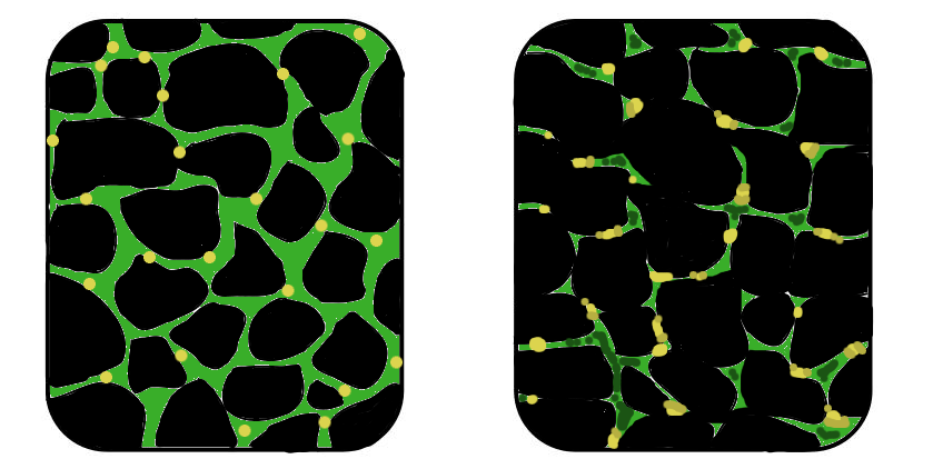
Distrofia muscular
📋 Origem dos dados
Esta página agrega dados de fontes públicas e oficiais. Dados sobre cobertura no SUS (PCDT, CEAF) são verificados ativamente por agente proativo (ver badge no infobox). Demais dados têm atribuição de fonte + data da última sincronização — clique para abrir o original.
- Doença rara (ontologia)
- fonte: Orphanet
- Identificador unificado
- fonte: MONDO
- Codificação WHO/SUS
- fonte: WHO ICD-10 / DATASUS
- CID-11 (futuro)
- fonte: WHO ICD-11
- NIH/GARD
- fonte: GARD (NIH)
- Indexação biomédica
- fonte: MeSH (NLM)
- Dado público estruturado
- fonte: Wikidata
- Medicamentos (literatura)
- fonte: Orphanet
- Reposicionamento
- fonte: Drug Repurposing Hub
- Ensaios clínicos
- fonte: ClinicalTrials.gov