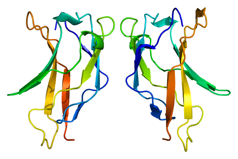
O fator de transcrição p65, também conhecido como subunidade p65 do fator nuclear NF-kappa-B, é uma proteína que em humanos é codificada pelo gene RELA.
Introdução
O que você precisa saber de cara
Visão geral
O ependimoma RELA fusão-positivo é um tipo de tumor do sistema nervoso central que se origina das células ependimárias, que revestem os ventrículos cerebrais e o canal central da medula espinhal. Ele é caracterizado por uma fusão genética específica envolvendo o gene RELA (fator de transcrição p65) e, na maioria dos casos, o gene ZFTA (proteína associada à translocação de dedos de zinco). Essa alteração molecular define um subtipo distinto de ependimoma, reconhecido pela classificação da Organização Mundial da Saúde (OMS). A doença pode se manifestar em diferentes faixas etárias, desde a infância até a idade adulta, e sua prevalência exata ainda não é conhecida.[1]
Sinais e sintomas
Os sinais e sintomas do ependimoma RELA fusão-positivo variam conforme a localização do tumor e a idade do paciente. Por ser um tumor intracraniano, os sintomas mais comuns incluem dores de cabeça persistentes, náuseas, vômitos (especialmente pela manhã), alterações visuais, convulsões e déficits neurológicos focais, como fraqueza em um lado do corpo ou dificuldade de equilíbrio. Em crianças, pode haver aumento do perímetro cefálico, irritabilidade e atraso no desenvolvimento. Como os sintomas podem ser inespecíficos, o diagnóstico precoce depende de exames de imagem e análise molecular.[1]
Causas genéticas
A causa genética principal do ependimoma RELA fusão-positivo é a fusão somática (adquirida, não hereditária) entre o gene RELA (fator de transcrição p65) e, mais frequentemente, o gene ZFTA (proteína associada à translocação de dedos de zinco). Essa fusão gera uma proteína quimérica que ativa de forma anormal a via de sinalização NF-κB, promovendo o crescimento tumoral. A doença não é herdada dos pais; trata-se de uma alteração genética que ocorre nas células do sistema nervoso durante a vida da pessoa.[1][3]
Diagnóstico
O diagnóstico do ependimoma RELA fusão-positivo é baseado em exames de imagem, como ressonância magnética (RM) do crânio, e na análise histopatológica do tumor após biópsia ou ressecção cirúrgica. A confirmação do subtipo molecular depende da identificação da fusão RELA-ZFTA por técnicas de biologia molecular, como sequenciamento de nova geração (NGS) ou PCR. Testes genéticos específicos para essa fusão estão disponíveis (código de teste genético: 336) e há 69 variantes reportadas no ClinVar relacionadas a esse diagnóstico. O código CID-10 associado é D43.0 (neoplasia de comportamento incerto do encéfalo).[1][3]
Tratamento e manejo
O tratamento do ependimoma RELA fusão-positivo geralmente envolve uma combinação de cirurgia para ressecção máxima segura do tumor, radioterapia e, em alguns casos, quimioterapia. A escolha do tratamento depende da localização, extensão do tumor, idade do paciente e condições clínicas. Não há medicamentos específicos aprovados exclusivamente para esse subtipo, mas a literatura científica menciona a carmustina como um fármaco que já foi estudado em contexto de reposicionamento (repurposing) para ependimomas. No Brasil, esse subtipo não possui cobertura específica pelo Sistema Único de Saúde (SUS) para procedimentos ou medicamentos direcionados.[1]
Tratamentos citados na literatura
A literatura científica, por meio de mineração automatizada de artigos (PubTator3), identificou associações entre o ependimoma RELA fusão-positivo e o seguinte fármaco: carmustina. É importante destacar que essas são associações observadas em publicações científicas e não representam uma recomendação formal de tratamento. O número de publicações que mencionam essa associação não está disponível no momento.[1]
Prognóstico e qualidade de vida
O prognóstico do ependimoma RELA fusão-positivo varia amplamente e depende de fatores como a extensão da ressecção cirúrgica, a idade do paciente e a resposta ao tratamento adjuvante. Em geral, tumores com fusão RELA-ZFTA tendem a ter um comportamento mais agressivo e maior taxa de recorrência em comparação com outros subtipos de ependimoma. O acompanhamento multidisciplinar com neurologia, oncologia, radioterapia e reabilitação é essencial para manejar as sequelas do tumor e do tratamento, visando a melhor qualidade de vida possível.[1]
Conteúdo informativo gerado e mantido automaticamente a partir de fontes oficiais (Orphanet, HPO, OMIM, SUS). Não substitui avaliação médica.
O fator de transcrição p65, também conhecido como subunidade p65 do fator nuclear NF-kappa-B, é uma proteína que em humanos é codificada pelo gene RELA.
Escala de raridade
<1/50kMuito rara
1/20kRara
1/10kPouco freq.
1/5kIncomum
1/2k
Encontrou um erro ou informação desatualizada? Sugira uma correção →
Entender a doença
Do básico ao detalhe, leia no seu ritmo
Preparando trilha educativa...
Sinais e sintomas
O que aparece no corpo e com que frequência cada sintoma acontece
Visão geral
O ependimoma RELA fusão-positivo é um tipo de tumor do sistema nervoso central que se origina das células ependimárias, que revestem os ventrículos cerebrais e o canal central da medula espinhal. Ele é caracterizado por uma fusão genética específica envolvendo o gene RELA (fator de transcrição p65) e, na maioria dos casos, o gene ZFTA (proteína associada à translocação de dedos de zinco). Essa alteração molecular define um subtipo distinto de ependimoma, reconhecido pela classificação da Organização Mundial da Saúde (OMS). A doença pode se manifestar em diferentes faixas etárias, desde a infância até a idade adulta, e sua prevalência exata ainda não é conhecida.[1]
Sinais e sintomas
Os sinais e sintomas do ependimoma RELA fusão-positivo variam conforme a localização do tumor e a idade do paciente. Por ser um tumor intracraniano, os sintomas mais comuns incluem dores de cabeça persistentes, náuseas, vômitos (especialmente pela manhã), alterações visuais, convulsões e déficits neurológicos focais, como fraqueza em um lado do corpo ou dificuldade de equilíbrio. Em crianças, pode haver aumento do perímetro cefálico, irritabilidade e atraso no desenvolvimento. Como os sintomas podem ser inespecíficos, o diagnóstico precoce depende de exames de imagem e análise molecular.[1]
Causas genéticas
A causa genética principal do ependimoma RELA fusão-positivo é a fusão somática (adquirida, não hereditária) entre o gene RELA (fator de transcrição p65) e, mais frequentemente, o gene ZFTA (proteína associada à translocação de dedos de zinco). Essa fusão gera uma proteína quimérica que ativa de forma anormal a via de sinalização NF-κB, promovendo o crescimento tumoral. A doença não é herdada dos pais; trata-se de uma alteração genética que ocorre nas células do sistema nervoso durante a vida da pessoa.[1][3]
Diagnóstico
O diagnóstico do ependimoma RELA fusão-positivo é baseado em exames de imagem, como ressonância magnética (RM) do crânio, e na análise histopatológica do tumor após biópsia ou ressecção cirúrgica. A confirmação do subtipo molecular depende da identificação da fusão RELA-ZFTA por técnicas de biologia molecular, como sequenciamento de nova geração (NGS) ou PCR. Testes genéticos específicos para essa fusão estão disponíveis (código de teste genético: 336) e há 69 variantes reportadas no ClinVar relacionadas a esse diagnóstico. O código CID-10 associado é D43.0 (neoplasia de comportamento incerto do encéfalo).[1][3]
Tratamento e manejo
O tratamento do ependimoma RELA fusão-positivo geralmente envolve uma combinação de cirurgia para ressecção máxima segura do tumor, radioterapia e, em alguns casos, quimioterapia. A escolha do tratamento depende da localização, extensão do tumor, idade do paciente e condições clínicas. Não há medicamentos específicos aprovados exclusivamente para esse subtipo, mas a literatura científica menciona a carmustina como um fármaco que já foi estudado em contexto de reposicionamento (repurposing) para ependimomas. No Brasil, esse subtipo não possui cobertura específica pelo Sistema Único de Saúde (SUS) para procedimentos ou medicamentos direcionados.[1]
Tratamentos citados na literatura
A literatura científica, por meio de mineração automatizada de artigos (PubTator3), identificou associações entre o ependimoma RELA fusão-positivo e o seguinte fármaco: carmustina. É importante destacar que essas são associações observadas em publicações científicas e não representam uma recomendação formal de tratamento. O número de publicações que mencionam essa associação não está disponível no momento.[1]
Prognóstico e qualidade de vida
O prognóstico do ependimoma RELA fusão-positivo varia amplamente e depende de fatores como a extensão da ressecção cirúrgica, a idade do paciente e a resposta ao tratamento adjuvante. Em geral, tumores com fusão RELA-ZFTA tendem a ter um comportamento mais agressivo e maior taxa de recorrência em comparação com outros subtipos de ependimoma. O acompanhamento multidisciplinar com neurologia, oncologia, radioterapia e reabilitação é essencial para manejar as sequelas do tumor e do tratamento, visando a melhor qualidade de vida possível.[1]
Conteúdo informativo gerado e mantido automaticamente a partir de fontes oficiais (Orphanet, HPO, OMIM, SUS). Não substitui avaliação médica.
Linha do tempo da pesquisa
Encontrou um erro ou informação desatualizada? Sugira uma correção →
Genética e causas
O que está alterado no DNA e como passa nas famílias
Genes associados
2 genes identificados com associação a esta condição.
NF-kappa-B is a pleiotropic transcription factor present in almost all cell types and is the endpoint of a series of signal transduction events that are initiated by a vast array of stimuli related to many biological processes such as inflammation, immunity, differentiation, cell growth, tumorigenesis and apoptosis. NF-kappa-B is a homo- or heterodimeric complex formed by the Rel-like domain-containing proteins RELA/p65, RELB, NFKB1/p105, NFKB1/p50, REL and NFKB2/p52. The heterodimeric RELA-NFKB
NucleusCytoplasm
Variantes genéticas (ClinVar)
69 variantes patogênicas registradas no ClinVar.
Vias biológicas (Reactome)
27 vias biológicas associadas aos genes desta condição.
Diagnóstico
Os sinais que médicos procuram e os exames que confirmam
Tratamento e manejo
Remédios, cuidados de apoio e o que precisa acompanhar
Onde tratar no SUS
Hospitais de referência no Brasil e o protocolo oficial do SUS (PCDT)
🇧🇷 Atendimento SUS — Ependimoma RELA fusão-positivo
Selecione um estado ou use sua localização para ver resultados.
Dados de DATASUS/CNES, SBGM, ABNeuro e Ministério da Saúde. Sempre confirme a disponibilidade diretamente com o estabelecimento.
Pesquisa ativa
Ensaios clínicos abertos e novidades científicas recentes
Pesquisa e ensaios clínicos
0 ensaios clínicos encontrados.
Publicações mais relevantes
Supratentorial ependymoma, zinc finger translocation-associated fusion positive, with extensive synaptophysin immunoreactivity arising from malignant transformation of clear cell ependymoma: A case report.
We describe a case of a supratentorial ependymoma, zinc finger translocation-associated (ZFTA) fusion positive with extensive synaptophysin immunoreactivity arising from malignant transformation of an ependymoma with clear cell features in a patient with long-term follow-up. A 55-year-old woman presented with seizures and ataxia 15 years after an initial resection of a clear cell ependymoma, Grade 2. Imaging demonstrated an enhancing right paracentral mass and the patient underwent biopsy and resection. Microscopic analysis showed regions of the tumor with morphological and immunohistochemical features typical of ependymoma, including perivascular pseudorosettes and focal dot- like epithelial membrane antigen positivity, as well as high-grade features. In addition, the neoplasm contained large nodular regions of clear cells exhibiting extensive synaptophysin immunoreactivity, suggestive of neural differentiation, and only focally positive immunoreactivity for glial markers. Electron microscopy showed poorly formed and ill-defined junctional complexes, but no cilia, microvilli, or dense granules were seen. Molecular profiling revealed the presence of a fusion between ZFTA (previously known as C11orf95) and RELA fusion. We report a case of extensive synaptophysin immunoreactivity in a ZFTA-RELA fusion-positive ependymoma that had undergone malignant transformation from a clear cell ependymoma and has long-term follow-up, contributing to the assessment of prognostic significance of synaptophysin immunoreactivity in supratentorial ependymoma, ZFTA fusion positive.
Ependymomas.
Ependymal neoplasms are a heterogenous group of neoplasms arising from the progenitors of the cells lining the ventricular system and the spinal central canal. During the last few years, significant novel data concerning oncogenesis, molecular characteristics and clinical correlations of these tumours have been collected, with a strong relevance for their pathological classification. The recently published 5th edition of WHO Classification of Central Nervous System Tumours integrates this novel knowledge and represents a substantial update compared to the previous edition. Concerning supratentorial ependymomas, the previous RELA fusion-positive ependymoma has been renamed into ZFTA fusion-positive and the novel YAP1 fusion-positive ependymoma subtype has been added. Posterior fossa ependymomas should now be allocated either to the Type A or Type B subtypes based on molecular profiling or using the H3 K27me3 immunohistochemical surrogate. Regarding spinal ependymomas, a novel subtype has been added based on a distinctive molecular trait, presence of MYCN amplification, and on the unfavourable outcome. Finally, myxopapillary ependymoma is now classified as a grade 2 tumour in accordance with its overall prognosis which mirrors that of conventional spinal ependymomas. The aim of this review is to present these changes and summarize the current diagnostic framework of ependymal tumours, according to the most recent updates.
Supratentorial extra-axial RELA fusion-positive ependymoma misdiagnosed as meningioma by intraoperative histological and cytological examinations: a case report.
Dura-attached supratentorial extra-axial ependymoma is a very rare type of tumor, with only nine reported cases. Preoperative diagnosis of dura-attached supratentorial extra-axial ependymoma is difficult and often radiologically misdiagnosed as a meningioma. We report a case of dura-attached supratentorial extra-axial ependymoma that was misdiagnosed using intraoperative histological and cytological examinations. A 26-year-old Japanese man with headache and nausea was referred to our medical facility. Magnetic resonance imaging revealed a cystic mass of 70 × 53 × 57 mm in the left temporoparietal lobe. A peritumoral band with hyperintensity on T2-weighted imaging was observed at the periphery of the lesion, suggesting an extra-axial lesion with no apparent connection to the ventricle. A dural tail sign was also noted on the gadolinium-enhanced T1-weighted image. Preoperative clinical diagnosis was meningioma. Proliferated tumor cells in sheets with intermingled branching vessels were observed in the frozen tissue. Perivascular rosettes were inconspicuous, and the tumor cells had rhabdoid cytoplasm. The tumor was intraoperatively diagnosed as a meningioma, suspected to be a rhabdoid meningioma. Perivascular rosettes were evident in the formalin-fixed paraffin-embedded tissues, suggesting ependymoma. The tumor cells had eosinophilic cytoplasm without a rhabdoid appearance. Anaplastic features, such as high tumor cellularity, increased mitotic activity, microvascular proliferation, and necrosis, were observed. Ependymal differentiation was confirmed on the basis of ultrastructural analysis. Molecular analysis detected C11orf95-RELA fusion gene. The final diagnosis was RELA fusion-positive ependymoma, World Health Organization grade III. Owing to its unusual location, dura-attached supratentorial extra-axial ependymomas are frequently misdiagnosed as meningiomas. Neuropathologists should take great precaution in intraoperatively diagnosing this rare subtype of ependymoma to avoid misdiagnosis of the lesion as other common dura-attached tumors.
Ependymoma with C11orf95-MAML2 fusion: presenting with granular cell and ganglion cell features.
C11orf95-RELA fusion or, less frequently, YAP1 fusion is recurrently detected in most cases of supratentorial ependymoma. Other fusions have rarely been reported in some cases of supratentorial ependymoma, and little is known about their pathological or clinical features. Here, we present a case of supratentorial ependymoma with unusual pathological findings and C11orf95-MAML2 fusion. A 23-year-old man was admitted to our hospital because of headache and vomiting. Magnetic resonance imaging revealed a cystic lesion in the right frontal lobe, and gross total resection of the tumor was performed. Pathologically, the tumor was mainly composed of typical ependymal lesions with perivascular pseudorosettes and contained some atypical lesions, with granular and ganglion cell features. The tumor was diagnosed as anaplastic ependymoma, which was classified as grade III on the World Health Organization scale, and found to be RELA fusion-positive in the DNA methylation analysis. However, the tumor was negative for C11orf95-RELA fusion, and RNA sequencing detected C11orf95-MAML2 fusion. The patient has not received adjuvant therapy and has remained alive without any evidence of disease for 30 months, suggesting that the prognosis might be better than that of typical C11orf95-RELA fusion-positive ependymoma.
RELA Fusion-Positive Ependymoma in a Child with Down Syndrome: A Case Report.
Down syndrome (DS) is the most common multiple malformation syndrome in humans and is associated with an increased risk of childhood malignancy, particularly leukemia. Incidence of brain tumors in patients with DS is limited to sporadic cases. We report the first case of a RELA fusion-positive ependymoma in a 3-year-old boy with DS. Imaging prompted by new left-sided hemiparesis demonstrated an 8-cm hemorrhagic right temporal-parietal mass. Subsequent image-complete resection confirmed a RELA fusion-positive anaplastic ependymoma with 90% OLIG2 staining. Postoperatively, the patient, unfortunately, experienced fatal recurrence and drop metastases with leptomeningeal involvement. To our knowledge, this is the first reported case of a confirmed RELA fusion-positive ependymoma in a child with DS. We discuss this finding in the context of intracranial tumors in children with DS, as well as the finding of 90% positive OLIG2 expression and its potential as a prognostic marker.
Publicações recentes
Supratentorial extra-axial RELA fusion-positive ependymoma misdiagnosed as meningioma by intraoperative histological and cytological examinations: a case report.
🥉 Relato de casoSupratentorial ependymoma, zinc finger translocation-associated fusion positive, with extensive synaptophysin immunoreactivity arising from malignant transformation of clear cell ependymoma: A case report.
RELA Fusion-Positive Ependymoma in a Child with Down Syndrome: A Case Report.
Ependymoma with C11orf95-MAML2 fusion: presenting with granular cell and ganglion cell features.
📚 EuropePMC7 artigos no totalmostrando 14
Supratentorial extra-axial RELA fusion-positive ependymoma misdiagnosed as meningioma by intraoperative histological and cytological examinations: a case report.
Journal of medical case reportsSupratentorial ependymoma, zinc finger translocation-associated fusion positive, with extensive synaptophysin immunoreactivity arising from malignant transformation of clear cell ependymoma: A case report.
Surgical neurology internationalRELA Fusion-Positive Ependymoma in a Child with Down Syndrome: A Case Report.
Pediatric neurosurgeryEpendymoma with C11orf95-MAML2 fusion: presenting with granular cell and ganglion cell features.
Brain tumor pathologyCortically based cystic supratentorial RELA fusion-positive ependymoma: a case report with unusual presentation and appearance and review of literature.
Radiology case reportsRELA fusion-positive ependymoma accompanied by extensive desmoplasia: a case report.
Brain tumor pathologyL1CAM High Expression Associates with Poor Prognosis in Glioma but Does Not Correlate with C11orf95-RELA Fusion.
BioMed research internationalIn vitro benchmarking of NF-κB inhibitors.
European journal of pharmacologyPreoperative and intraoperative perfusion magnetic resonance imaging in a RELA fusion-positive anaplastic ependymoma: A case report.
Surgical neurology internationalDNA methylation-based classification of ependymomas in adulthood: implications for diagnosis and treatment.
Neuro-oncologyA Simplified Overview of World Health Organization Classification Update of Central Nervous System Tumors 2016.
Journal of neurosciences in rural practiceWHO 2016 Classification of gliomas.
Neuropathology and applied neurobiologyThe 2016 WHO Classification of Tumours of the Central Nervous System: The Major Points of Revision.
Neurologia medico-chirurgicaThe 2016 World Health Organization Classification of Tumors of the Central Nervous System: a summary.
Acta neuropathologicaAssociações
Organizações que acompanham esta doença — pra ter apoio e orientação
Ainda não temos associações cadastradas para Ependimoma RELA fusão-positivo.
É de uma associação que acompanha esta doença? Fale com a gente →
Comunidades
Grupos ativos de quem convive com esta doença aqui no Raras
Ainda não existe comunidade no Raras para Ependimoma RELA fusão-positivo
Pacientes, familiares e cuidadores se organizam em comunidades pra compartilhar experiências, fazer perguntas e se apoiar. Você pode ser o primeiro.
Tire suas dúvidas
Perguntas, dicas e experiências compartilhadas aqui na página
Participe da discussão
Faça login para postar dúvidas, compartilhar experiências e interagir com especialistas.
Fazer loginDoenças relacionadas
Doenças com sintomas parecidos — ajudam quem ainda está buscando diagnóstico
Ainda não achamos doenças com sintomas parecidos o suficiente.
Referências e fontes
Bases de dados externas citadas neste artigo
Publicações científicas
Artigos indexados no PubMed ligados a esta doença no grafo RarasNet — título, periódico e PMID direto da fonte, sem intermediação de IA.
- Supratentorial ependymoma, zinc finger translocation-associated fusion positive, with extensive synaptophysin immunoreactivity arising from malignant transformation of clear cell ependymoma: A case report.
- Ependymomas.
- Supratentorial extra-axial RELA fusion-positive ependymoma misdiagnosed as meningioma by intraoperative histological and cytological examinations: a case report.
- Ependymoma with C11orf95-MAML2 fusion: presenting with granular cell and ganglion cell features.
- RELA Fusion-Positive Ependymoma in a Child with Down Syndrome: A Case Report.
Bases de dados e fontes oficiais
Identificadores e referências canônicas usadas para montar este verbete.
- ORPHA:530792(Orphanet)
- MONDO:0033980(MONDO)
- GARD:22210(GARD (NIH))
- Variantes catalogadas(ClinVar)
- Busca completa no PubMed(PubMed)
Dados compilados pelo RarasNet a partir de fontes abertas (Orphanet, OMIM, MONDO, PubMed/EuropePMC, ClinicalTrials.gov, DATASUS, PCDT/MS). Este conteúdo é informativo e não substitui avaliação médica.
Conteúdo mantido por Agente Raras · Médicos e pesquisadores podem colaborar
Ependimoma RELA fusão-positivo
📋 Origem dos dados
Esta página agrega dados de fontes públicas e oficiais. Dados sobre cobertura no SUS (PCDT, CEAF) são verificados ativamente por agente proativo (ver badge no infobox). Demais dados têm atribuição de fonte + data da última sincronização — clique para abrir o original.
- Doença rara (ontologia)
- fonte: Orphanet
- Identificador unificado
- fonte: MONDO
- Codificação WHO/SUS
- fonte: WHO ICD-10 / DATASUS
- NIH/GARD
- fonte: GARD (NIH)
- Reposicionamento
- fonte: Drug Repurposing Hub