Meduloblastoma clássico é um tumor cerebral maligno agressivo que se origina no cerebelo, mais comum em crianças. Caracteriza-se por proliferação celular rápida e potencial de metástase.
Introdução
O que você precisa saber de cara
Visão geral
O meduloblastoma clássico é uma variante histológica do meduloblastoma, um tumor maligno de origem embrionária. Ele geralmente se localiza na linha média do cérebro e ocorre com mais frequência em crianças. Os sintomas podem variar, incluindo dores de cabeça, náuseas, vômitos e ataxia (falta de coordenação dos movimentos).[1]
Sinais e sintomas
Os sintomas do meduloblastoma clássico são variáveis e podem incluir dores de cabeça, náuseas, vômitos e ataxia. Esses sinais decorrem principalmente do aumento da pressão intracraniana causada pelo tumor.[1]
Causas genéticas
Diagnóstico
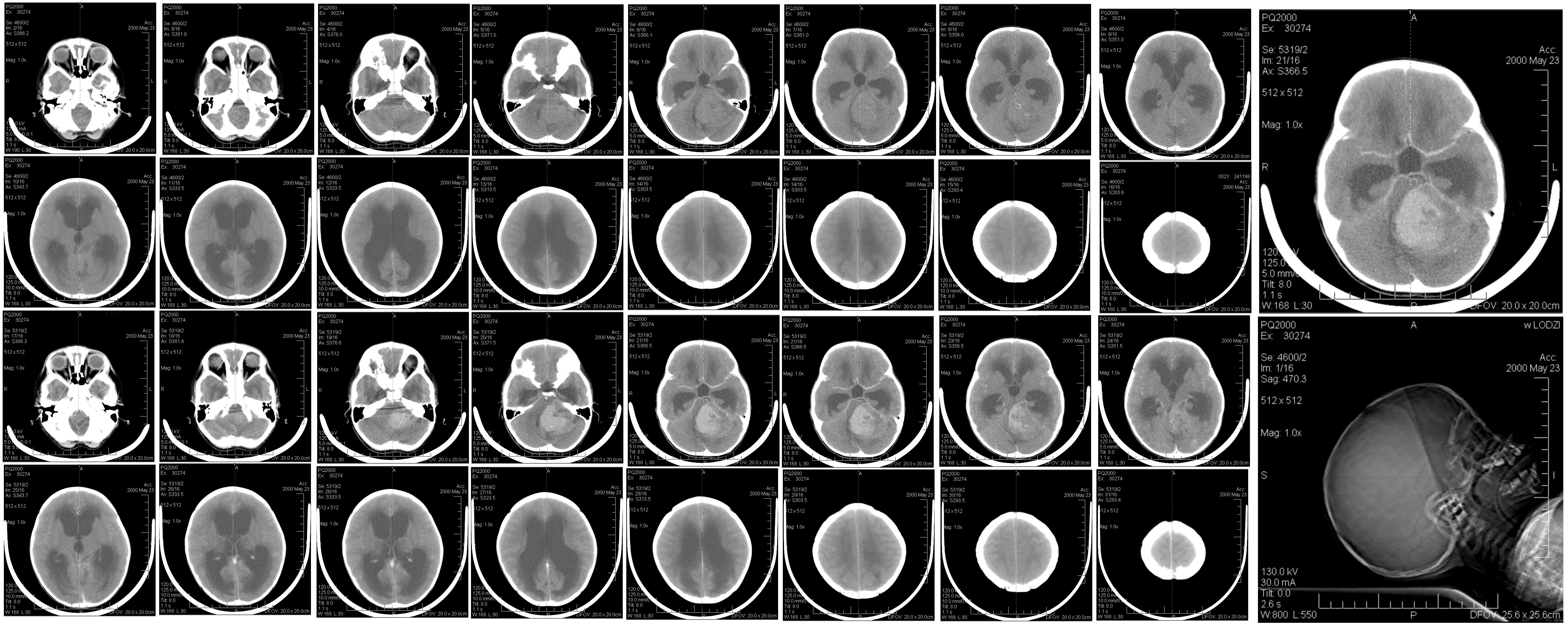
O diagnóstico do meduloblastoma clássico é baseado em exames de imagem (como ressonância magnética) e confirmado por análise histopatológica do tumor após biópsia ou ressecção cirúrgica. Testes genéticos podem ser realizados para auxiliar na classificação do tumor e no planejamento do tratamento, embora não exista um teste genético específico para esta variante isoladamente.[1][2][4]
Tratamento e manejo
O tratamento do meduloblastoma clássico geralmente envolve uma combinação de cirurgia, radioterapia e quimioterapia. O plano de tratamento é individualizado, levando em conta a idade do paciente, o estágio da doença e outros fatores. No Brasil, o meduloblastoma clássico não possui cobertura específica pelo Sistema Único de Saúde (SUS) para procedimentos ou medicamentos padronizados, sendo o acesso ao tratamento realizado por meio de protocolos clínicos e judicialização, quando necessário.[1][2]
Tratamentos citados na literatura
A literatura científica menciona os seguintes fármacos em associação com o meduloblastoma clássico, com base em mineração de textos (PubTator3): carmustina e trofosfamida. É importante destacar que essas são associações encontradas em publicações científicas e não representam uma recomendação de tratamento. Consulte sempre seu médico para decisões terapêuticas.[1][2]
Prognóstico e qualidade de vida
O prognóstico do meduloblastoma clássico depende de vários fatores, incluindo a extensão da doença no momento do diagnóstico, a idade do paciente e a resposta ao tratamento. O acompanhamento multidisciplinar é essencial para manejar os efeitos colaterais do tratamento e promover a melhor qualidade de vida possível.[1][2]
Conteúdo informativo gerado e mantido automaticamente a partir de fontes oficiais (Orphanet, HPO, OMIM, SUS). Não substitui avaliação médica.
Meduloblastoma clássico é um tumor cerebral maligno agressivo que se origina no cerebelo, mais comum em crianças. Caracteriza-se por proliferação celular rápida e potencial de metástase.
Encontrou um erro ou informação desatualizada? Sugira uma correção →
Entender a doença
Do básico ao detalhe, leia no seu ritmo
Preparando trilha educativa...
Sinais e sintomas
O que aparece no corpo e com que frequência cada sintoma acontece
Visão geral
O meduloblastoma clássico é uma variante histológica do meduloblastoma, um tumor maligno de origem embrionária. Ele geralmente se localiza na linha média do cérebro e ocorre com mais frequência em crianças. Os sintomas podem variar, incluindo dores de cabeça, náuseas, vômitos e ataxia (falta de coordenação dos movimentos).[1]
Sinais e sintomas
Os sintomas do meduloblastoma clássico são variáveis e podem incluir dores de cabeça, náuseas, vômitos e ataxia. Esses sinais decorrem principalmente do aumento da pressão intracraniana causada pelo tumor.[1]
Causas genéticas
Diagnóstico
O diagnóstico do meduloblastoma clássico é baseado em exames de imagem (como ressonância magnética) e confirmado por análise histopatológica do tumor após biópsia ou ressecção cirúrgica. Testes genéticos podem ser realizados para auxiliar na classificação do tumor e no planejamento do tratamento, embora não exista um teste genético específico para esta variante isoladamente.[1][2][4]
Tratamento e manejo
O tratamento do meduloblastoma clássico geralmente envolve uma combinação de cirurgia, radioterapia e quimioterapia. O plano de tratamento é individualizado, levando em conta a idade do paciente, o estágio da doença e outros fatores. No Brasil, o meduloblastoma clássico não possui cobertura específica pelo Sistema Único de Saúde (SUS) para procedimentos ou medicamentos padronizados, sendo o acesso ao tratamento realizado por meio de protocolos clínicos e judicialização, quando necessário.[1][2]
Tratamentos citados na literatura
A literatura científica menciona os seguintes fármacos em associação com o meduloblastoma clássico, com base em mineração de textos (PubTator3): carmustina e trofosfamida. É importante destacar que essas são associações encontradas em publicações científicas e não representam uma recomendação de tratamento. Consulte sempre seu médico para decisões terapêuticas.[1][2]
Prognóstico e qualidade de vida
O prognóstico do meduloblastoma clássico depende de vários fatores, incluindo a extensão da doença no momento do diagnóstico, a idade do paciente e a resposta ao tratamento. O acompanhamento multidisciplinar é essencial para manejar os efeitos colaterais do tratamento e promover a melhor qualidade de vida possível.[1][2]
Conteúdo informativo gerado e mantido automaticamente a partir de fontes oficiais (Orphanet, HPO, OMIM, SUS). Não substitui avaliação médica.
Linha do tempo da pesquisa
Encontrou um erro ou informação desatualizada? Sugira uma correção →
Genética e causas
O que está alterado no DNA e como passa nas famílias
Nenhum gene associado encontrado
Os dados genéticos desta condição ainda estão sendo catalogados.
Diagnóstico
Os sinais que médicos procuram e os exames que confirmam
Tratamento e manejo
Remédios, cuidados de apoio e o que precisa acompanhar
Onde tratar no SUS
Hospitais de referência no Brasil e o protocolo oficial do SUS (PCDT)
🇧🇷 Atendimento SUS — Meduloblastoma clássico
Selecione um estado ou use sua localização para ver resultados.
Dados de DATASUS/CNES, SBGM, ABNeuro e Ministério da Saúde. Sempre confirme a disponibilidade diretamente com o estabelecimento.
Pesquisa ativa
Ensaios clínicos abertos e novidades científicas recentes
Ensaios em destaque
Pesquisa e ensaios clínicos
0 ensaios clínicos encontrados.
Publicações mais relevantes
CD4 T cells correlate with better prognosis in medulloblastoma.
T cells and tumor-associated macrophages (TAMs) are critical immune components within the brain tumor microenvironment (TME), yet their precise roles in medulloblastoma remains unclear. In this study, we examined the infiltration characteristics of T cells in medulloblastoma tissues and analyzed the correlation between T cells and the clinical outcomes of medulloblastoma patients. Additionally, we further investigated the relationship between T cells and TAMs. We enrolled a total of 72 patients diagnosed with medulloblastoma and subsequently detected the T cell makers and programmed death 1/programmed death-ligand 1 (PD-1/PD-L1) in paraffin-embedded sections using multiple immunofluorescence staining method. The correlation between T cell infiltration, clinical characteristics and prognosis were analyzed. Finally, we used Spearman correlation analysis to evaluate the correlation between T cells and TAMs. The median age at diagnosis of 72 patients (54 boys, 18 girls) was 7.5 years (range: 0.8-18 years). These patients included 43 cases of classic medulloblastoma (CMB), 24 cases of desmoplastic/nodular medulloblastoma (DNMB), 2 cases of medulloblastoma with extensive nodularity (MBEN) and 3 cases of large-cell/anaplastic medulloblastoma (LCA). The molecular subgroups consisted of 3 wingless (WNT), 29 sonic hedgehog (SHH) and 40 non-WNT/non-SHH cases. Twenty-five cases presented with metastasis at diagnosis, while 47 cases were without metastasis. Thirteen cases exhibited with high-risk genetic abnormalities. The total T cells (P = 0.031) and CD4 T cells (P = 0.045) were significantly elevated in the SHH subgroup compared to those in the non-WNT/non-SHH subgroup. Patients with increased CD4 T cells had better 5-year PFS (P = 0.000) and OS (P = 0.001), while patients without metastasis showed better 5-year PFS (P = 0.031) and OS (P = 0.015). Multivariate analysis showed that CD4 T cells were an independent prognostic factor affecting both the 5-year PFS (P = 0.004, HR = 0.230, 95% CI = 0.085-0.662) and OS (P = 0.017, HR = 0.180, 95% CI = 0.044-0.739). Additionally, it was observed that CD4 T cells exhibited a positive correlation with Mtotal (total macrophages) (P < 0.05, r = 0.249) and Mmix (M1/M2 mixed phenotype macrophages) (P < 0.01, r = 0.325), and CD3+CD8+PD-1+ cells showed a positive correlation with Mmix (P < 0.05, r = 0.258). The increase in CD4 T cells predicts a better prognosis in medulloblastoma patients, particularly within the SHH and non-WNT/non-SHH subgroups, and they may serve as a potential therapeutic target for medulloblastoma. Additionally, there may be a potential interaction between CD4 T cells and TAMs that warrants further investigation.
Acute Toxicities of Proton Craniospinal Irradiation in Pediatric Medulloblastoma: A Pediatric Proton/Photon Consortium Registry (PPCR) Study.
Medulloblastoma is one of the most common malignant brain tumors in children, and 75% of patients achieve long-term survival. There are limited studies reporting acute toxicity for pediatric patients receiving proton therapy for craniospinal irradiation (CSI) in medulloblastoma, and these are limited by their retrospective nature, modest cohort sizes, and lack of a comparator group. We analyzed data from the Pediatric Proton/Photon Consortium Registry. Patients with a diagnosis of medulloblastoma, receiving CSI with doses of either 23.4 Gy or 36 to 39.6 Gy, and with toxicity data at baseline and completion of treatment, were identified for inclusion. A total of 272 patients were included for analysis. All patients received proton therapy. The median age of patients was 8 years (range 3-22 years), and 67.6% were male. Most patients were of good performance status with eastern cooperative oncology group (ECOG) 0 or 1, 36.8% and 31.6%, respectively. In total, 68.8% of patients had classic medulloblastoma; 76.8% had M0 disease; and 62.9% of patients had standard-risk disease.Acute toxicities were reported as grade 1 or higher. The most common toxicities occurring during treatment were skin (90.3%), gastrointestinal (71.6%), hematological (54.9%), and mouth (32.0%). Toxicity rates were lower than in the published literature. All patients completed treatment. Our findings suggest that proton CSI has an acceptable degree of acute toxicity in the treatment of pediatric medulloblastoma. Future studies would benefit from toxicity grading, toxicity timing, and a photon comparator group.
Tumor-associated macrophages correlate with better outcome in SHH medulloblastoma.
Tumor-associated macrophages (TAMs) constitute a significant proportion of the immune cell population within brain tumors. The polarization of macrophages exerts an important influence on the tumor microenvironment (TME). Nevertheless, the specific role of TAMs in sonic hedgehog (SHH) medulloblastoma remains unclear. To investigate the polarization characteristics and effects of TAMs in SHH medulloblastoma, we evaluated the infiltration of M1 and M2 macrophages in SHH medulloblastoma tissues and analyzed the correlation between TAMs recruitment and the clinical outcome of SHH medulloblastoma patients. We enrolled a total of 42 patients diagnosed with SHH medulloblastoma. Using multiple immunofluorescence staining on paraffin-embedded sections, we detected the activated phenotype (M1/M2) by monoclonal antibodies for CD68, HLA-DR and CD163. Subsequently, we analyzed the correlation between TAMs and clinical characteristics as well as prognostic factors. The median age of 42 patients (31 boys, 11 girls) was 5.3 years (range: 0.8-15.1 years). All patients had confirmed pathological types, including 4 cases of classic medulloblastoma (CMB), 33 cases of desmoplastic/nodular medulloblastoma (DNMB), 3 cases of medulloblastoma with extensive nodularity (MBEN), and 2 cases of large-cell/anaplastic medulloblastoma (LCA). Thirteen cases presented with metastasis at diagnosis, while twenty-nine cases were without metastasis. Four cases had high-risk genetic abnormalities. Different proportions of macrophages were found in the collected medulloblastoma tissues, and large amounts of CD68+HLA-DR+CD163+ cells were found. The study revealed that Mtotal (total macrophages) and Mmix (CD68+HLA-DR+CD163+ cells) were significantly higher in group of patients <5 years old (P < 0.05), and Mtotal in non-metastatic group were significantly higher than that in metastatic group (P = 0.043). M2 macrophages in CMB group were significantly higher than that in DNMB/MBEN group (P = 0.036), M1 macrophages were significantly higher in children without high-risk genetic abnormalities (P = 0.007). Five-year PFS was significantly poorer in patients ≥5 years old and metastatic group (P < 0.05). High Mtotal group had a better 5-year PFS (P = 0.000), whereas high M2 group had both better 5-year PFS and OS (P = 0.001, P = 0.001). Multivariate analysis showed that Mtotal and M2 macrophages were independent prognostic factors for 5-year PFS, and M2 macrophages were an independent prognostic factor for 5-year OS. The increase in total macrophages and M2 macrophages predicts a better outcome of SHH medulloblastoma. TAMs especially M2 macrophages might be a therapeutic target for SHH medulloblastoma.
Slow-growing WNT medulloblastoma with atypical magnetic resonance imaging findings: illustrative case.
Medulloblastomas, with four molecular subgroups, are generally rapid-growing tumors with significant contrast enhancement and well-defined margins. However, each subgroup's clinical features, including disease time course and imaging characteristics, are not well defined. The authors describe the case of a 15-year-old female who presented with a 7-month history of impaired left-hand movement and was found to have a lesion on the dorsal side of the fourth ventricle. T2-weighted magnetic resonance imaging (MRI) at the patient's first presentation showed diffuse hyperintense signal without apparent mass, and gadolinium-enhanced T1-weighted imaging showed very slight contrast enhancement. In 1 month, her symptoms progressed, and follow-up MRI revealed an increase in the size of the lesion, showing greater diffusion restriction and contrast enhancement. She underwent gross-total resection, and pathology was consistent with classic medulloblastoma. Genetic analysis of the tumor confirmed the wingless (WNT) molecular subgroup. Adjuvant chemotherapy and proton beam therapy were performed. At the 18-month follow-up, MRI showed no recurrence of disease. Slow-growing medulloblastoma is very rare and not known to be associated with a specific molecular subgroup. Here, the authors report a case of slow-growing WNT medulloblastoma, indicating that slow growth may be a feature of this subgroup.
Pediatric synchronous multifocal and disseminated cerebrospinal classic medulloblastoma revealed by bilateral decreased visual acuity: a case report.
Medulloblastoma (MB) is a rapidly growing malignant solid tumor that arises from stem cells located in the subependymal germinal matrix or outer granular layer of the cerebellum. It represents 15 to 30% of pediatric brain tumors and less than 1% of primary brain tumors. The reason for the high incidence of MB in children compared to adults is the embryonic origin of the tumor. In typical cases, MB manifests as a solitary lesion in the fourth ventricle or in the cerebellar parenchyma; cases of synchronous multifocal and disseminated MB are quite rare in patients without familial tumor syndromes. To date, only 7 cases in adults and a single pediatric case with Gorlin syndrome have been described previously. Here, the authors report a new case of synchronous multifocal classic cerebrospinal histologically confirmed MB in a 10-year-old male patient revealed by bilateral decreased visual acuity without any other localizing neurological signs. The authors will proceed with a review of the current literature regarding this rare entity.
Publicações recentes
CD4 T cells correlate with better prognosis in medulloblastoma.
Acute Toxicities of Proton Craniospinal Irradiation in Pediatric Medulloblastoma: A Pediatric Proton/Photon Consortium Registry (PPCR) Study.
Tumor-associated macrophages correlate with better outcome in SHH medulloblastoma.
Slow-growing WNT medulloblastoma with atypical magnetic resonance imaging findings: illustrative case.
Pediatric synchronous multifocal and disseminated cerebrospinal classic medulloblastoma revealed by bilateral decreased visual acuity: a case report.
📚 EuropePMC7 artigos no totalmostrando 17
CD4 T cells correlate with better prognosis in medulloblastoma.
Frontiers in oncologyAcute Toxicities of Proton Craniospinal Irradiation in Pediatric Medulloblastoma: A Pediatric Proton/Photon Consortium Registry (PPCR) Study.
International journal of particle therapyTumor-associated macrophages correlate with better outcome in SHH medulloblastoma.
Frontiers in oncologySlow-growing WNT medulloblastoma with atypical magnetic resonance imaging findings: illustrative case.
Journal of neurosurgery. Case lessonsPediatric synchronous multifocal and disseminated cerebrospinal classic medulloblastoma revealed by bilateral decreased visual acuity: a case report.
Child's nervous system : ChNS : official journal of the International Society for Pediatric NeurosurgeryCerbellar medulloblastoma in the elderly: Case report and review of the literature.
International journal of surgery case reportsDefining the Spectrum, Treatment and Outcome of Patients With Genetically Confirmed Gorlin Syndrome From the HIT-MED Cohort.
Frontiers in oncologyMolecular Characterization of Medulloblastoma in a Patient with Neurofibromatosis Type 1: Case Report and Literature Review.
Diagnostics (Basel, Switzerland)Medulloblastoma of the adult: results from a multicenter retrospective study by AINO (Italian Association of Neuro-Oncology) and SIN (Italian Society of Neurology).
Neurological sciences : official journal of the Italian Neurological Society and of the Italian Society of Clinical NeurophysiologyNonmetastatic Medulloblastoma of Early Childhood: Results From the Prospective Clinical Trial HIT-2000 and An Extended Validation Cohort.
Journal of clinical oncology : official journal of the American Society of Clinical OncologyTwenty years experience in treating childhood medulloblastoma: Between the past and the present.
Cancer radiotherapie : journal de la Societe francaise de radiotherapie oncologiqueChildhood Medulloblastoma Revisited.
Topics in magnetic resonance imaging : TMRIDNA polymerase β deficiency in the p53 null cerebellum leads to medulloblastoma formation.
Biochemical and biophysical research communications[Medulloblastoma. Pathology].
Neuro-ChirurgieAdded value of diffusion weighted imaging in pediatric central nervous system embryonal tumors surveillance.
OncotargetIdentification of a novel inherited ALK variant M1199L in the WNT type of medulloblastoma.
Folia neuropathologicaResolution of precocious puberty following resection of fourth ventricular medulloblastoma: case report.
Journal of neurosurgery. PediatricsAssociações
Organizações que acompanham esta doença — pra ter apoio e orientação
Ainda não temos associações cadastradas para Meduloblastoma clássico.
É de uma associação que acompanha esta doença? Fale com a gente →
Comunidades
Grupos ativos de quem convive com esta doença aqui no Raras
Ainda não existe comunidade no Raras para Meduloblastoma clássico
Pacientes, familiares e cuidadores se organizam em comunidades pra compartilhar experiências, fazer perguntas e se apoiar. Você pode ser o primeiro.
Tire suas dúvidas
Perguntas, dicas e experiências compartilhadas aqui na página
Participe da discussão
Faça login para postar dúvidas, compartilhar experiências e interagir com especialistas.
Fazer loginDoenças relacionadas
Doenças com sintomas parecidos — ajudam quem ainda está buscando diagnóstico
Ainda não achamos doenças com sintomas parecidos o suficiente.
Referências e fontes
Bases de dados externas citadas neste artigo
Publicações científicas
Artigos indexados no PubMed ligados a esta doença no grafo RarasNet — título, periódico e PMID direto da fonte, sem intermediação de IA.
- CD4 T cells correlate with better prognosis in medulloblastoma.
- Acute Toxicities of Proton Craniospinal Irradiation in Pediatric Medulloblastoma: A Pediatric Proton/Photon Consortium Registry (PPCR) Study.
- Tumor-associated macrophages correlate with better outcome in SHH medulloblastoma.
- Slow-growing WNT medulloblastoma with atypical magnetic resonance imaging findings: illustrative case.
- Pediatric synchronous multifocal and disseminated cerebrospinal classic medulloblastoma revealed by bilateral decreased visual acuity: a case report.Child's nervous system : ChNS : official journal of the International Society for Pediatric Neurosurgery· 2022· PMID 35590112mais citado
Bases de dados e fontes oficiais
Identificadores e referências canônicas usadas para montar este verbete.
- ORPHA:251867(Orphanet)
- MONDO:0016712(MONDO)
- GARD:17216(GARD (NIH))
- Busca completa no PubMed(PubMed)
- Q56013880(Wikidata)
Dados compilados pelo RarasNet a partir de fontes abertas (Orphanet, OMIM, MONDO, PubMed/EuropePMC, ClinicalTrials.gov, DATASUS, PCDT/MS). Este conteúdo é informativo e não substitui avaliação médica.
Conteúdo mantido por Agente Raras · Médicos e pesquisadores podem colaborar
Meduloblastoma clássico
📋 Origem dos dados
Esta página agrega dados de fontes públicas e oficiais. Dados sobre cobertura no SUS (PCDT, CEAF) são verificados ativamente por agente proativo (ver badge no infobox). Demais dados têm atribuição de fonte + data da última sincronização — clique para abrir o original.
- Doença rara (ontologia)
- fonte: Orphanet
- Identificador unificado
- fonte: MONDO
- Genética mendeliana
- fonte: OMIM
- Codificação WHO/SUS
- fonte: WHO ICD-10 / DATASUS
- CID-11 (futuro)
- fonte: WHO ICD-11
- NIH/GARD
- fonte: GARD (NIH)
- Dado público estruturado
- fonte: Wikidata
- Reposicionamento
- fonte: Drug Repurposing Hub