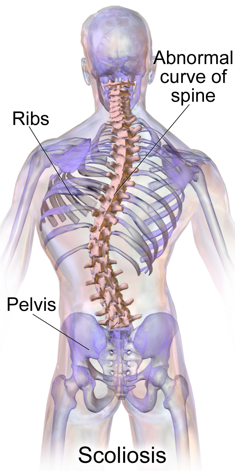
Escoliose é uma condição médica na qual a coluna vertebral apresenta uma curvatura irregular no plano coronal. A curvatura geralmente tem formato de S ou C em três dimensões. Em alguns casos, o grau da curvatura é estável, enquanto em outros, aumenta com o tempo. A escoliose leve normalmente não causa problemas, mas casos mais graves podem afetar a respiração e os movimentos. A dor está geralmente presente em adultos e pode piorar com a idade. À medida que a condição progride, ela pode alterar a vida de uma pessoa e, portanto, também pode ser considerada uma deficiência.
Introdução
O que você precisa saber de cara
Visão geral
A siringomielia idiopática é uma malformação rara do sistema nervoso central, caracterizada pela presença de uma cavidade alongada, preenchida por líquido, dentro do parênquima da medula espinhal ou do canal central, sem uma causa identificável. Essa condição não está associada a síndromes genéticas conhecidas.[1]
Sinais e sintomas
A siringomielia idiopática geralmente está associada a dor, distúrbios sensoriais e/ou musculoesqueléticos. No entanto, também pode ser um achado incidental e assintomático, descoberto durante exames de imagem realizados por outros motivos.[1]
Causas genéticas
Diagnóstico
O diagnóstico da siringomielia idiopática é baseado em exames de imagem, como a ressonância magnética da coluna vertebral, que demonstra a presença da cavidade (siringe) na medula espinhal. Testes genéticos, como o sequenciamento completo do exoma (WES), podem ser utilizados para excluir outras causas, mas não há um teste genético específico para confirmar a condição.[1][3]
Tratamento e manejo
O manejo da siringomielia idiopática é individualizado e pode incluir acompanhamento clínico regular, fisioterapia e tratamento da dor. No Brasil, o Sistema Único de Saúde (SUS) oferece cobertura mínima para essa condição, incluindo atendimento em reabilitação para doenças raras. Não há medicamentos específicos aprovados para o tratamento da siringomielia idiopática.[1]
Prognóstico e qualidade de vida
O prognóstico varia amplamente. Muitos pacientes apresentam sintomas estáveis ou de progressão lenta, enquanto outros podem ter um curso mais grave. O diagnóstico precoce e o acompanhamento multidisciplinar são importantes para preservar a função neurológica e a qualidade de vida.[1]
Conteúdo informativo gerado e mantido automaticamente a partir de fontes oficiais (Orphanet, HPO, OMIM, SUS). Não substitui avaliação médica.
Escoliose é uma condição médica na qual a coluna vertebral apresenta uma curvatura irregular no plano coronal. A curvatura geralmente tem formato de S ou C em três dimensões. Em alguns casos, o grau da curvatura é estável, enquanto em outros, aumenta com o tempo. A escoliose leve normalmente não causa problemas, mas casos mais graves podem afetar a respiração e os movimentos. A dor está geralmente presente em adultos e pode piorar com a idade. À medida que a condição progride, ela pode alterar a vida de uma pessoa e, portanto, também pode ser considerada uma deficiência.
Encontrou um erro ou informação desatualizada? Sugira uma correção →
Entender a doença
Do básico ao detalhe, leia no seu ritmo
Preparando trilha educativa...
Sinais e sintomas
O que aparece no corpo e com que frequência cada sintoma acontece
Visão geral
A siringomielia idiopática é uma malformação rara do sistema nervoso central, caracterizada pela presença de uma cavidade alongada, preenchida por líquido, dentro do parênquima da medula espinhal ou do canal central, sem uma causa identificável. Essa condição não está associada a síndromes genéticas conhecidas.[1]
Sinais e sintomas
A siringomielia idiopática geralmente está associada a dor, distúrbios sensoriais e/ou musculoesqueléticos. No entanto, também pode ser um achado incidental e assintomático, descoberto durante exames de imagem realizados por outros motivos.[1]
Causas genéticas
Diagnóstico
O diagnóstico da siringomielia idiopática é baseado em exames de imagem, como a ressonância magnética da coluna vertebral, que demonstra a presença da cavidade (siringe) na medula espinhal. Testes genéticos, como o sequenciamento completo do exoma (WES), podem ser utilizados para excluir outras causas, mas não há um teste genético específico para confirmar a condição.[1][3]
Tratamento e manejo
O manejo da siringomielia idiopática é individualizado e pode incluir acompanhamento clínico regular, fisioterapia e tratamento da dor. No Brasil, o Sistema Único de Saúde (SUS) oferece cobertura mínima para essa condição, incluindo atendimento em reabilitação para doenças raras. Não há medicamentos específicos aprovados para o tratamento da siringomielia idiopática.[1]
Prognóstico e qualidade de vida
O prognóstico varia amplamente. Muitos pacientes apresentam sintomas estáveis ou de progressão lenta, enquanto outros podem ter um curso mais grave. O diagnóstico precoce e o acompanhamento multidisciplinar são importantes para preservar a função neurológica e a qualidade de vida.[1]
Conteúdo informativo gerado e mantido automaticamente a partir de fontes oficiais (Orphanet, HPO, OMIM, SUS). Não substitui avaliação médica.
Linha do tempo da pesquisa
Encontrou um erro ou informação desatualizada? Sugira uma correção →
Genética e causas
O que está alterado no DNA e como passa nas famílias
Nenhum gene associado encontrado
Os dados genéticos desta condição ainda estão sendo catalogados.
Diagnóstico
Os sinais que médicos procuram e os exames que confirmam
Tratamento e manejo
Remédios, cuidados de apoio e o que precisa acompanhar
Onde tratar no SUS
Hospitais de referência no Brasil e o protocolo oficial do SUS (PCDT)
🇧🇷 Atendimento SUS — Siringomielia, idiopática
Selecione um estado ou use sua localização para ver resultados.
Dados de DATASUS/CNES, SBGM, ABNeuro e Ministério da Saúde. Sempre confirme a disponibilidade diretamente com o estabelecimento.
Pesquisa ativa
Ensaios clínicos abertos e novidades científicas recentes
Pesquisa e ensaios clínicos
Nenhum ensaio clínico registrado para esta condição.
Publicações mais relevantes
Therapeutic lumbar puncture as a potential treatment for symptomatic paediatric syringomyelia: A single centre cohort study.
Idiopathic syringomyelia represents a group of patients with a syrinx without an underlying pathological cause. Patients may present with back pain, leg pain and/or neurological dysfunction. We aim to examine the clinico-radiological outcomes of paediatric patients presenting with idiopathic syringomyelia managed with a therapeutic lumbar puncture. All paediatric patients with idiopathic syringomyelia who underwent a lumbar puncture over a 10-year time period were identified and included. Pre- and post-procedural clinico-radiological features were obtained from electronic patient records. Procedural details, including opening pressure and drainage volume, were obtained from electronic operative records. 31 patients underwent a therapeutic lumbar puncture. Median age at time of first lumbar puncture was 12 years (range 2-17). 7/31 (23%) patients had significant long-term improvement or resolution in their symptoms following a single lumbar puncture. 13/31 (42%) patients had temporary improvement of their symptoms and underwent further lumbar punctures. 3/31 (10%) patients had resolution of their symptoms after more than one lumbar puncture. 8/31 (26%) patients did not experience any improvement in their symptoms. Symptomatic improvement was more likely in those with a high opening pressure of equal to or greater than 24cmH2O (OR 13 (1.3 - 126.3) P = 0.02). Median length of follow up was 3 yrs (range 6 months - 9 years). Complications occurred in 9/31 (29%) patients, with 8 of these experiencing a low pressure headache, and 1 patient experiencing temporary back pain following the lumbar puncture. Lumbar puncture may be a safe and effective treatment for symptomatic idiopathic syringomyelia in the paediatric population. It appears to be more effective in those with a higher opening pressure.
Idiopathic Syringomyelia: Diagnostic Value of Cranial Morphometric Parameters.
Background: Identifying the etiological factors of syringomyelia, which can cause progressive neurological deficits in the spinal cord, is critically important for both diagnosis and treatment. This study aimed to assess the cranial morphometric features of patients with idiopathic syringomyelia by conducting comparative analyses with individuals diagnosed with Chiari Type I, Chiari Type I accompanied by syringomyelia, and healthy controls, in order to elucidate the potential structural contributors to the pathogenesis of idiopathic syringomyelia. Methods: In this retrospective and comparative study, a total of 172 patients diagnosed with Chiari Type I and/or syringomyelia between 2016 and 2024, along with 156 radiologically normal individuals, were included. The participants were categorized into four groups: healthy controls, Chiari Type I, Chiari Type I with syringomyelia, and idiopathic syringomyelia (defined as syringomyelia without an identifiable cause). Midline sagittal T1-weighted MR images were used to obtain quantitative measurements of the posterior fossa, cerebellum, intracranial area, and foramen magnum. All measurements were stratified and statistically analyzed by sex. Results: In cases with idiopathic syringomyelia, both the posterior fossa area and the cerebellum/posterior fossa ratio differed significantly from those of healthy controls. In male patients, the foramen magnum diameter was significantly larger in the Chiari + syringomyelia group compared with the idiopathic group. A significant correlation was found between the degree of tonsillar descent and selected morphometric parameters in female subjects, whereas no such correlation was observed in males. Both Chiari groups exhibited significantly smaller posterior fossa dimensions compared with the healthy and idiopathic groups, indicating greater neural crowding. Additionally, in Chiari Type I patients, increasing degrees of tonsillar descent were associated with a decreased incidence of syringomyelia. Conclusions: Anatomical variations such as a reduced posterior fossa area or altered foramen magnum diameter may contribute to the pathogenesis of idiopathic syringomyelia. Cranial morphometric analysis appears to offer diagnostic value in these cases. Further prospective, multicenter studies incorporating advanced neuroimaging modalities, particularly those assessing cerebrospinal fluid dynamics, are warranted to better understand the mechanisms underlying syringomyelia of unknown etiology.
Partially calcified giant hemorrhagic syringomyelia and hematomyelia.
Syringomyelia is a rare condition characterized by the formation of a fluid-filled cyst within the spinal cord, leading to myelopathy. In addition, the pathological enlargement of the central canal is referred to as hydromyelia or cleft-like syrinx. We present a case of idiopathic syringomyelia and hematomyelia in a 50-year-old female patient with a 5-year follow-up on her disease progression. Magnetic resonance imaging (MRI) images revealed low-signal intensity on T1 and high-signal intensity on T2, with elevated hemorrhagic signal intensity on T1 and low peripheral signal intensity on T2. A fluid-filled lesion measuring 12 × 36 mm was observed between the C7 and Th3 vertebrae, with separation from some of the detailed components. No contrast enhancement was noted following IV contrast administration. Based on the MRI findings, a diagnosis consistent with giant hemorrhagic syringomyelia was established. Subsequently, a neurosurgical intervention was performed, resulting in a reduction in the size of the syringomyelia and a moderate improvement in the patient's symptom profile.
A Novel Clinical Insight Into Idiopathic Syringomyelia With Occult Arachnoid Webs: Neuropathological Features, Differential Diagnosis, and Surgical Strategy.
Idiopathic syringomyelia (IS) associated with occult arachnoid pathology is a relatively rare condition characterized by a subtle onset, atypical clinical manifestations, and significant diagnostic and therapeutic challenges. This study aims to evaluate the radiographic and clinicopathological features of IS to improve surgical management and patient outcomes. In this study, clinical and radiologic data were retrospectively extracted from a single-center syringomyelia database (N=1,039) spanning December 2020 to March 2025. Among these, 15 patients diagnosed with IS underwent preoperative magnetic resonance imaging and myelography to identify the responsible spinal segments precisely. Comprehensive perioperative assessments and clinical outcomes were collected. During surgery, the subarachnoid space (SAS) was thoroughly explored, with complete removal of thickened and adherent arachnoid tissue to restore normal cerebrospinal fluid (CSF) circulation. Additionally, clinical data, pathological features, and surgical outcomes of IS were compared to those of posttraumatic delayed syringomyelia (PTDS) to evaluate potential differences. In this series, all patients underwent preoperative myelography, revealing varying degrees of SAS obstruction. For IS cases that received precise and comprehensive arachnoid lysis, overall postoperative outcomes were favorable. Intraoperative pathology confirmed that all IS cases were characterized by noninfectious, nonacute inflammation. The preoperative maximal syrinx/cord ratio averaged 0.70±0.07 (range, 0.54-0.88), while the syrinx resolution rate varied from 12.2% to 100%, with a mean improvement of 29.6%. Patients with PTDS exhibited a relatively higher incidence of hypesthesia and a greater syrinx tension index. However, no significant differences were observed between IS and PTDS in terms of syrinx length, deviation, or location. Notably, the IS group demonstrated significantly better postoperative syrinx resolution and improvement in syringomyelia-related symptoms compared to the PTDS group. While both IS and PTDS share a common underlying mechanism of arachnoid adhesions, they differ significantly in pathological features, treatment approaches, and clinical outcomes. In cases of IS, thorough spinal arachnoid lysis at the affected segment could restore normal spinal cord pulsation and CSF circulation, leading to effective syrinx resolution and a favorable long-term prognosis.
Paediatric idiopathic syringomyelia - a follow-up of radiological and clinical outcomes into adulthood.
Idiopathic syringomyelia (IS) is defined by the presence of a spinal syrinx without identifiable primary pathology. The natural history, clinical implications, and radiological management of IS in the paediatric patient remain poorly understood, with existing literature providing limited long-term data. This study aims to evaluate the long-term clinical and radiological outcomes of paediatric patients with IS, focusing on understanding predictive factors of clinical or radiological progression, and the correlation between clinical and radiological evolution. This retrospective study included 28 paediatric patients diagnosed with IS, all with a minimum of three years of radiological follow-up. Cases of secondary syrinx caused by upstream pathology were excluded. Clinical and imaging data were analysed to assess the radiological change of the syrinx and change in symptoms. Correlations between radiological and clinical features were explored. The median radiological and clinical follow-up durations were 6.84 years and 7.66 years, respectively. A reduction in syrinx size (≥ 1 mm in width or anterior-posterior diameter) was observed in 53.6% of patients, with no significant association with gender, age at diagnosis, initial syrinx size, or scoliosis. Back pain occurred in 25% of patients; other modes of presentation include urological disturbance and neurological deficits. All patients remained clinically asymptomatic, stable or improved during follow-up. Radiological changes did not correlate with clinical outcomes. No predictors were found for radiological or clinical outcomes. IS in paediatric patients follow a predominantly benign course, with radiological changes showing little clinical relevance. Repeated interval imaging appears unnecessary in stable cases, and management should prioritise clinical symptoms. This study provides the largest long-term dataset to date, supporting a conservative approach to IS management.
Publicações recentes
Therapeutic lumbar puncture as a potential treatment for symptomatic paediatric syringomyelia: A single centre cohort study.
A Novel Clinical Insight Into Idiopathic Syringomyelia With Occult Arachnoid Webs: Neuropathological Features, Differential Diagnosis, and Surgical Strategy.
Idiopathic Syringomyelia: Diagnostic Value of Cranial Morphometric Parameters.
Partially calcified giant hemorrhagic syringomyelia and hematomyelia.
Paediatric idiopathic syringomyelia - a follow-up of radiological and clinical outcomes into adulthood.
📚 EuropePMC34 artigos no totalmostrando 34
Therapeutic lumbar puncture as a potential treatment for symptomatic paediatric syringomyelia: A single centre cohort study.
Child's nervous system : ChNS : official journal of the International Society for Pediatric NeurosurgeryA Novel Clinical Insight Into Idiopathic Syringomyelia With Occult Arachnoid Webs: Neuropathological Features, Differential Diagnosis, and Surgical Strategy.
NeurospineIdiopathic Syringomyelia: Diagnostic Value of Cranial Morphometric Parameters.
Brain sciencesPartially calcified giant hemorrhagic syringomyelia and hematomyelia.
Journal of clinical imaging sciencePaediatric idiopathic syringomyelia - a follow-up of radiological and clinical outcomes into adulthood.
Child's nervous system : ChNS : official journal of the International Society for Pediatric NeurosurgeryRadiometric and Morphologic Analysis of Arnold Chiari Type-I Malformation and Idiopathic Syringomyelia: A Case Series from Pakistan.
Pakistan journal of medical sciencesNeuro-cranio-vertebral syndrome related to coccygeal dislocation: A preliminary study.
World neurosurgery: XThe Pathogenesis of Chiari Malformation and Syringomyelia: A Case Report and Systematic Review of Current Theories.
CureusCentral or axial atlantoaxial dislocation and craniovertebral junction alterations: a review of 393 patients treated over 12 years.
Neurosurgical focusDorsal arachnoid web: A rare cause of syringomyelia and myelopathy.
NeurocirugiaThe Management of Idiopathic and Refractory Syringomyelia.
Advances and technical standards in neurosurgeryPattern of Syringomyelia in Presumed Idiopathic and Congenital Scoliosis.
Asian spine journalSpinal cord subependymoma mimicking syringomyelia in a child: a case report.
Child's nervous system : ChNS : official journal of the International Society for Pediatric NeurosurgeryClinical Image of a Spinal Ependymoma Discovered 8 Years after Initial Misdiagnosis as an Idiopathic Syringomyelia.
World neurosurgeryWhat Is the Clinical Importance of a Chiari-I Malformation in a Patient with Syringomyelia and Surgically Treated Scoliosis?: Commentary on an article by Haining Tan, MD, et al.: "Surgical Scoliosis Correction in Chiari-I Malformation with Syringomyelia Versus Idiopathic Syringomyelia".
The Journal of bone and joint surgery. American volumeMINIMALLY INVASIVE TREATMENT OF IDIOPATHIC SYRINGOMYELIA USING MYRINGOTOMY T-TUBES: A CASE REPORT AND TECHNICAL NOTE.
Acta clinica CroaticaSurgical Scoliosis Correction in Chiari-I Malformation with Syringomyelia Versus Idiopathic Syringomyelia.
The Journal of bone and joint surgery. American volumeThe Filum disease and the Neuro-Cranio-vertebral syndrome: definition, clinical picture and imaging features.
BMC neurologyComparison of Radiological Features and Clinical Characteristics in Scoliosis Patients With Chiari I Malformation and Idiopathic Syringomyelia: A Matched Study.
SpineSpinal subarachnoid space tapering in patients with syringomyelia.
The neuroradiology journalClinical manifestations and radiological characteristics in patients with idiopathic syringomyelia and scoliosis.
European spine journal : official publication of the European Spine Society, the European Spinal Deformity Society, and the European Section of the Cervical Spine Research SocietySyrinx to Subarachnoid Shunting for Syringomyelia.
World neurosurgeryIdiopathic Syringomyelia in a Military Helicopter Pilot.
Aerospace medicine and human performanceCell Therapy as a New Approach to the Treatment of Posttraumatic Syringomyelia.
World neurosurgeryTreatment of "idiopathic" syrinx by atlantoaxial fixation: Report of an experience with nine cases.
Journal of craniovertebral junction & spineDorsal Arachnoid Web and Scalpel Sign: A Diagnostic Imaging Entity.
Turkish neurosurgerySyringomyelia secondary to "occult" dorsal arachnoid webs: Report of two cases with review of literature.
Journal of craniovertebral junction & spineCervical spinal canal narrowing in idiopathic syringomyelia.
NeuroradiologyPulse wave myelopathy: An update of an hypothesis highlighting the similarities between syringomyelia and normal pressure hydrocephalus.
Medical hypothesesComparison of the scoliosis curve patterns and MRI syrinx cord characteristics of idiopathic syringomyelia versus Chiari I malformation.
European spine journal : official publication of the European Spine Society, the European Spinal Deformity Society, and the European Section of the Cervical Spine Research SocietyTreatment of selected syringomyelias with syringo-pleural shunt: the experience with a consecutive 26 cases.
Clinical neurology and neurosurgeryClinical Applications of Cine Balanced Steady-State Free Precession MRI for the Evaluation of the Subarachnoid Spaces.
Clinical neuroradiologyFast dynamic imaging technique to identify obstructive lesions in the CSF space: report of 2 cases.
Journal of neurosurgery. PediatricsClinical-radiological improvement following low-tech surgical treatment of an extensive cervical-medullary idiopathic syringomyelia in a low-resource African neurosurgical practice.
Neurosurgical reviewAssociações
Organizações que acompanham esta doença — pra ter apoio e orientação
Ainda não temos associações cadastradas para Siringomielia, idiopática.
É de uma associação que acompanha esta doença? Fale com a gente →
Comunidades
Grupos ativos de quem convive com esta doença aqui no Raras
Ainda não existe comunidade no Raras para Siringomielia, idiopática
Pacientes, familiares e cuidadores se organizam em comunidades pra compartilhar experiências, fazer perguntas e se apoiar. Você pode ser o primeiro.
Tire suas dúvidas
Perguntas, dicas e experiências compartilhadas aqui na página
Participe da discussão
Faça login para postar dúvidas, compartilhar experiências e interagir com especialistas.
Fazer loginDoenças relacionadas
Doenças com sintomas parecidos — ajudam quem ainda está buscando diagnóstico
Ainda não achamos doenças com sintomas parecidos o suficiente.
Referências e fontes
Bases de dados externas citadas neste artigo
Publicações científicas
Artigos indexados no PubMed ligados a esta doença no grafo RarasNet — título, periódico e PMID direto da fonte, sem intermediação de IA.
- Therapeutic lumbar puncture as a potential treatment for symptomatic paediatric syringomyelia: A single centre cohort study.Child's nervous system : ChNS : official journal of the International Society for Pediatric Neurosurgery· 2026· PMID 41820679mais citado
- Idiopathic Syringomyelia: Diagnostic Value of Cranial Morphometric Parameters.
- Partially calcified giant hemorrhagic syringomyelia and hematomyelia.
- A Novel Clinical Insight Into Idiopathic Syringomyelia With Occult Arachnoid Webs: Neuropathological Features, Differential Diagnosis, and Surgical Strategy.
- Paediatric idiopathic syringomyelia - a follow-up of radiological and clinical outcomes into adulthood.Child's nervous system : ChNS : official journal of the International Society for Pediatric Neurosurgery· 2025· PMID 40670849mais citado
Bases de dados e fontes oficiais
Identificadores e referências canônicas usadas para montar este verbete.
- ORPHA:99858(Orphanet)
- MONDO:0020510(MONDO)
- GARD:19693(GARD (NIH))
- Busca completa no PubMed(PubMed)
- Q55789425(Wikidata)
Dados compilados pelo RarasNet a partir de fontes abertas (Orphanet, OMIM, MONDO, PubMed/EuropePMC, ClinicalTrials.gov, DATASUS, PCDT/MS). Este conteúdo é informativo e não substitui avaliação médica.
Conteúdo mantido por Agente Raras · Médicos e pesquisadores podem colaborar
Siringomielia, idiopática
📋 Origem dos dados
Esta página agrega dados de fontes públicas e oficiais. Dados sobre cobertura no SUS (PCDT, CEAF) são verificados ativamente por agente proativo (ver badge no infobox). Demais dados têm atribuição de fonte + data da última sincronização — clique para abrir o original.
- Doença rara (ontologia)
- fonte: Orphanet
- Identificador unificado
- fonte: MONDO
- Codificação WHO/SUS
- fonte: WHO ICD-10 / DATASUS
- CID-11 (futuro)
- fonte: WHO ICD-11
- NIH/GARD
- fonte: GARD (NIH)
- Dado público estruturado
- fonte: Wikidata