Coroidite serpiginosa, também conhecida como coroidopatia peripapilar helicoidal geográfica (CPHG), é uma doença inflamatória bilateral rara, crônica, progressiva e recorrente que envolve o epitélio pigmentar da retina (EPR), as coriocapilares e a coroide. Afeta igualmente homens e mulheres adultos entre a segunda e a sétima décadas de vida.
Introdução
O que você precisa saber de cara
Visão geral
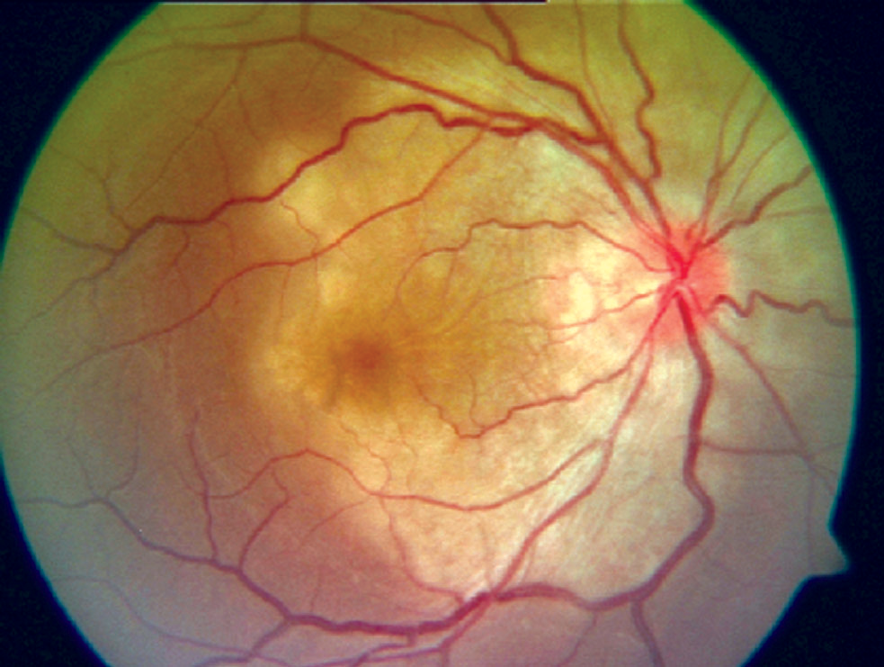
A coroidite em serpentina é uma doença inflamatória rara que afeta a parte posterior do olho (coroide e retina). Ela recebe esse nome porque as lesões oculares se espalham de forma semelhante a uma serpente. A condição geralmente se desenvolve entre os 30 e 70 anos de idade e pode levar à perda de visão, especialmente quando a mácula (região central da retina) é afetada.[1]
Sinais e sintomas
As pessoas afetadas apresentam lesões nos olhos que duram de semanas a meses e causam cicatrização do tecido ocular. A recorrência dessas lesões é comum. A perda de visão pode ocorrer em um ou ambos os olhos quando a mácula está envolvida.[1]
Causas genéticas
Diagnóstico
O diagnóstico é baseado no exame oftalmológico detalhado, incluindo a observação das lesões típicas em formato de serpente. Exames de imagem como angiografia fluoresceínica e verde de indocianina podem auxiliar na avaliação. Não existem testes genéticos específicos disponíveis para essa condição.[1][3][4]
Tratamento e manejo
Tratamentos citados na literatura
A literatura científica (fonte: PubTator3) menciona associações entre a coroidite em serpentina e diversos medicamentos, com base no número de publicações. Essas informações são mineradas de estudos e não representam recomendações de tratamento. Os fármacos mais citados incluem: Esteroides (81 publicações), Prednisona (49), Verde de Indocianina (31), Fluoresceína (28), Prednisolona (24), Azatioprina (21), Ciclosporina (17), Penicilinas (17), Bevacizumabe (16) e Ácido Micofenólico (16).[4]
Prognóstico e qualidade de vida
O prognóstico varia conforme a extensão do envolvimento da mácula e a frequência das recorrências. A perda de visão pode ser significativa se a mácula for afetada, mas o tratamento adequado pode ajudar a controlar a inflamação e preservar a visão. O acompanhamento oftalmológico regular é fundamental para a qualidade de vida.[1]
Conteúdo informativo gerado e mantido automaticamente a partir de fontes oficiais (Orphanet, HPO, OMIM, SUS). Não substitui avaliação médica.
Coroidite serpiginosa, também conhecida como coroidopatia peripapilar helicoidal geográfica (CPHG), é uma doença inflamatória bilateral rara, crônica, progressiva e recorrente que envolve o epitélio pigmentar da retina (EPR), as coriocapilares e a coroide. Afeta igualmente homens e mulheres adultos entre a segunda e a sétima décadas de vida.
Encontrou um erro ou informação desatualizada? Sugira uma correção →
Entender a doença
Do básico ao detalhe, leia no seu ritmo
Preparando trilha educativa...
Sinais e sintomas
O que aparece no corpo e com que frequência cada sintoma acontece
Visão geral
A coroidite em serpentina é uma doença inflamatória rara que afeta a parte posterior do olho (coroide e retina). Ela recebe esse nome porque as lesões oculares se espalham de forma semelhante a uma serpente. A condição geralmente se desenvolve entre os 30 e 70 anos de idade e pode levar à perda de visão, especialmente quando a mácula (região central da retina) é afetada.[1]
Sinais e sintomas
As pessoas afetadas apresentam lesões nos olhos que duram de semanas a meses e causam cicatrização do tecido ocular. A recorrência dessas lesões é comum. A perda de visão pode ocorrer em um ou ambos os olhos quando a mácula está envolvida.[1]
Causas genéticas
Diagnóstico
O diagnóstico é baseado no exame oftalmológico detalhado, incluindo a observação das lesões típicas em formato de serpente. Exames de imagem como angiografia fluoresceínica e verde de indocianina podem auxiliar na avaliação. Não existem testes genéticos específicos disponíveis para essa condição.[1][3][4]
Tratamento e manejo
Tratamentos citados na literatura
A literatura científica (fonte: PubTator3) menciona associações entre a coroidite em serpentina e diversos medicamentos, com base no número de publicações. Essas informações são mineradas de estudos e não representam recomendações de tratamento. Os fármacos mais citados incluem: Esteroides (81 publicações), Prednisona (49), Verde de Indocianina (31), Fluoresceína (28), Prednisolona (24), Azatioprina (21), Ciclosporina (17), Penicilinas (17), Bevacizumabe (16) e Ácido Micofenólico (16).[4]
Prognóstico e qualidade de vida
O prognóstico varia conforme a extensão do envolvimento da mácula e a frequência das recorrências. A perda de visão pode ser significativa se a mácula for afetada, mas o tratamento adequado pode ajudar a controlar a inflamação e preservar a visão. O acompanhamento oftalmológico regular é fundamental para a qualidade de vida.[1]
Conteúdo informativo gerado e mantido automaticamente a partir de fontes oficiais (Orphanet, HPO, OMIM, SUS). Não substitui avaliação médica.
Linha do tempo da pesquisa
Encontrou um erro ou informação desatualizada? Sugira uma correção →
Genética e causas
O que está alterado no DNA e como passa nas famílias
Nenhum gene associado encontrado
Os dados genéticos desta condição ainda estão sendo catalogados.
Diagnóstico
Os sinais que médicos procuram e os exames que confirmam
Tratamento e manejo
Remédios, cuidados de apoio e o que precisa acompanhar
Onde tratar no SUS
Hospitais de referência no Brasil e o protocolo oficial do SUS (PCDT)
🇧🇷 Atendimento SUS — Coroidite em serpentina
Selecione um estado ou use sua localização para ver resultados.
Dados de DATASUS/CNES, SBGM, ABNeuro e Ministério da Saúde. Sempre confirme a disponibilidade diretamente com o estabelecimento.
Pesquisa ativa
Ensaios clínicos abertos e novidades científicas recentes
Ensaios em destaque
Pesquisa e ensaios clínicos
2 ensaios clínicos encontrados.
Publicações mais relevantes
NON-INFECTIOUS POSTERIOR UVEITIDES - Atypicals, Variants, and Masquerades: the jungle of differential diagnosis.
Non-infectious posterior and panuveitides (NIPUs) comprise a heterogeneous group of inflammatory disorders of the outer retina and choroid, historically referred to as "white dot syndromes." Recent consensus efforts by the Multimodal Imaging in Uveitis (MUV) Task Force have established standardized diagnostic criteria for the major NIPUs, including multiple evanescent white dot syndrome (MEWDS), multifocal choroiditis and panuveitis/punctate inner choroiditis (MFCPU/PIC), acute posterior multifocal placoid pigment epitheliopathy (APMPPE), serpiginous choroiditis, and birdshot chorioretinopathy (BSCR). Nevertheless, a substantial proportion of cases deviate from classical presentations and fall into diagnostic "grey zones", blurring boundaries between diseases entities and complicating both differential diagnosis and management. This review aims to describe the broad spectrum of atypical, variant, and secondary forms of NIPUs as well as masquerade syndromes. Atypical MEWDS includes bilateral presentations or complicated courses, while MFCPU/PIC with outer retinal atrophy emerges as a notable entity with unclear therapeutic implications. Inflammatory reactions resembling both MEWDS and MFCPU/PIC may also occur as secondary phenomena, triggered by other chorioretinal disorders, most notably inherited retinal diseases (IRDs). Placoid chorioretinopathies, including APMPPE, persistent placoid maculopathy, serpiginous choroiditis, and relentless placoid chorioretinitis, are often distinguished only a posteriori based on disease course, but likely represent a continuum of disorders unified by choroidal ischemia. Atypical presentations of BSCR may feature extensive outer retinal damage, mimicking IRDs. Equally important is the consideration of masquerade syndromes in all suspected cases of NIPUs, as they can present with similar features yet require entirely different treatments. Infectious masquerades include tuberculosis-associated serpiginous-like choroiditis, acute syphilitic posterior placoid chorioretinopathy, and West Nile virus chorioretinitis, whereas vitreoretinal lymphoma is the most frequent neoplastic masquerade. In conclusion, integrating clinical context with high-quality multimodal imaging remains essential to navigate the jungle of differential diagnosis in NIPUs, while future studies should aim to link imaging phenotypes with immune and molecular biomarkers to refine classification and guide targeted therapies.
Sarcoid Uveitis with Choroidal Involvement.
To investigate characteristics of choroidal involvement in sarcoid uveitis. Included were patients with confirmed sarcoidosis and uveitis with an eye examination at the Tays Eye Center at Tampere University Hospital between January 2014 and January 2021. Choroidal involvement was found in 31 of 97 sarcoid uveitis patients (32%), and 5 of them were detected only after reviewing the ocular images for this study. None of our patients presented with vasculitis. Choroidal nodules were found in 10%, multifocal choroiditis in 90% and serpiginous choroiditis in 3% of patients with choroidal involvement. They had a higher rate of chronic course of uveitis (71% vs 29%, p < 0.001), bilateral uveitis (90% vs 58%, p = 0.001), vitreal snowballs (65% vs 15%, p < 0.001), and macular edema (36% vs 9%, p = 0.001). Choroidal involvement is common and may be underdiagnosed in sarcoid uveitis. Therefore, we recommend carefully looking for choroidal involvement not only with slit-lamp but also with ocular imaging in patients with uveitis.
Atypical presentation of macular serpiginous choroiditis masquerading as age related macular degeneration.
To describe a case of macular serpiginous choroiditis (SC) masquerading as age related macular degeneration. Multimodal imaging (MMI), including ultra-widefield fundus autofluorescence and fluorescein and indocyanine green angiography werer performed. Cross-sectional and en face optical coherence tomography (OCT) and OCT angiography (OCTA) facilitated the correct diagnosis of this masquerade condition. A 57-year-old woman, with a history of long-standing vision loss in the left eye (OS), presented with acute vision loss in the right eye (OD). Color fundus photography showed a focal macular lesion central OD and an irregular atrophic macular scar central OS. OCT OD displayed large drusenoid lesions in the macula OD and a macular scar OS. OCTA showed evidence of inner choroidal ischemia central OD corresponding to the drusenoid lesions without shadow artifact OD. A diagnosis of macular serpiginous choroiditis was rendered OD and the patient was started on immunomodulatory therapy (IMT) including oral steroids and increasing dosages of mycophenolate therapy. After three months of follow-up, the visual acuity remarkably improved, the drusenoid lesions completely resolved on OCT, and choroidal ischemia completely regressed on OCTA OD. Placoid disorders can masquerade as AMD. We present an unusual case of macular serpiginous choroiditis with new onset drusenoid lesions driven by inner choroidal ischemia on OCTA and with resolution following IMT. OCTA can be an invaluable tool to correctly diagnose placoid conditions which is essential to guide therapeutic considerations.
En Face Optical Coherence Tomography and OCT Angiography in the Pathoanatomy of Inflammatory Macular Disease.
To review the pivotal role of en face optical coherence tomography (OCT) and OCT angiography (OCTA) in elucidating the pathoanatomy, diagnostic markers, and clinical management of inflammatory macular diseases. These noninvasive modalities provide depth-resolved, layer-specific visualization of structural and vascular alterations, enhancing our understanding of disease mechanisms and facilitating monitoring of disease activity and therapeutic response. This perspective article synthesizes recent advances in multimodal imaging (MMI) with emphasis on en face OCT and OCTA across the spectrum of posterior noninfectious and infectious uveitic disorders. Representative diseases include multiple evanescent white dot syndrome (MEWDS), punctate inner choroidopathy (PIC), and idiopathic multifocal choroiditis (iMFC), acute zonal occult outer retinopathy (AZOOR), acute posterior multifocal placoid pigment epitheliopathy (APMPPE) and its chronic variants, serpiginous choroiditis (SC), acute syphilitic posterior placoid chorioretinopathy (ASPPC), and Vogt-Koyanagi-Harada (VKH) disease. Imaging signatures were reviewed in relation to pathophysiology, differential diagnosis, and clinical management. En face OCT delineates disease-specific topographic patterns: "dots and spots" in MEWDS, hyperreflective plaques in PIC/iMFC, trizonal maps in AZOOR, sharply demarcated placoid lesions in APMPPE, geographic patterns in SC, and concentric hyperreflective "fingerprint" rings in VKH. OCTA provides complementary vascular insights, distinguishing preserved vs impaired choriocapillaris (CC) flow and revealing disease-driven ischemia. MEWDS typically demonstrates preserved CC perfusion, in contrast to the flow deficits of APMPPE, relentless placoid chorioretinitis, and SC. In PIC/iMFC, OCTA enables detection of inflammatory choroidal neovascularization (iCNV), while in ASPPC and VKH, it captures reversible or persistent CC flow voids following appropriate systemic therapy. Structural-vascular correlation permits differentiation between ischemic and nonischemic disorders, recognition of masquerades, and monitoring of subclinical disease activity. En face OCT and OCTA have redefined the diagnostic and management landscape of inflammatory macular diseases by providing rapid, reproducible, and layer-specific structural-vascular information. Their disease-specific signatures enhance accuracy, support treatment decisions, and improve monitoring of progression and complications such as inflammatory CNV. Integration of these tools into routine multimodal imaging protocols is essential, while future research should prioritize standardization of acquisition and interpretation criteria, quantitative biomarkers, and expanded use of widefield technologies to capture peripheral and longitudinal disease dynamics.
Relentless placoid chorioretinitis.
Relentless placoid chorioretinitis (RPC) is a rare, vision-threatening posterior uveitis that predominantly affects young adults. The hallmark clinical findings are numerous scattered placoid chorioretinal lesions involving the midperipheral and far peripheral fundus as well as the posterior pole and a persistent or recurrent course resulting in lesions at different chronological stages, with fresh creamy placoid lesions and healing pigmented lesions being present at the same time. Although RPC is frequently described as a disease that is intermediate between acute posterior multifocal placoid pigment epitheliopathy (APMPPE) and serpiginous choroiditis, it more closely resembles APMPPE. It is important to identify those patients with RPC who present with an APMPPE phenotype so that appropriate immunomodulatory therapy is instituted without delay, as most cases of RPC are refractory to corticosteroid monotherapy. Examination findings may help differentiate RPC and APMPPE. Multimodal imaging, including ultra-widefield imaging, and selective investigations aid in distinguishing RPC from other placoid diseases and types of posterior uveitis.
Publicações recentes
Ultra-Widefield Indocyanine Green Angiography in Uveitis.
Noninfectious Posterior Uveitides - Atypicals, Variants, and Masquerades: The Jungle of Differential Diagnosis.
Sarcoid Uveitis with Choroidal Involvement.
Atypical presentation of macular serpiginous choroiditis masquerading as age related macular degeneration.
En Face Optical Coherence Tomography and OCT Angiography in the Pathoanatomy of Inflammatory Macular Disease.
📚 EuropePMC149 artigos no totalmostrando 108
NON-INFECTIOUS POSTERIOR UVEITIDES - Atypicals, Variants, and Masquerades: the jungle of differential diagnosis.
American journal of ophthalmologySarcoid Uveitis with Choroidal Involvement.
Clinical ophthalmology (Auckland, N.Z.)Atypical presentation of macular serpiginous choroiditis masquerading as age related macular degeneration.
American journal of ophthalmology case reportsEn Face Optical Coherence Tomography and OCT Angiography in the Pathoanatomy of Inflammatory Macular Disease.
American journal of ophthalmologyOpposing pathophysiological concepts in multiple evanescent white dot syndrome: The great MEWDS schism.
Saudi journal of ophthalmology : official journal of the Saudi Ophthalmological SocietyBilateral ampiginous choroiditis after COVID-19: a report of two cases.
Romanian journal of ophthalmologyThe Role of Adding Intravitreal Dexamethasone Implant to the Standard Management of Serpiginous Choroiditis for Achieving Rapid Remission: A Case Report.
International medical case reports journalRelentless Placoid Chorioretinitis: A Differential Diagnosis and Management Approach in a Challenging Case.
CureusMinimum Imaging Sets for Diagnosis, Activity Assessment, and Complications in Noninfectious Posterior Uveitis - Multimodal Imaging in Uveitis (MUV) Task Force Report 9.
American journal of ophthalmologyNoninfectious Uveitis Syndromes.
Advances in experimental medicine and biologyRelentless placoid chorioretinitis.
Survey of ophthalmologySystematic Review of Clinical Utility of Multimodal Imaging in Noninfectious Posterior Uveitis: MUV Project Report 3.
American journal of ophthalmologyClinical and Imaging Features Aiding the Differentiation of Acute Posterior Multifocal Placoid Pigmented Epitheliopathy from Other Placoid Diseases.
Ocular immunology and inflammationEvidence and Consensus-Based Imaging Guidelines in Serpiginous Choroiditis-Multimodal Imaging in Uveitis (MUV) Task Force Report 4.
American journal of ophthalmologyManagement of Tubercular Retinal Vasculitis: A Case Report From an Endemic Region.
CureusAn update of multimodal imaging in white dot syndrome.
Oman journal of ophthalmologyChoroidal vasculitis as a biomarker of inflammation of the choroid. Indocyanine Green Angiography (ICGA) spearheading for diagnosis and follow-up, an imaging tutorial.
Journal of ophthalmic inflammation and infectionSerpiginous Choroiditis After COVID-19 Infection.
Journal of vitreoretinal diseasesLong-Term Outcomes of Adalimumab Treatment in Conventional Treatment-Resistant Serpiginous Choroiditis.
Ocular immunology and inflammationRare Case of Tubercular Serpiginous-Like Choroiditis.
CureusDiagnostic Challenges in Inflammatory Choroidal Neovascularization.
Medicina (Kaunas, Lithuania)A retrospective study describing the effective exchange of total blood plasma for disease control in the exacerbation of serpiginous choroiditis.
Therapeutic apheresis and dialysis : official peer-reviewed journal of the International Society for Apheresis, the Japanese Society for Apheresis, the Japanese Society for Dialysis TherapyBeauty of Black and White: Autofluorescence-aided Differentiation of Serpiginous Choroiditis from Tubercular Serpiginous-Like Choroiditis.
Nepalese journal of ophthalmology : a biannual peer-reviewed academic journal of the Nepal Ophthalmic Society : NEPJOPHClinical association and visual morbidity in uveitis with systemic diseases: An analysis from a tertiary ophthalmic center.
Oman journal of ophthalmologyTREATMENT OF SERPIGINOUS CHOROIDITIS WITH SUBTENON TRIAMCINOLONE IN CONJUNCTION WITH SYSTEMIC STEROIDS: A CASE REPORT.
Retinal cases & brief reportsChallenges in posterior uveitis-tips and tricks for the retina specialist.
Journal of ophthalmic inflammation and infectionDexamethasone intravitreal implant for macular edema and some other rare indications in uveitis.
Medicine internationalBlue-Light Fundus Autofluorescence (BAF), an Essential Modality for the Evaluation of Inflammatory Diseases of the Photoreceptors: An Imaging Narrative.
Diagnostics (Basel, Switzerland)[Acute posterior multifocal placoid pigment epitheliopathy, serpiginous choroiditis and related diseases].
Journal francais d'ophtalmologieRelentless placoid chorioretinitis: A review of four cases in pediatric and young adult patients with a discussion of therapeutic strategies.
Frontiers in pediatricsSequential MFN2-Related Optic Neuropathies in a Patient With Serpiginous Choroiditis.
Journal of neuro-ophthalmology : the official journal of the North American Neuro-Ophthalmology SocietySustained Control of Serpiginous Choroiditis with the Fluocinolone Acetonide 0.18 mg Intravitreal Implant.
Case reports in ophthalmological medicineTubercular serpiginous choroiditis.
Journal of ophthalmic inflammation and infectionLong-term outcomes of small-incision cataract surgery in patients with uveitis.
Indian journal of ophthalmologyBenefits and Limitations of OCT-A in the Diagnosis and Follow-Up of Posterior Intraocular Inflammation in Current Clinical Practice: A Valuable Tool or a Deceiver?
Diagnostics (Basel, Switzerland)Tubercular DNA PCR of ocular fluids and blood in cases of presumed ocular tuberculosis: a pilot study.
Therapeutic advances in ophthalmologyMechanisms, Pathophysiology and Current Immunomodulatory/Immunosuppressive Therapy of Non-Infectious and/or Immune-Mediated Choroiditis.
Pharmaceuticals (Basel, Switzerland)[Serpiginous-Like Choroiditis: literature Review].
Revista de la Facultad de Ciencias Medicas (Cordoba, Argentina)Diagnosis and Treatment of Primary Inflammatory Choriocapillaropathies (PICCPs): A Comprehensive Overview.
Medicina (Kaunas, Lithuania)Tuberculosis-related serpiginous choroiditis: aggressive therapy with dual concomitant combination of multiple anti-tubercular and multiple immunosuppressive agents is needed to halt the progression of the disease.
Journal of ophthalmic inflammation and infectionCommentary: Importance of ocular imaging in macular serpiginous choroiditis.
Indian journal of ophthalmologyClinical profile, multimodal imaging, and treatment response in macular serpiginous choroiditis.
Indian journal of ophthalmologyROLE OF OPTICAL COHERENCE TOMOGRAPHY ANGIOGRAPHY IN DETECTING AND MONITORING INFLAMMATORY CHOROIDAL NEOVASCULARIZATION.
Retina (Philadelphia, Pa.)Clinicopathology of non-infectious choroiditis: evolution of its appraisal during the last 2-3 decades from "white dot syndromes" to precise classification.
Journal of ophthalmic inflammation and infectionSerpiginous Choroiditis Complicated with Choroidal Neovascular Membrane Detected using Optical Coherence Tomography Angiography: A Case Series and Literature Review.
Turkish journal of ophthalmologyLongitudinal study of serpiginous choroiditis and serpiginous like choroiditis using wide field OCT angiography.
European journal of ophthalmologyClassification of Non-Infectious and/or Immune Mediated Choroiditis: A Brief Overview of the Essentials.
Diagnostics (Basel, Switzerland)Classification Criteria for Serpiginous Choroiditis.
American journal of ophthalmologyReview of the Current Literature and Our Experience on the Value of OCT-angiography in White Dot Syndromes.
Ocular immunology and inflammationChlorambucil combination therapy in refractory serpiginous choroiditis: A cure?
American journal of ophthalmology case reportsSwept source OCTA reveals a link between choriocapillaris blood flow and vision loss in a case of tubercular serpiginous-like choroiditis.
American journal of ophthalmology case reportsPlacoid lesions of the retina: progress in multimodal imaging and clinical perspective.
The British journal of ophthalmologyUnilateral macular serpiginous-like choroiditis as the initial manifestation of presumed ocular tuberculosis.
International journal of retina and vitreous[Fundus imaging features of purified protein derivative and T-spot positive tubercular serpiginous-like choroiditis].
[Zhonghua yan ke za zhi] Chinese journal of ophthalmologySerpiginous choroiditis presenting after SARS-CoV-2 infection: A new immunological trigger?
European journal of ophthalmologyThe impact of impending / onset of vision loss on depression, anxiety, and vision-related quality of life in Birdshot-Retinochoroiditis and Serpiginous Choroiditis.
PloS one[Unilateral serpiginous choroiditis secondary to ocular tuberculosis].
Journal francais d'ophtalmologieSerpiginous Choroiditis Presenting in Association With Clostridium difficile Infection and Ulcerative Colitis.
Journal of vitreoretinal diseasesEvaluation of change in the vascular density of choriocapillaris on optical coherence tomography angiography in eyes with serpiginous choroiditis.
Indian journal of ophthalmologyA Novel Method of Quantifying the Choriocapillaris in Normal and Post-inflammatory Eyes.
Ocular immunology and inflammationOptical coherence tomography angiography (OCTA) findings in Serpiginous Choroiditis.
BMC ophthalmologyMacular serpiginous choroiditis secondary to tuberculosis.
Journal francais d'ophtalmologie[Placoid pigment epitheliopathy and serpiginous choroiditis].
Journal francais d'ophtalmologiePlacoid pigment epitheliopathy and serpiginous choroiditis.
Journal francais d'ophtalmologie[White dot syndromes : Principles, diagnostics, and treatment].
Der Ophthalmologe : Zeitschrift der Deutschen Ophthalmologischen GesellschaftTextural Properties of Choriocapillaris on OCTA in Healed Inflammatory Choriocapillaropathies.
Ophthalmic surgery, lasers & imaging retinaCase Report: A Rare Variant of Macular Serpiginous Choroiditis.
Optometry and vision science : official publication of the American Academy of OptometryRelentless Placoid Chorioretinitis: A Case Series of Successful Tapering of Systemic Immunosuppressants Achieved with Adalimumab.
Case reports in ophthalmologyLimited efficacy of adalimumab in the acute phase of serpiginous choroiditis refractory to corticosteroid and cyclosporine, a case report.
BMC ophthalmologyCyclosporine and prednisolone combination therapy as a potential therapeutic strategy for relentless placoid chorioretinitis.
American journal of ophthalmology case reportsCommentary: Serpiginous choroiditis-so near yet so far.
Indian journal of ophthalmologyEnigma of serpiginous choroiditis.
Indian journal of ophthalmologyChoroidal neovascularisation secondary to serpiginous choroiditis: The value of OCT-angiography in diagnosis and response to therapy with aflibercept.
Archivos de la Sociedad Espanola de OftalmologiaMacular serpiginous choroiditis - case report.
Romanian journal of ophthalmologySimilarities and differences between three different types of white dot syndrome and the therapeutic possibilities.
Romanian journal of ophthalmologyAn update on inflammatory choroidal neovascularization: epidemiology, multimodal imaging, and management.
Journal of ophthalmic inflammation and infectionSwept-Source OCT Angiography of Serpiginous Choroiditis.
Ophthalmology. RetinaMultimodal Imaging to Monitor Recurrent Serpiginous Choroiditis.
OphthalmologyOptical Coherence Tomography Features of Tuberculous Serpiginous-like Choroiditis and Serpiginous Choroiditis.
Biomedical and environmental sciences : BESQuantitative polymerase chain reaction analysis of serpiginous choroiditis with biopsy-proven testicular tuberculosis.
Indian journal of ophthalmologyThe Role of Dexamethasone Implant in the Management of Tubercular Uveitis.
Ocular immunology and inflammationVITREOUS TREPONEMAL ANTIBODY AS A SUPPLEMENTARY TEST TO SEROLOGY FOR THE CONFIRMATION OF SYPHILITIC CHORIORETINITIS.
Retinal cases & brief reportsA Rare Case of Infectious Multifocal Serpiginoid Choroiditis.
Acta clinica CroaticaUltra-Wide Field Imaging in Paradoxical Worsening of Tubercular Multifocal Serpiginoid Choroiditis after the Initiation of Anti-Tubercular Therapy.
Ocular immunology and inflammationSwept-source optical coherence tomography angiography in serpiginous choroiditis.
The British journal of ophthalmologyForme fruste focal choroidal excavation in a case of serpiginous choroiditis.
Clinical & experimental optometryMultimodal Evaluation of Patients with Acute Posterior Multifocal Placoid Pigment Epitheliopathy and Serpiginous Choroiditis.
Ocular immunology and inflammationThe Incidence of Ocular Tuberculosis in Australia Over the Past 10 Years (2006-2015).
Ophthalmic epidemiologyTreatment Results in Serpiginous Choroiditis and Multifocal Serpiginoid Choroiditis Associated with Latent Tuberculosis.
Turkish journal of ophthalmologyMulti-modal imaging and anatomic classification of the white dot syndromes.
International journal of retina and vitreousMultimodal Imaging Including Optical Coherence Tomography Angiography in Serpiginous Choroiditis.
Ocular immunology and inflammationObservation and Clinical Pattern in Patients with White Dot Syndromes: The Role of Color Photography in Monitoring Ocular Changes in Long-Term Observation.
Medical science monitor : international medical journal of experimental and clinical researchParadoxical Worsening of Tubercular Serpiginous-Like Choroiditis after Initiation of Antitubercular Therapy.
Turkish journal of ophthalmologySERPIGINOUS CHOROIDITIS IMAGED BY OPTICAL COHERENCE TOMOGRAPHY ANGIOGRAPHY.
Retinal cases & brief reportsEfficacy of reduced dose of intravitreal triamcinolone acetonide in a case of active serpiginous choroiditis.
Indian journal of ophthalmologyShort-term Intensive Immunosuppression: A Randomized, Three-arm Study of Intravenous Pulse Methylprednisolone and Cyclophosphamide in Macular Serpiginous Choroiditis.
Ocular immunology and inflammationMultimodal Imaging of Serpiginous Choroiditis.
Optometry and vision science : official publication of the American Academy of OptometryTreatment of Serpiginous Choroiditis with Chlorambucil: A Report of 17 Patients.
Ocular immunology and inflammationRelentless placoid chorioretinitis – A case report.
Srpski arhiv za celokupno lekarstvoDexamethasone intravitreal implant in serpiginous choroiditis.
The British journal of ophthalmologyENHANCED DEPTH IMAGING OPTICAL COHERENCE TOMOGRAPHY FEATURES IN AREAS OF CHORIOCAPILLARIS HYPOPERFUSION.
Retina (Philadelphia, Pa.)A Case of Ampiginous Choroiditis.
Case reports in ophthalmologySimultaneous Single Dexamethasone Implant and Ranibizumab Injection in a Case with Active Serpiginous Choroiditis and Choroidal Neovascular Membrane.
Case reports in ophthalmology[A form of macular serpiginous choroiditis].
Journal francais d'ophtalmologiePreservation of macular structure and function after intravitreal aflibercept for choroidal neovascularization associated with serpiginous choroiditis.
Acta ophthalmologicaReal-time and nested polymerase chain reaction in the diagnosis of multifocal serpiginoid choroiditis caused by Mycobacterium tuberculosis - a case report.
Journal of ophthalmic inflammation and infection[Macular serpiginous choroiditis].
The Pan African medical journalPulse cyclophosphamide therapy in the management of patients with macular serpiginous choroidopathy.
Indian journal of ophthalmologyAssociações
Organizações que acompanham esta doença — pra ter apoio e orientação
Ainda não temos associações cadastradas para Coroidite em serpentina.
É de uma associação que acompanha esta doença? Fale com a gente →
Comunidades
Grupos ativos de quem convive com esta doença aqui no Raras
Ainda não existe comunidade no Raras para Coroidite em serpentina
Pacientes, familiares e cuidadores se organizam em comunidades pra compartilhar experiências, fazer perguntas e se apoiar. Você pode ser o primeiro.
Tire suas dúvidas
Perguntas, dicas e experiências compartilhadas aqui na página
Participe da discussão
Faça login para postar dúvidas, compartilhar experiências e interagir com especialistas.
Fazer loginDoenças relacionadas
Doenças com sintomas parecidos — ajudam quem ainda está buscando diagnóstico
Ainda não achamos doenças com sintomas parecidos o suficiente.
Referências e fontes
Bases de dados externas citadas neste artigo
Publicações científicas
Artigos indexados no PubMed ligados a esta doença no grafo RarasNet — título, periódico e PMID direto da fonte, sem intermediação de IA.
- NON-INFECTIOUS POSTERIOR UVEITIDES - Atypicals, Variants, and Masquerades: the jungle of differential diagnosis.
- Sarcoid Uveitis with Choroidal Involvement.
- Atypical presentation of macular serpiginous choroiditis masquerading as age related macular degeneration.
- En Face Optical Coherence Tomography and OCT Angiography in the Pathoanatomy of Inflammatory Macular Disease.
- Relentless placoid chorioretinitis.
- Ultra-Widefield Indocyanine Green Angiography in Uveitis.
Bases de dados e fontes oficiais
Identificadores e referências canônicas usadas para montar este verbete.
- ORPHA:35686(Orphanet)
- MONDO:0018152(MONDO)
- GARD:31(GARD (NIH))
- Busca completa no PubMed(PubMed)
- Q7455142(Wikidata)
Dados compilados pelo RarasNet a partir de fontes abertas (Orphanet, OMIM, MONDO, PubMed/EuropePMC, ClinicalTrials.gov, DATASUS, PCDT/MS). Este conteúdo é informativo e não substitui avaliação médica.
Conteúdo mantido por Agente Raras · Médicos e pesquisadores podem colaborar
Coroidite em serpentina
📋 Origem dos dados
Esta página agrega dados de fontes públicas e oficiais. Dados sobre cobertura no SUS (PCDT, CEAF) são verificados ativamente por agente proativo (ver badge no infobox). Demais dados têm atribuição de fonte + data da última sincronização — clique para abrir o original.
- Doença rara (ontologia)
- fonte: Orphanet
- Identificador unificado
- fonte: MONDO
- Codificação WHO/SUS
- fonte: WHO ICD-10 / DATASUS
- CID-11 (futuro)
- fonte: WHO ICD-11
- NIH/GARD
- fonte: GARD (NIH)
- Dado público estruturado
- fonte: Wikidata