A vasculite, caracterizada por lesões inflamatórias na parede dos vasos, pode ser causada por diferentes vírus.
Introdução
O que você precisa saber de cara
A vasculite, caracterizada por lesões inflamatórias na parede dos vasos, pode ser causada por diferentes vírus.
Escala de raridade
<1/50kMuito rara
1/20kRara
1/10kPouco freq.
1/5kIncomum
1/2k
Encontrou um erro ou informação desatualizada? Sugira uma correção →
Entender a doença
Do básico ao detalhe, leia no seu ritmo
Preparando trilha educativa...
Sinais e sintomas
O que aparece no corpo e com que frequência cada sintoma acontece
Partes do corpo afetadas
+ 22 sintomas em outras categorias
Características mais comuns
Os sintomas variam de pessoa para pessoa. Abaixo estão as 57 características clínicas mais associadas, ordenadas por frequência.
Linha do tempo da pesquisa
Encontrou um erro ou informação desatualizada? Sugira uma correção →
Genética e causas
O que está alterado no DNA e como passa nas famílias
Nenhum gene associado encontrado
Os dados genéticos desta condição ainda estão sendo catalogados.
Diagnóstico
Os sinais que médicos procuram e os exames que confirmam
Tratamento e manejo
Remédios, cuidados de apoio e o que precisa acompanhar
Onde tratar no SUS
Hospitais de referência no Brasil e o protocolo oficial do SUS (PCDT)
🇧🇷 Atendimento SUS — Vasculite vírica não HBV não HCV
Selecione um estado ou use sua localização para ver resultados.
Dados de DATASUS/CNES, SBGM, ABNeuro e Ministério da Saúde. Sempre confirme a disponibilidade diretamente com o estabelecimento.
Pesquisa ativa
Ensaios clínicos abertos e novidades científicas recentes
Pesquisa e ensaios clínicos
Nenhum ensaio clínico registrado para esta condição.
Publicações mais relevantes
More Than Just Mosquito Bites: A Travel-Associated Case of Cutaneous Small-Vessel Vasculitis.
A 39-year-old man presented with a bilateral lower limb pruritic and purpuric rash following travel to Chongqing, China. He was initially treated for cellulitis abroad. On arrival, his rash was noted, and the initial workup revealed raised inflammatory markers with a CRP of 252 mg/L and an ESR of 80 mm/hr. Examination of the lower limb reveals symmetrical, non-blanchable purpuric plaques with post-inflammatory hyperpigmentation. He was admitted for further inpatient treatment. Serological workup for autoimmune vasculitis was negative. Urine phase contrast microscopy showed 12% dysmorphic red blood cells, below the threshold typically seen in glomerular bleeding. A punch biopsy revealed minimal, superficial perivascular lymphocytic infiltrate without classic vasculitic features, likely due to timing post-acute inflammation. The clinical workup and examination findings support a diagnosis of post-infectious cutaneous small-vessel vasculitis (CSVV). This case demonstrates the classic distribution and morphology of CSVV. Early recognition of CSVV facilitates prompt systemic evaluation and discontinuation of potential triggers and avoids unnecessary antibiotic use.
Delayed cerebral infarction in pneumococcal meningitis due to postinfectious vasculitis: A case report.
Delayed cerebral infarction (DCI) is a rare but devastating complication of bacterial meningitis that is characterized by an initial good recovery followed by sudden deterioration several days after presentation. The potential mechanisms underlying DCI encompass postinfectious vasculitis, vasospasm, and cerebral thrombosis. Sequential magnetic resonance angiography and characteristic high-resolution magnetic resonance patterns are helpful in discerning the cause of DCI. A right-handed 45-year-old man with nasal leakage of cerebrospinal fluid from a car accident was admitted to the local hospital for fever, vomiting, and impaired consciousness. Post-infectious vasculitis. Meropenem (1 g 3 times/day), vancomycin (0.5 g 4 times/day), and dexamethasone (10 mg/day, gradually reduced to withdraw) were administered immediately after the patient was diagnosed with Streptococcus pneumoniae meningitis. After the patient was diagnosed with DCI, we applied nimodipine (10 mg 3 times/day) to prevent vasospasm, clopidogrel (75 mg/day), and heparin to counteract thrombosis, and prednisone (60 mg/day) was applied to reduce the inflammatory reaction. On the 52nd day, the patient's magnetic resonance angiography reexamination indicated a continuous expansion of the cerebral infarction area and a worsening of cerebrovascular occlusion. After the prednisone dose was increased, the consciousness disorder ceased to progress. Early detection and treatment are very important for DCI, the sequential magnetic resonance imaging and high-resolution magnetic resonance imaging are conducive to identify the etiology of DCI and providing guidance for treatment. The delayed tapering of glucocorticoids appears to be beneficial for alleviating the severity of DCI.
Post-infectious vasculitis secondary to Campylobacter coli infection.
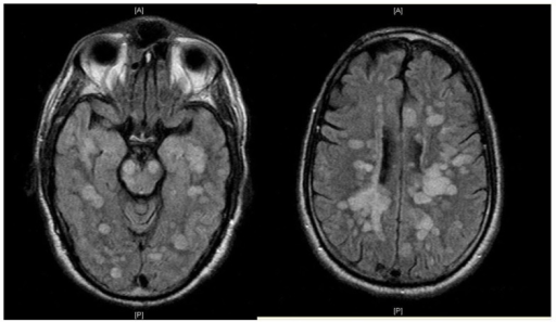
Magnetic Resonance Vessel Wall Imaging in Central Nervous System Vasculitides: A Case Series.
We aim to report 3 cases of central nervous system (CNS) vasculitides, in which high-resolution magnetic resonance vessel wall imaging (HR-VWI) findings were instrumental in the diagnosis and management. Case 1: A 41-year-old obese, smoker female with arterial hypertension presented with recurrent transient ischemic attacks. Computed topography angiography demonstrated bilateral middle cerebral artery (MCA) stenosis. HR-VWI revealed uniform enhancement and thickening of the arterial wall, suggestive of MCA vasculitis. The patient reported chronic calf rash that was biopsied and revealed unspecified connective tissue disease. With immunomodulation, patient remained asymptomatic and 6-month surveillance HR-VWI showed improved MCA stenoses.Case 2: A 56-year-old male with herpes simplex virus 1 encephalitis was treated with antiviral therapy and improved clinically. Two months later, the brain magnetic resonance imaging revealed new temporo-parietal edema and distal MCA hyperintense vessels. HR-VWI showed MCA concentric smooth contrast enhancement, that was attributed to postinfectious vasculitis and had resolved on follow-up HR-VWI.Case 3: A 41-year-old male presented with 1-week of headache and encephalopathy. Brain magnetic resonance imaging revealed punctate multifocal acute ischemic infarcts and no contrast-enhancement. HR-VWI showed multifocal diffuse enhancement of distal cerebral vasculature. Patient subsequently developed branch retinal artery occlusion and hearing loss and was diagnosed with Susac syndrome. No recurrent symptoms were noted after immunotherapy initiation. In these 3 cases, HR-VWI identified distinctive vascular inflammatory changes, which were crucial to guide the etiological workup, positive diagnosis, surveillance neuroimaging, and targeted treatment. HR-VWI is an important diagnostic tool in CNS vasculitides, by providing nuanced information about arterial wall integrity and pathology.
Pneumococcal Meningitis Complicated by Cerebral Vasculitis, Abscess, Hydrocephalus, and Hearing Loss.
Intracranial abscesses, postinfectious vasculitis, and hydrocephalus are rare complications of Streptococcus pneumoniae (S. pneumoniae) meningitis, and to our knowledge, there have been no case reports where all these 3 complications occurred in a single patient with Streptococcus pneumoniae meningitis. Here, we report a case of a 48-year-old male who developed postinfectious vasculitis, abscess, hydrocephalus, and hearing loss after S. pneumoniae meningitis. Clinicians ought to be aware of the possible adverse outcomes of S. pneumoniae meningitis and the limitations of current treatment options.
Publicações recentes
Delayed cerebral infarction in pneumococcal meningitis due to postinfectious vasculitis: A case report.
Magnetic Resonance Vessel Wall Imaging in Central Nervous System Vasculitides: A Case Series.
Pneumococcal Meningitis Complicated by Cerebral Vasculitis, Abscess, Hydrocephalus, and Hearing Loss.
📚 EuropePMC1 artigos no totalmostrando 6
More Than Just Mosquito Bites: A Travel-Associated Case of Cutaneous Small-Vessel Vasculitis.
CureusDelayed cerebral infarction in pneumococcal meningitis due to postinfectious vasculitis: A case report.
MedicinePost-infectious vasculitis secondary to Campylobacter coli infection.
International journal of infectious diseases : IJID : official publication of the International Society for Infectious DiseasesMagnetic Resonance Vessel Wall Imaging in Central Nervous System Vasculitides: A Case Series.
The neurologistPneumococcal Meningitis Complicated by Cerebral Vasculitis, Abscess, Hydrocephalus, and Hearing Loss.
Case reports in infectious diseasesStreptococcus Pneumoniae Intracranial Abscess and Post-Infectious Vasculitis.
Infectious disease reportsAssociações
Organizações que acompanham esta doença — pra ter apoio e orientação
Ainda não temos associações cadastradas para Vasculite vírica não HBV não HCV.
É de uma associação que acompanha esta doença? Fale com a gente →
Comunidades
Grupos ativos de quem convive com esta doença aqui no Raras
Ainda não existe comunidade no Raras para Vasculite vírica não HBV não HCV
Pacientes, familiares e cuidadores se organizam em comunidades pra compartilhar experiências, fazer perguntas e se apoiar. Você pode ser o primeiro.
Tire suas dúvidas
Perguntas, dicas e experiências compartilhadas aqui na página
Participe da discussão
Faça login para postar dúvidas, compartilhar experiências e interagir com especialistas.
Fazer loginDoenças relacionadas
Doenças com sintomas parecidos — ajudam quem ainda está buscando diagnóstico
Referências e fontes
Bases de dados externas citadas neste artigo
Publicações científicas
Artigos indexados no PubMed ligados a esta doença no grafo RarasNet — título, periódico e PMID direto da fonte, sem intermediação de IA.
- More Than Just Mosquito Bites: A Travel-Associated Case of Cutaneous Small-Vessel Vasculitis.
- Delayed cerebral infarction in pneumococcal meningitis due to postinfectious vasculitis: A case report.
- Post-infectious vasculitis secondary to Campylobacter coli infection.International journal of infectious diseases : IJID : official publication of the International Society for Infectious Diseases· 2023· PMID 36828235mais citado
- Magnetic Resonance Vessel Wall Imaging in Central Nervous System Vasculitides: A Case Series.
- Pneumococcal Meningitis Complicated by Cerebral Vasculitis, Abscess, Hydrocephalus, and Hearing Loss.
Bases de dados e fontes oficiais
Identificadores e referências canônicas usadas para montar este verbete.
- ORPHA:48435(Orphanet)
- MONDO:0018837(MONDO)
- GARD:18835(GARD (NIH))
- Busca completa no PubMed(PubMed)
- Q55788379(Wikidata)
Dados compilados pelo RarasNet a partir de fontes abertas (Orphanet, OMIM, MONDO, PubMed/EuropePMC, ClinicalTrials.gov, DATASUS, PCDT/MS). Este conteúdo é informativo e não substitui avaliação médica.
Conteúdo mantido por Agente Raras · Médicos e pesquisadores podem colaborar
Vasculite vírica não HBV não HCV
📋 Origem dos dados
Esta página agrega dados de fontes públicas e oficiais. Dados sobre cobertura no SUS (PCDT, CEAF) são verificados ativamente por agente proativo (ver badge no infobox). Demais dados têm atribuição de fonte + data da última sincronização — clique para abrir o original.
- Doença rara (ontologia)
- fonte: Orphanet
- Identificador unificado
- fonte: MONDO
- Codificação WHO/SUS
- fonte: WHO ICD-10 / DATASUS
- CID-11 (futuro)
- fonte: WHO ICD-11
- NIH/GARD
- fonte: GARD (NIH)
- Dado público estruturado
- fonte: Wikidata