Um perineurioma intraneural é um tumor benigno raro dentro da bainha de um único nervo que cresce, mas geralmente não recorre nem metastatiza. Essas lesões são compostas apenas por células perineuriais, clonadas de uma única célula. Elas são distintas de schwannoma e neurofibroma.
Introdução
O que você precisa saber de cara
Visão geral
O perineurioma intraneural é um tumor benigno (grau I da Organização Mundial da Saúde, OMS) que se origina dentro do endoneuro, a camada mais interna que envolve as fibras nervosas. A doença é caracterizada pela formação de estruturas chamadas 'bulbos de pseudo-cebola', resultantes da proliferação das células perineurais. Por ser um tumor de crescimento lento e baixo potencial de agressividade, geralmente não se espalha para outras partes do corpo.[1]
Sinais e sintomas
Os sintomas do perineurioma intraneural podem variar de acordo com a localização e o tamanho do tumor. Os sinais mais comuns incluem fraqueza muscular progressiva, perda de sensibilidade (como formigamento ou dormência) e dor no trajeto do nervo afetado. Em alguns casos, pode haver atrofia (diminuição do volume) dos músculos inervados pelo nervo comprometido. A doença pode se manifestar na infância, adolescência ou idade adulta.[1]
Causas genéticas
Até o momento, não há um gene específico identificado como causa direta do perineurioma intraneural. A doença não é hereditária, ou seja, não é transmitida de pais para filhos. Estudos genéticos estão em andamento para compreender melhor os mecanismos moleculares envolvidos no desenvolvimento desse tumor.[1][3]
Diagnóstico
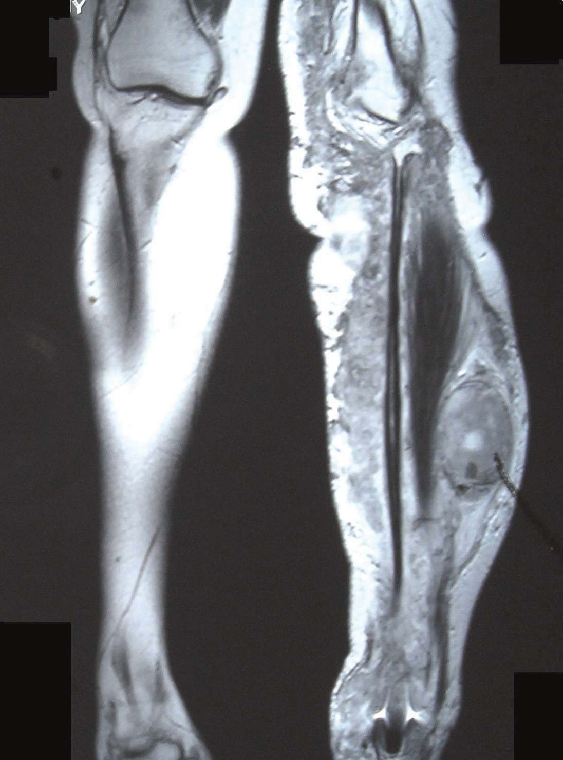
O diagnóstico do perineurioma intraneural é baseado na combinação de exames de imagem, como ressonância magnética (RM), e na análise histopatológica (biópsia) do tecido tumoral. Na microscopia, a presença dos 'bulbos de pseudo-cebola' formados por células perineurais é característica. Exames genéticos não são rotineiramente utilizados para o diagnóstico, pois não há um gene causal conhecido. O código CID-10 associado à doença é D36.1 (tumor benigno do tecido conjuntivo e outros tecidos moles).[1][2]
Tratamento e manejo
O tratamento do perineurioma intraneural é individualizado e depende da localização, tamanho do tumor e dos sintomas apresentados. A principal abordagem é a ressecção cirúrgica (remoção) do tumor, sempre que possível, com o objetivo de preservar a função do nervo. Em alguns casos, quando a cirurgia apresenta riscos elevados de dano neurológico, pode-se optar pelo acompanhamento clínico com exames de imagem periódicos. Não há medicamentos específicos aprovados para o tratamento dessa doença. No Brasil, o perineurioma intraneural não possui cobertura específica pelo Sistema Único de Saúde (SUS) para procedimentos ou medicamentos.[1]
Prognóstico e qualidade de vida
O prognóstico para pacientes com perineurioma intraneural é geralmente favorável, por se tratar de um tumor benigno e de crescimento lento. A qualidade de vida depende do grau de comprometimento neurológico antes do tratamento. A remoção cirúrgica completa pode levar à melhora dos sintomas, mas sequelas como fraqueza residual ou dormência podem persistir. O acompanhamento médico regular é importante para monitorar possíveis recidivas (reaparecimento) locais do tumor.[1]
Conteúdo informativo gerado e mantido automaticamente a partir de fontes oficiais (Orphanet, HPO, OMIM, SUS). Não substitui avaliação médica.
Um perineurioma intraneural é um tumor benigno raro dentro da bainha de um único nervo que cresce, mas geralmente não recorre nem metastatiza. Essas lesões são compostas apenas por células perineuriais, clonadas de uma única célula. Elas são distintas de schwannoma e neurofibroma.
Escala de raridade
<1/50kMuito rara
1/20kRara
1/10kPouco freq.
1/5kIncomum
1/2k
Encontrou um erro ou informação desatualizada? Sugira uma correção →
Entender a doença
Do básico ao detalhe, leia no seu ritmo
Preparando trilha educativa...
Sinais e sintomas
O que aparece no corpo e com que frequência cada sintoma acontece
Visão geral
O perineurioma intraneural é um tumor benigno (grau I da Organização Mundial da Saúde, OMS) que se origina dentro do endoneuro, a camada mais interna que envolve as fibras nervosas. A doença é caracterizada pela formação de estruturas chamadas 'bulbos de pseudo-cebola', resultantes da proliferação das células perineurais. Por ser um tumor de crescimento lento e baixo potencial de agressividade, geralmente não se espalha para outras partes do corpo.[1]
Sinais e sintomas
Os sintomas do perineurioma intraneural podem variar de acordo com a localização e o tamanho do tumor. Os sinais mais comuns incluem fraqueza muscular progressiva, perda de sensibilidade (como formigamento ou dormência) e dor no trajeto do nervo afetado. Em alguns casos, pode haver atrofia (diminuição do volume) dos músculos inervados pelo nervo comprometido. A doença pode se manifestar na infância, adolescência ou idade adulta.[1]
Causas genéticas
Até o momento, não há um gene específico identificado como causa direta do perineurioma intraneural. A doença não é hereditária, ou seja, não é transmitida de pais para filhos. Estudos genéticos estão em andamento para compreender melhor os mecanismos moleculares envolvidos no desenvolvimento desse tumor.[1][3]
Diagnóstico
O diagnóstico do perineurioma intraneural é baseado na combinação de exames de imagem, como ressonância magnética (RM), e na análise histopatológica (biópsia) do tecido tumoral. Na microscopia, a presença dos 'bulbos de pseudo-cebola' formados por células perineurais é característica. Exames genéticos não são rotineiramente utilizados para o diagnóstico, pois não há um gene causal conhecido. O código CID-10 associado à doença é D36.1 (tumor benigno do tecido conjuntivo e outros tecidos moles).[1][2]
Tratamento e manejo
O tratamento do perineurioma intraneural é individualizado e depende da localização, tamanho do tumor e dos sintomas apresentados. A principal abordagem é a ressecção cirúrgica (remoção) do tumor, sempre que possível, com o objetivo de preservar a função do nervo. Em alguns casos, quando a cirurgia apresenta riscos elevados de dano neurológico, pode-se optar pelo acompanhamento clínico com exames de imagem periódicos. Não há medicamentos específicos aprovados para o tratamento dessa doença. No Brasil, o perineurioma intraneural não possui cobertura específica pelo Sistema Único de Saúde (SUS) para procedimentos ou medicamentos.[1]
Prognóstico e qualidade de vida
O prognóstico para pacientes com perineurioma intraneural é geralmente favorável, por se tratar de um tumor benigno e de crescimento lento. A qualidade de vida depende do grau de comprometimento neurológico antes do tratamento. A remoção cirúrgica completa pode levar à melhora dos sintomas, mas sequelas como fraqueza residual ou dormência podem persistir. O acompanhamento médico regular é importante para monitorar possíveis recidivas (reaparecimento) locais do tumor.[1]
Conteúdo informativo gerado e mantido automaticamente a partir de fontes oficiais (Orphanet, HPO, OMIM, SUS). Não substitui avaliação médica.
Linha do tempo da pesquisa
Encontrou um erro ou informação desatualizada? Sugira uma correção →
Genética e causas
O que está alterado no DNA e como passa nas famílias
Nenhum gene associado encontrado
Os dados genéticos desta condição ainda estão sendo catalogados.
Diagnóstico
Os sinais que médicos procuram e os exames que confirmam
Tratamento e manejo
Remédios, cuidados de apoio e o que precisa acompanhar
Onde tratar no SUS
Hospitais de referência no Brasil e o protocolo oficial do SUS (PCDT)
🇧🇷 Atendimento SUS — Perineurioma intraneural
Selecione um estado ou use sua localização para ver resultados.
Dados de DATASUS/CNES, SBGM, ABNeuro e Ministério da Saúde. Sempre confirme a disponibilidade diretamente com o estabelecimento.
Pesquisa ativa
Ensaios clínicos abertos e novidades científicas recentes
Pesquisa e ensaios clínicos
Nenhum ensaio clínico registrado para esta condição.
Publicações mais relevantes
The PERISCOPE Cohort: A Retrospective Study of Clinicopathological and TRAF7 Genetic Findings in Intraneural Perineurioma.
Intraneural perineurioma (INP) is a rare, benign peripheral nerve sheath tumour that typically presents in adolescence or early adulthood as a slowly progressive, motor-predominant mononeuropathy or plexopathy. Although its clinicoradiological and histopathological features are well characterised, the genetic basis remains incompletely defined. We retrospectively analysed 10 patients with histologically confirmed INP diagnosed between February 2015 and December 2024. Demographic and clinical data, MRI/MR neurography findings and histopathology (immunohistochemistry and electron microscopy) were analysed. Targeted Sanger sequencing of TRAF7 exons 17-18 (WD40 domain) was performed. Interphase FISH with an EWSR1 (22q12) probe was performed on archival FFPE tissue in a subset. All patients exhibited progressive motor deficits, with at least one muscle group graded ≤ 2 on the MRC scale. Sensory symptoms were present in 8/10 and pain in 4/10. MRI demonstrated fusiform nerve enlargement and homogeneous gadolinium enhancement in all cases, with T2 hyperintensity in 9/10. A pathogenic TRAF7 p.His521Arg variant was identified in 2/9 evaluable tumours (22.2%). Tendon transfer was performed in 7/10 patients as a reconstructive strategy to improve motor function, resulting in heterogeneous functional outcomes. The MRI triad of fusiform enlargement, T2 hyperintensity and homogeneous enhancement strongly supports INP diagnosis and may obviate biopsy in typical cases. Our hotspot-limited assay detected TRAF7 mutations in only 22.2%, underscoring methodological limitations and probable genetic heterogeneity. Despite an indolent imaging appearance, INP frequently causes severe functional impairment requiring reconstructive surgery. Early recognition, structured functional monitoring and risk-adapted intervention are essential to optimise outcomes.
An Unsuspected Intraneural Perineurioma in a Pediatric Patient: A Case Report.
Perineuriomas are rare tumors arising from perineurial cells that form the protective layer surrounding peripheral nerve fascicles. Four types of perineuriomas have been described: (i) intraneural, (ii) soft tissue (extraneural), (iii) sclerosing, and (iv) mucosal. Intraneural perineuriomas are rarely reported nerve sheath tumors that primarily affect the peripheral nerves of the upper and lower extremities. In this report, we present a pediatric case in which the diagnosis of perineurioma was not suspected until lesional tissue was obtained, and the final pathologic diagnosis was made. The patient is a 17-year-old girl who presented with a three-year history of symptoms involving the left upper extremity, including weakness and cramping, which became progressively worse over time. Diagnostic workup included magnetic resonance imaging (MRI), which showed enlargement and contrast enhancement of two of the left brachial plexus nerve trunks, suggestive of an inflammatory or infectious etiology, with schwannoma or neurofibroma also listed as less likely possibilities. An electromyogram (EMG) showed findings concerning for an anterior horn cell process, including amyotrophic lateral sclerosis (ALS). Nerve conduction studies (NCS) demonstrated axonal findings only in motor nerves, and needle EMG demonstrated denervation and fasciculations in multiple muscles. An initial biopsy of the brachial plexus was performed but was non-diagnostic. Ultimately, resection of the involved nerve trunks was performed. The diagnosis of intraneural perineurioma was not suspected preoperatively and was made only after histologic and immunohistochemical examination.
The Role of Ultrasound in the Diagnosis of Intraneural Perineurioma.
Reports on imaging-based diagnosis of intraneural perineurioma remain limited. This study aimed to summarize the role of ultrasound in diagnosing intraneural perineuriomas to enhance clinical awareness and diagnostic accuracy among clinicians and ultrasonographers. A retrospective study was conducted on 11 patients diagnosed with intraneural perineurioma through surgery or biopsy at our hospital between June 2015 and June 2022. All patients underwent ultrasonography, and their ultrasonographic characteristics were analyzed. All patients presented with isolated mononeuropathy. Sonography revealed fusiform enlargement of the affected nerve. The length of the lesion ranged from 1.5 to 20.0 (5.2 ± 5.4) cm. The intraneural perineuriomas were hypoechoic relative to skeletal muscle. The internal homogeneity was heterogeneous, without posterior acoustic enhancement. The lesion was centrally located within the nerve. The inner epineurium was blurred with a continuous fascicular structure. Two patients had hyperechoic calcifications with posterior acoustic shadows in their lesions. Vascularization assessment using color or power Doppler imaging revealed blood flow in all the tumors. The cross-sectional area of the affected nerves was at least twice as large as that of the contralateral side, with some patients exhibiting up to an eight-fold increase. High-frequency ultrasonography provides important evidence for the clinical diagnosis of intraneural perineurioma. When combined with magnetic resonance imaging and electrophysiological studies, ultrasound can aid in clinical decision-making and facilitate long-term monitoring. Given its accessibility and diagnostic potential, ultrasonography can be one of the preferred imaging examinations for evaluating intraneural perineuriomas.
Intraneural Pseudoperineuriomatous Proliferations and Traumatic Neuromas: A Retrospective Multicenter Study of Clinicopathological Characteristics.
Described in 2013, intraneural pseudoperineuriomatous proliferations (IPP) present perineurial cells concentrically surrounding the axon-Schwann cell complexes, forming pseudo-onion bulbs. Different from intraneural perineuriomas, rare neoplasms with differentiation of perineural cells, IPP are reactive, associated with fibrosis, and frequently diagnosed histologically as traumatic neuroma (TN). The aim of this study was to characterize IPP by exploring its clinicopathological features and differentiating it from the main neural lesions that are part of the histopathological differential diagnosis through a retrospective study in six Brazilian Oral Pathology laboratories. Cases diagnosed as IPP, TN and intraneural perineuriomas were selected from the archives of the participating centers. Data on age, sex, race, symptoms, site, size, and clinical features and diagnosis were obtained from histopathological reports. Hematoxylin and eosin-stained slides were then re-evaluated by two examiners. Finally, statistical tests were performed to assess the association between clinical, pathological and demographic characteristics (p < 0.05). After reclassification, 152 TN, 48 IPP and no case of intraneural perineurioma were diagnosed. Clinically, IPP and TN are similar, but IPP affects younger individuals, presents less reported pain, and is more commonly found on the tongue, while TN is frequently observed on the lip, alveolar ridge, and mental foramen. Both lesions typically present as fibrous nodules, often clinically misdiagnosed as fibrous hyperplasia. IPP is fibrous in all cases, more superficial in the mucosa, less frequently associated with adipose tissue and inflammation. These features may assist clinical dentists and pathologists in differentiating lesions. Although histologically similar, pathologists should note the perineural cell proliferation in IPP to avoid confusion with TN (a common reactive lesion) or intraneural perineurioma (a rare neoplastic lesion).
Acral Cutaneous Intraneural Pseudoperineurioma.
Several cutaneous neurovascular stromal lesions are clinically and pathologically ill-defined entities. They are known by different nomenclatures, often unrecognized, misinterpreted, and confused with other skin lesions. Reports have documented cases of palmar and oral lesions in both children and adults. There is uncertainty regarding their true prevalence, clinicopathologic characteristics, and classification. Our aim is to highlight the salient histopathologic, histochemical, and immunohistochemical features of acral nodular tumors showing perineuriomatous differentiation. We found 3 teenagers (0.2%) [2 females, 1 male, average age: 13 years] with hand nodules out of 1331 patients with cutaneous and oral polypoid lesions. They were isolated, asymptomatic, nontraumatic, subcentimetric, palmar, digital nodules with an average duration of 5 years. They demonstrated dermal-based anomalous growths composed of thick tortuous neurovascular bundles and collagenous fibrovascular stroma. Masson trichome demarcated micronodular and plexiform neurovascular bundles showing concentric onion-bulb whorls ensheathed in collagenous fibrovascular stroma. Elastic fibers were absent. Alcian blue demonstrated intraneural mucinous alteration and loose interstitial myxoid mesenchyme. CD31, ERG, and smooth muscle actin highlighted small intraneural capillary-sized, and larger venous and arteriolar interstitial vasculatures. CD34 decorated the interstitial mesenchyme. S100, SOX10, and neurofilament revealed sparse neural components, whereas EMA and GLUT1 highlighted prominent perineurial components within the neurovascular bundles and onion-bulb micronodules. The findings suggest that cutaneous intraneural pseudoperineurioma nodules may represent a distinct clinicopathologic entity among traumatic neuromas, resembling cutaneous intraneural perineurioma. Further validation studies are necessary because of the small size of the case series and the lack of molecular genetic studies.
Publicações recentes
An Unsuspected Intraneural Perineurioma in a Pediatric Patient: A Case Report.
The PERISCOPE Cohort: A Retrospective Study of Clinicopathological and TRAF7 Genetic Findings in Intraneural Perineurioma.
The Role of Ultrasound in the Diagnosis of Intraneural Perineurioma.
Intraneural Pseudoperineuriomatous Proliferations and Traumatic Neuromas: A Retrospective Multicenter Study of Clinicopathological Characteristics.
Acral Cutaneous Intraneural Pseudoperineurioma.
📚 EuropePMC66 artigos no totalmostrando 45
An Unsuspected Intraneural Perineurioma in a Pediatric Patient: A Case Report.
CureusThe PERISCOPE Cohort: A Retrospective Study of Clinicopathological and TRAF7 Genetic Findings in Intraneural Perineurioma.
European journal of neurologyThe Role of Ultrasound in the Diagnosis of Intraneural Perineurioma.
Muscle & nerveIntraneural Pseudoperineuriomatous Proliferations and Traumatic Neuromas: A Retrospective Multicenter Study of Clinicopathological Characteristics.
Head and neck pathologyAcral Cutaneous Intraneural Pseudoperineurioma.
The American Journal of dermatopathologyA Rare Case of Perineurioma of the Posterior Interosseous Nerve: A Case Report.
CureusUtility of LEF1 to differentiate desmoid fibromatosis from its histologic mimics.
Virchows Archiv : an international journal of pathologyTreatment of childhood intraneural perineurioma: A case report and literature review.
HeliyonCase report: Intraneural perineurioma in dogs: a case series and brief literature review.
Frontiers in veterinary scienceWhat is new in intraneural perineurioma?
Acta neurochirurgicaRare peripheral nerve tumor of the median nerve.
Hand surgery & rehabilitationPlexal intraneural perineurioma: an analysis of the clinicoradiologic presentation of this rare variant.
Acta neurochirurgicaA Rare case of Ulnar Nerve Intraneural Perineurioma in an elderly gentleman.
JPRAS openNerve conduction assessment and magnetic resonance imaging for the diagnosis of localized hypertrophic neuropathy of the sciatic nerve and the lumbo-sacral plexus.
Clinical neurology and neurosurgeryClinical Features, Natural History, and Outcomes of Intraneural Perineuriomas: A Systematic Review of the Literature.
World neurosurgeryPerineurioma: A Rare Entity of Peripheral Nerve Sheath Tumors.
Journal of neurological surgery. Part A, Central European neurosurgeryIntraneural perineurioma of the median nerve at the elbow: A clinical case.
Hand surgery & rehabilitationHybrid Schwannoma/Perineurioma: Morphologic Variations and Genetic Profiles.
Applied immunohistochemistry & molecular morphology : AIMM[Informative value of ultrasound in the diagnosis of intraneural perineurioma].
Zhurnal voprosy neirokhirurgii imeni N. N. Burdenko[Intraneural perineurioma of the radial nerve in the spiral canal as the cause of a 6-year progressive paresis of hand and finger extension].
Der NervenarztIntraneural perineurioma of the mandible: case series of a rare entity.
Oral surgery, oral medicine, oral pathology and oral radiologyFacial Nerve Intraneural Perineurioma Masquerading as a Schwannoma.
JAMA otolaryngology-- head & neck surgeryIntraneural perineurioma with thenar atrophy presenting as congenital absence of thenar muscles.
Clinical anatomy (New York, N.Y.)Association of intraneural perineurioma with neurofibromatosis type 2.
Acta neurochirurgicaLimb Undergrowth in Intraneural Perineuriomas: An Under-Recognized Association.
World neurosurgeryIntraneural perineurioma in neurofibromatosis type 2 with molecular analysis.
Clinical neuropathologyModern treatment of perineuriomas: a case-series and systematic review.
BMC neurologyIntraneural perineurioma: a retrospective study of 19 patients.
The Pan African medical journalPerineurioma of the colon: an uncommon tumor with an unusual location. Report of a case and review of the literature.
PathologicaClinical and Radiological Follow-up of Intraneural Perineuriomas.
NeurosurgeryIntraosseous intraneural perineurioma derived from the inferior alveolar nerve with an abnormality of chromosome 22 and expression of the BCR-ABL fusion gene: report of a case and review of recent literature.
World journal of surgical oncologyAnesthesia for a parturient with intraneural perineurioma: A case report.
MedicineClinicoradiological features of intraneural perineuriomas obviate the need for tissue diagnosis.
Journal of neurosurgeryIntraneural perineuriomas: diagnostic value of magnetic resonance neurography.
Journal of the peripheral nervous system : JPNSTeaching NeuroImages: Sonographic detection of intraneural perineurioma in therapy-refractory carpal tunnel syndrome.
NeurologyCan Intraneural Perineuriomas Occur Intradurally? An Anatomic Perspective.
NeurosurgeryOral perineurioma: clinicopathologic features from two cases and review of literature.
Oral surgery, oral medicine, oral pathology and oral radiologyIntratemporal Intraneural Perineurioma of the Facial Nerve.
Otology & neurotology : official publication of the American Otological Society, American Neurotology Society [and] European Academy of Otology and NeurotologyCase report: Intraneural perineurioma of the sciatic nerve in an adolescent - strategies for revealing the diagnosis.
Clinical case reportsDiagnosis and outcome of childhood perineurioma.
Child's nervous system : ChNS : official journal of the International Society for Pediatric NeurosurgeryNerve ultrasound and 3D-MR neurography suggestive of intraneural perineurioma.
Neurology[Sciatic nerve intraneural perineurioma].
Annales de pathologieCase report of bilateral relapsing-remitting sciatic nerve palsy during two pregnancies.
BMC research notesThe almost-invisible perineurioma.
Neurosurgical focusIntraneural perineurioma of the digital nerve: a case report.
The Journal of hand surgery, European volumeAssociações
Organizações que acompanham esta doença — pra ter apoio e orientação
Ainda não temos associações cadastradas para Perineurioma intraneural.
É de uma associação que acompanha esta doença? Fale com a gente →
Comunidades
Grupos ativos de quem convive com esta doença aqui no Raras
Ainda não existe comunidade no Raras para Perineurioma intraneural
Pacientes, familiares e cuidadores se organizam em comunidades pra compartilhar experiências, fazer perguntas e se apoiar. Você pode ser o primeiro.
Tire suas dúvidas
Perguntas, dicas e experiências compartilhadas aqui na página
Participe da discussão
Faça login para postar dúvidas, compartilhar experiências e interagir com especialistas.
Fazer loginDoenças relacionadas
Doenças com sintomas parecidos — ajudam quem ainda está buscando diagnóstico
Ainda não achamos doenças com sintomas parecidos o suficiente.
Referências e fontes
Bases de dados externas citadas neste artigo
Publicações científicas
Artigos indexados no PubMed ligados a esta doença no grafo RarasNet — título, periódico e PMID direto da fonte, sem intermediação de IA.
- The PERISCOPE Cohort: A Retrospective Study of Clinicopathological and TRAF7 Genetic Findings in Intraneural Perineurioma.
- An Unsuspected Intraneural Perineurioma in a Pediatric Patient: A Case Report.
- The Role of Ultrasound in the Diagnosis of Intraneural Perineurioma.
- Intraneural Pseudoperineuriomatous Proliferations and Traumatic Neuromas: A Retrospective Multicenter Study of Clinicopathological Characteristics.
- Acral Cutaneous Intraneural Pseudoperineurioma.
Bases de dados e fontes oficiais
Identificadores e referências canônicas usadas para montar este verbete.
- ORPHA:100003(Orphanet)
- MONDO:0015032(MONDO)
- GARD:10921(GARD (NIH))
- Busca completa no PubMed(PubMed)
- Q18556440(Wikidata)
Dados compilados pelo RarasNet a partir de fontes abertas (Orphanet, OMIM, MONDO, PubMed/EuropePMC, ClinicalTrials.gov, DATASUS, PCDT/MS). Este conteúdo é informativo e não substitui avaliação médica.
Conteúdo mantido por Agente Raras · Médicos e pesquisadores podem colaborar
Perineurioma intraneural
📋 Origem dos dados
Esta página agrega dados de fontes públicas e oficiais. Dados sobre cobertura no SUS (PCDT, CEAF) são verificados ativamente por agente proativo (ver badge no infobox). Demais dados têm atribuição de fonte + data da última sincronização — clique para abrir o original.
- Doença rara (ontologia)
- fonte: Orphanet
- Identificador unificado
- fonte: MONDO
- Codificação WHO/SUS
- fonte: WHO ICD-10 / DATASUS
- CID-11 (futuro)
- fonte: WHO ICD-11
- NIH/GARD
- fonte: GARD (NIH)
- Dado público estruturado
- fonte: Wikidata