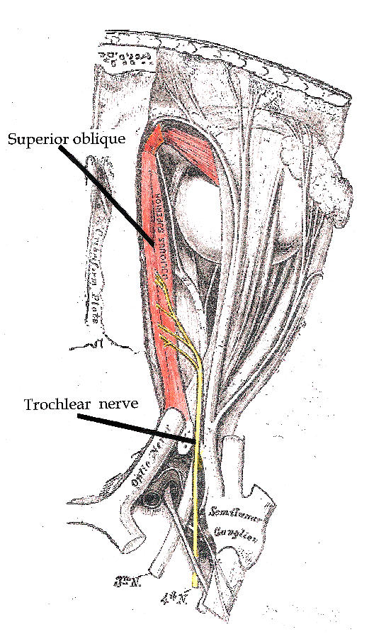
O nervo troclear, também conhecido como quarto par craniano, nervo craniano IV ou NC IV, é um nervo craniano que inerva um único músculo: o músculo oblíquo superior do olho. Diferente da maioria dos outros nervos cranianos, o nervo troclear é exclusivamente um nervo motor.
Introdução
O que você precisa saber de cara
Visão geral
A paralisia do nervo troclear congênita é uma condição rara presente desde o nascimento (neonatal) que afeta o movimento dos olhos. O nervo troclear (quarto par craniano) controla um dos músculos que movimenta o globo ocular para baixo e para dentro. Quando esse nervo não funciona adequadamente desde o nascimento, a pessoa pode ter dificuldade em olhar para baixo ou girar o olho afetado.[1]
Sinais e sintomas
Os sintomas geralmente estão presentes desde o período neonatal. A principal manifestação é a limitação do movimento do olho para baixo, especialmente quando o olho está voltado para dentro (adução). Isso pode causar visão dupla (diplopia) ao olhar para baixo, além de uma posição anormal da cabeça (torcicolo compensatório) para evitar a visão dupla. Em muitos casos, a condição é unilateral (afeta apenas um olho).[1]
Causas genéticas
Diagnóstico
O diagnóstico é baseado no exame clínico oftalmológico, que avalia os movimentos oculares e a posição da cabeça. Exames de imagem, como ressonância magnética, podem ser solicitados para descartar outras causas. Testes genéticos estão disponíveis (1 teste genético registrado), mas, como os genes específicos não são conhecidos, o diagnóstico é predominantemente clínico.[1][3]
Tratamento e manejo
O tratamento é focado no alívio dos sintomas e na correção do alinhamento ocular. Pode incluir o uso de prismas em óculos para reduzir a visão dupla, ou cirurgia de estrabismo para reposicionar os músculos oculares. O acompanhamento com oftalmologista especializado em estrabismo é essencial. Não há medicamentos aprovados especificamente para essa condição.[1]
Prognóstico e qualidade de vida
Com tratamento adequado, a maioria das pessoas consegue melhorar o alinhamento ocular e reduzir a visão dupla, mantendo boa qualidade de vida. A condição não afeta a expectativa de vida. O prognóstico visual é geralmente bom, embora possa ser necessário acompanhamento ao longo da vida para ajustes no tratamento.[1]
Conteúdo informativo gerado e mantido automaticamente a partir de fontes oficiais (Orphanet, HPO, OMIM, SUS). Não substitui avaliação médica.
O nervo troclear, também conhecido como quarto par craniano, nervo craniano IV ou NC IV, é um nervo craniano que inerva um único músculo: o músculo oblíquo superior do olho. Diferente da maioria dos outros nervos cranianos, o nervo troclear é exclusivamente um nervo motor.
Encontrou um erro ou informação desatualizada? Sugira uma correção →
Entender a doença
Do básico ao detalhe, leia no seu ritmo
Preparando trilha educativa...
Sinais e sintomas
O que aparece no corpo e com que frequência cada sintoma acontece
Visão geral
A paralisia do nervo troclear congênita é uma condição rara presente desde o nascimento (neonatal) que afeta o movimento dos olhos. O nervo troclear (quarto par craniano) controla um dos músculos que movimenta o globo ocular para baixo e para dentro. Quando esse nervo não funciona adequadamente desde o nascimento, a pessoa pode ter dificuldade em olhar para baixo ou girar o olho afetado.[1]
Sinais e sintomas
Os sintomas geralmente estão presentes desde o período neonatal. A principal manifestação é a limitação do movimento do olho para baixo, especialmente quando o olho está voltado para dentro (adução). Isso pode causar visão dupla (diplopia) ao olhar para baixo, além de uma posição anormal da cabeça (torcicolo compensatório) para evitar a visão dupla. Em muitos casos, a condição é unilateral (afeta apenas um olho).[1]
Causas genéticas
Diagnóstico
O diagnóstico é baseado no exame clínico oftalmológico, que avalia os movimentos oculares e a posição da cabeça. Exames de imagem, como ressonância magnética, podem ser solicitados para descartar outras causas. Testes genéticos estão disponíveis (1 teste genético registrado), mas, como os genes específicos não são conhecidos, o diagnóstico é predominantemente clínico.[1][3]
Tratamento e manejo
O tratamento é focado no alívio dos sintomas e na correção do alinhamento ocular. Pode incluir o uso de prismas em óculos para reduzir a visão dupla, ou cirurgia de estrabismo para reposicionar os músculos oculares. O acompanhamento com oftalmologista especializado em estrabismo é essencial. Não há medicamentos aprovados especificamente para essa condição.[1]
Prognóstico e qualidade de vida
Com tratamento adequado, a maioria das pessoas consegue melhorar o alinhamento ocular e reduzir a visão dupla, mantendo boa qualidade de vida. A condição não afeta a expectativa de vida. O prognóstico visual é geralmente bom, embora possa ser necessário acompanhamento ao longo da vida para ajustes no tratamento.[1]
Conteúdo informativo gerado e mantido automaticamente a partir de fontes oficiais (Orphanet, HPO, OMIM, SUS). Não substitui avaliação médica.
Linha do tempo da pesquisa
Encontrou um erro ou informação desatualizada? Sugira uma correção →
Genética e causas
O que está alterado no DNA e como passa nas famílias
Nenhum gene associado encontrado
Os dados genéticos desta condição ainda estão sendo catalogados.
Diagnóstico
Os sinais que médicos procuram e os exames que confirmam
Tratamento e manejo
Remédios, cuidados de apoio e o que precisa acompanhar
Onde tratar no SUS
Hospitais de referência no Brasil e o protocolo oficial do SUS (PCDT)
🇧🇷 Atendimento SUS — Paralisia do nervo troclear congênita
Centros de Referência SUS
24 centros habilitados pelo SUS para Paralisia do nervo troclear congênita
Centros para Paralisia do nervo troclear congênita
Detalhes dos centros
Hospital Universitário Prof. Edgard Santos (HUPES)
R. Dr. Augusto Viana, s/n - Canela, Salvador - BA, 40110-060 · CNES 0003808
Serviço de Referência
Hospital Infantil Albert Sabin
R. Tertuliano Sales, 544 - Vila União, Fortaleza - CE, 60410-794 · CNES 2407876
Serviço de Referência
Hospital de Apoio de Brasília (HAB)
AENW 3 Lote A Setor Noroeste - Plano Piloto, Brasília - DF, 70684-831 · CNES 0010456
Serviço de Referência
Hospital Estadual Infantil e Maternidade Alzir Bernardino Alves (HIABA)
Av. Min. Salgado Filho, 918 - Soteco, Vila Velha - ES, 29106-010 · CNES 6631207
Serviço de Referência
Hospital das Clínicas da UFG
Rua 235 QD. 68 Lote Área, Nº 285, s/nº - Setor Leste Universitário, Goiânia - GO, 74605-050 · CNES 2338424
Serviço de Referência
Hospital Universitário da UFJF
R. Catulo Breviglieri, Bairro - s/n - Santa Catarina, Juiz de Fora - MG, 36036-110 · CNES 2297442
Atenção Especializada
Hospital das Clínicas da UFMG
Av. Prof. Alfredo Balena, 110 - Santa Efigênia, Belo Horizonte - MG, 30130-100 · CNES 2280167
Serviço de Referência
Hospital Universitário Julio Müller (HUJM)
R. Luis Philippe Pereira Leite, s/n - Alvorada, Cuiabá - MT, 78048-902 · CNES 2726092
Atenção Especializada
Hospital Universitário João de Barros Barreto
R. dos Mundurucus, 4487 - Guamá, Belém - PA, 66073-000 · CNES 2337878
Serviço de Referência
Hospital Universitário Lauro Wanderley (HULW)
R. Tabeliao Estanislau Eloy, 585 - Castelo Branco, João Pessoa - PB, 58050-585 · CNES 0002470
Atenção Especializada
Instituto de Medicina Integral Prof. Fernando Figueira (IMIP)
R. dos Coelhos, 300 - Boa Vista, Recife - PE, 50070-902 · CNES 0000647
Serviço de Referência
Hospital Pequeno Príncipe
R. Des. Motta, 1070 - Água Verde, Curitiba - PR, 80250-060 · CNES 3143805
Serviço de Referência
Hospital Universitário Regional de Maringá (HUM)
Av. Mandacaru, 1590 - Parque das Laranjeiras, Maringá - PR, 87083-240 · CNES 2216108
Atenção Especializada
Hospital de Clínicas da UFPR
R. Gen. Carneiro, 181 - Alto da Glória, Curitiba - PR, 80060-900 · CNES 2364980
Serviço de Referência
Hospital Universitário Pedro Ernesto (HUPE-UERJ)
Blvd. 28 de Setembro, 77 - Vila Isabel, Rio de Janeiro - RJ, 20551-030 · CNES 2280221
Serviço de Referência
Instituto Nacional de Saúde da Mulher, da Criança e do Adolescente Fernandes Figueira (IFF/Fiocruz)
Av. Rui Barbosa, 716 - Flamengo, Rio de Janeiro - RJ, 22250-020 · CNES 2269988
Serviço de Referência
Hospital São Lucas da PUCRS
Av. Ipiranga, 6690 - Jardim Botânico, Porto Alegre - RS, 90610-000 · CNES 2232928
Serviço de Referência
Hospital de Clínicas de Porto Alegre (HCPA)
Rua Ramiro Barcelos, 2350 Bloco A - Av. Protásio Alves, 211 - Bloco B e C - Santa Cecília, Porto Alegre - RS, 90035-903 · CNES 2237601
Serviço de Referência
Hospital Universitário da UFSC (HU-UFSC)
R. Profa. Maria Flora Pausewang - Trindade, Florianópolis - SC, 88036-800 · CNES 2560356
Serviço de Referência
Hospital das Clínicas da FMUSP
R. Dr. Ovídio Pires de Campos, 225 - Cerqueira César, São Paulo - SP, 05403-010 · CNES 2077485
Serviço de Referência
Hospital de Base de São José do Rio Preto
Av. Brg. Faria Lima, 5544 - Vila Sao Jose, São José do Rio Preto - SP, 15090-000 · CNES 2079798
Atenção Especializada
Hospital de Clínicas da UNICAMP
R. Vital Brasil, 251 - Cidade Universitária, Campinas - SP, 13083-888 · CNES 2748223
Serviço de Referência
Hospital de Clínicas de Ribeirão Preto (HCRP-USP)
R. Ten. Catão Roxo, 3900 - Vila Monte Alegre, Ribeirão Preto - SP, 14015-010 · CNES 2082187
Serviço de Referência
UNIFESP / Hospital São Paulo
R. Napoleão de Barros, 715 - Vila Clementino, São Paulo - SP, 04024-002 · CNES 2688689
Serviço de Referência
Dados de DATASUS/CNES, SBGM, ABNeuro e Ministério da Saúde. Sempre confirme a disponibilidade diretamente com o estabelecimento.
Pesquisa ativa
Ensaios clínicos abertos e novidades científicas recentes
Ensaios em destaque
🟢 Recrutando agora
1 pesquisa recrutando participantes. Converse com seu médico sobre a possibilidade de participar.
Outros ensaios clínicos
0 ensaios clínicos encontrados.
Publicações mais relevantes
Prognostic Factors for Successful Surgical Outcomes in Trochlear Nerve Palsy: A Retrospective Study and Literature Review.
Trochlear nerve palsy is a common cause of double vision, particularly vertical diplopia. Surgery might be necessary if the condition does not improve independently. The success of the surgery can vary based on the method employed, and clear factors to predict its effectiveness are not evident. This study evaluates surgical techniques, success rates, and prognostic factors for trochlear nerve palsy at a tertiary hospital. A retrospective chart review was conducted on patients undergoing strabismus surgery for trochlear nerve palsy at Phramongkutklao Hospital between April 2012 and July 2024. Collected data included demographics, visual acuity, stereopsis, etiology, preoperative angles, surgical methods, and postoperative outcomes. A literature review regarding surgical success and prognostic factors was also conducted. Seventy-two cases were included, with 79.2% involving decompensated congenital trochlear nerve palsy. The overall surgical success rate was 76.39%, and inferior oblique myectomy was the most common and effective procedure (44.4% of cases). Based on multivariate logistic regression analysis, a preoperative hypertropia of ≤15 prism diopters was the significant factor for predicting successful outcomes in this study (OR 5.13, 95% CI 1.19-22.18). Inferior oblique muscle surgery effectively addresses small-angle deviations in trochlear nerve palsy. A <15 prism diopters vertical deviation strongly predicted positive surgical outcomes in this study. Further studies are needed to compare surgical techniques and explore additional prognostic factors to optimize long-term outcomes and improve patient care. Trochlear nerve palsy can lead to double vision, and surgery is required if there is no spontaneous improvement. The success of surgical procedures varies based on technique, and there are limited known predictors for which patients may achieve optimal results. When the condition does not resolve independently, surgery is necessary to rectify eye misalignment. This study explored various surgical methods, their success rates, and the factors affecting favorable outcomes from trochlear nerve palsy surgery. A review of medical records for 72 patients who underwent surgery for trochlear nerve palsy at Phramongkutklao Hospital between 2012 and 2024 showed that congenital causes were the most prevalent, accounting for 79.2% of cases. The findings indicated that 76.39% of the surgeries were successful, with inferior oblique myectomy being the most effective and frequently performed procedure. The primary predictor for a successful surgery was having a preoperative eye misalignment, specifically hypertropia, of ≤15 prism diopters. Notably, inferior oblique surgery successfully addresses small-angle deviations, with preoperative hypertropia remarkably predicting the surgical success. Further studies are necessary to compare surgical methods and explore other factors that may enhance long-term outcomes.
Postoperative outcomes for unilateral congenital trochlear nerve palsy: A retrospective cohort study.
Congenital trochlear nerve palsy is the most common cause of vertical strabismus. The goal of this study was to investigate surgical outcomes after superior oblique tendon plication with or without inferior oblique recession in children and adults with unilateral congenital trochlear nerve palsy. Data and outcomes were collected in patients with a diagnosis of unilateral congenital superior oblique palsy during a retrospective single-center study conducted at the University Hospital of Tours. A reproducible, standard ophthalmological and oculomotor examination was performed pre- and postoperatively at 1 year, including presence or absence of diplopia, vertical and horizontal deviations, and compensatory head posture. Surgical success, defined as an endpoint including absence of diplopia in primary position, absence of head tilt, and vertical deviation at distance fixation<5 prism diopters (PD), was analyzed. A total of fifty-seven patients (median [IQR] age of 11 years [5-42]) were analyzed. Patients experienced a significant reduction in vertical distance and near deviations (p<0.001), compensatory head tilt (p < 0.001), and diplopia after surgery (p < 0.001). Surgical success was higher in adults (17/24, 70.8%) than in children (15/33, 45.5%), although this did not reach statistical significance (p=0.0657). This study suggests that plication of the superior oblique muscle tendon, with or without recession of the inferior oblique muscle, can be effective in treating unilateral congenital trochlear nerve palsy. Further studies are necessary to compare surgical procedures and investigate their efficacy in adults compared to children in the short and long term.
Imaging of Cranial Nerves III, IV, VI in Congenital Cranial Dysinnervation Disorders.
Congenital cranial dysinnervation disorders are a group of diseases caused by abnormal development of cranial nerve nuclei or their axonal connections, resulting in aberrant innervation of the ocular and facial musculature. Its diagnosis could be facilitated by the development of high resolution thin-section magnetic resonance imaging. The purpose of this review is to describe the method to visualize cranial nerves III, IV, and VI and to present the imaging findings of congenital cranial dysinnervation disorders including congenital oculomotor nerve palsy, congenital trochlear nerve palsy, Duane retraction syndrome, Möbius syndrome, congenital fibrosis of the extraocular muscles, synergistic divergence, and synergistic convergence.
Imaging demonstration of trochlear nerve agenesis in superior oblique palsy emerging during the later life.
Congenital trochlear palsy may manifest with sudden vertical diplopia due to decompensation during the later life, which may bring a diagnostic challenge. Two men with vertical diplopia for several years after age of 50 were referred with persisting or suddenly aggravating diplopia. Findings were consistent with unilateral superior oblique palsy (SOP) in both patients with a contraversive head tilt. Facial asymmetry was suggestive of a congenital cause in a patient. High resolution magnetic resonance image (MRI)s disclosed atrophic superior oblique and absent trochlear nerve in the side of SOP in both patients. Imaging demonstration of superior oblique atrophy and absent trochlear nerve may aid in diagnosis of congenital SOP presenting sudden vertical diplopia during the later life due to delayed decompensation.
Publicações recentes
Prognostic Factors for Successful Surgical Outcomes in Trochlear Nerve Palsy: A Retrospective Study and Literature Review.
Postoperative outcomes for unilateral congenital trochlear nerve palsy: A retrospective cohort study.
Imaging of Cranial Nerves III, IV, VI in Congenital Cranial Dysinnervation Disorders.
Imaging demonstration of trochlear nerve agenesis in superior oblique palsy emerging during the later life.
Primary position and listing's law in acquired and congenital trochlear nerve palsy.
📚 EuropePMC3 artigos no totalmostrando 4
Prognostic Factors for Successful Surgical Outcomes in Trochlear Nerve Palsy: A Retrospective Study and Literature Review.
Clinical ophthalmology (Auckland, N.Z.)Postoperative outcomes for unilateral congenital trochlear nerve palsy: A retrospective cohort study.
Journal francais d'ophtalmologieImaging of Cranial Nerves III, IV, VI in Congenital Cranial Dysinnervation Disorders.
Korean journal of ophthalmology : KJOImaging demonstration of trochlear nerve agenesis in superior oblique palsy emerging during the later life.
Clinical neurology and neurosurgeryAssociações
Organizações que acompanham esta doença — pra ter apoio e orientação
Ainda não temos associações cadastradas para Paralisia do nervo troclear congênita.
É de uma associação que acompanha esta doença? Fale com a gente →
Comunidades
Grupos ativos de quem convive com esta doença aqui no Raras
Ainda não existe comunidade no Raras para Paralisia do nervo troclear congênita
Pacientes, familiares e cuidadores se organizam em comunidades pra compartilhar experiências, fazer perguntas e se apoiar. Você pode ser o primeiro.
Tire suas dúvidas
Perguntas, dicas e experiências compartilhadas aqui na página
Participe da discussão
Faça login para postar dúvidas, compartilhar experiências e interagir com especialistas.
Fazer loginDoenças relacionadas
Doenças com sintomas parecidos — ajudam quem ainda está buscando diagnóstico
Ainda não achamos doenças com sintomas parecidos o suficiente.
Referências e fontes
Bases de dados externas citadas neste artigo
Publicações científicas
Artigos indexados no PubMed ligados a esta doença no grafo RarasNet — título, periódico e PMID direto da fonte, sem intermediação de IA.
- Prognostic Factors for Successful Surgical Outcomes in Trochlear Nerve Palsy: A Retrospective Study and Literature Review.
- Postoperative outcomes for unilateral congenital trochlear nerve palsy: A retrospective cohort study.
- Imaging of Cranial Nerves III, IV, VI in Congenital Cranial Dysinnervation Disorders.
- Imaging demonstration of trochlear nerve agenesis in superior oblique palsy emerging during the later life.
- Primary position and listing's law in acquired and congenital trochlear nerve palsy.
Bases de dados e fontes oficiais
Identificadores e referências canônicas usadas para montar este verbete.
- ORPHA:98686(Orphanet)
- MONDO:0700463(MONDO)
- Busca completa no PubMed(PubMed)
- Q5160431(Wikidata)
Dados compilados pelo RarasNet a partir de fontes abertas (Orphanet, OMIM, MONDO, PubMed/EuropePMC, ClinicalTrials.gov, DATASUS, PCDT/MS). Este conteúdo é informativo e não substitui avaliação médica.
Conteúdo mantido por Agente Raras · Médicos e pesquisadores podem colaborar
Paralisia do nervo troclear congênita
📋 Origem dos dados
Esta página agrega dados de fontes públicas e oficiais. Dados sobre cobertura no SUS (PCDT, CEAF) são verificados ativamente por agente proativo (ver badge no infobox). Demais dados têm atribuição de fonte + data da última sincronização — clique para abrir o original.
- Doença rara (ontologia)
- fonte: Orphanet
- Identificador unificado
- fonte: MONDO
- Codificação WHO/SUS
- fonte: WHO ICD-10 / DATASUS
- CID-11 (futuro)
- fonte: WHO ICD-11
- Indexação biomédica
- fonte: MeSH (NLM)
- Dado público estruturado
- fonte: Wikidata