Introdução
O que você precisa saber de cara
Visão geral
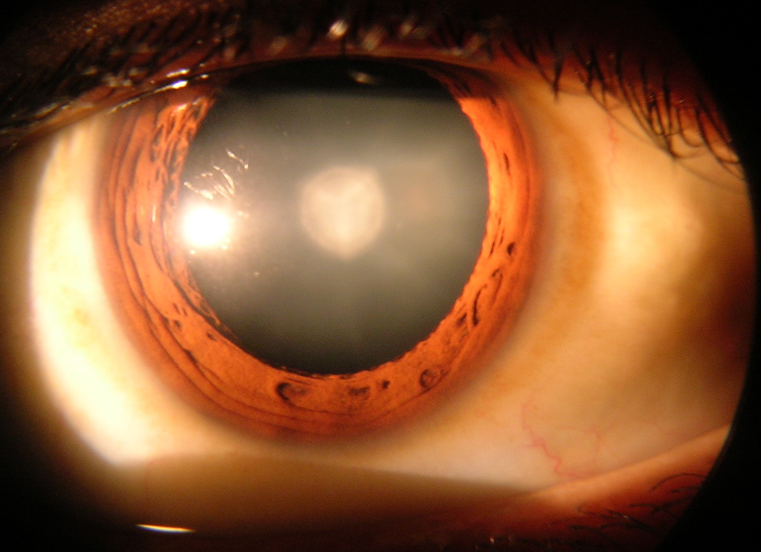
A catarata parcial de início precoce é uma condição rara que afeta a transparência do cristalino do olho, presente desde o nascimento ou nos primeiros meses de vida. Diferente da catarata comum do envelhecimento, nesta condição a opacidade é parcial, ou seja, não atinge todo o cristalino, e surge precocemente. A doença pode comprometer a visão de forma variável, desde uma leve redução da acuidade visual até perda visual significativa.[1][3]
Sinais e sintomas
Os principais sinais e sintomas incluem: catarata (opacificação do cristalino), catarata do desenvolvimento, catarata nuclear (opacidade na região central do cristalino), catarata nuclear difusa, catarata pulverulenta (aspecto de pó fino), catarata polar anterior e catarata polar posterior. Também podem ocorrer lenticone (protuberância do cristalino), lenticone anterior, alta miopia, ambliopia (olho preguiçoso), distrofia retiniana e acuidade visual levemente reduzida, podendo evoluir para perda visual.[1][3]
Causas genéticas
A catarata parcial de início precoce tem causas genéticas. Mutações em diversos genes estão associadas à condição, incluindo: CRYAA (cadeia A da alfa-cristalina), BFSP2 (faquinina), PITX3 (homeobox 3 da hipófise), CRYGS (gama-cristalina S), CRYGB (gama-cristalina B), VIM (vimentina), NHS (regulador de remodelação de actina NHS), CRYGC (gama-cristalina C), CRYBA4 (beta-cristalina A4), UNC45B (proteína unc-45 homólogo B), CRYGD (gama-cristalina D) e CRYBA2 (beta-cristalina A2). Esses genes estão envolvidos na estrutura e função do cristalino.[1][4]
Diagnóstico
O diagnóstico é baseado na avaliação clínica oftalmológica, que identifica a opacidade parcial do cristalino desde o nascimento ou infância. Exames genéticos podem auxiliar na confirmação diagnóstica. No Brasil, estão disponíveis pelo Sistema Único de Saúde (SUS) os seguintes procedimentos: cariótipo com bandas G, Q ou R; pesquisa de microdeleções/microduplicações por FISH; sequenciamento completo do exoma (WES); dosagem de alfa-fetoproteína; e atendimento em reabilitação para doenças raras. Atualmente, há 336 testes genéticos disponíveis e 507 variantes registradas no ClinVar para essa condição.[1][4]
Tratamento e manejo
O tratamento é individualizado e pode incluir correção cirúrgica da catarata quando há comprometimento visual significativo, além de medidas para tratar a ambliopia (como uso de óculos ou tampão ocular) e acompanhamento oftalmológico regular. Não há medicamentos específicos aprovados para reverter a opacidade do cristalino. O manejo deve ser realizado por equipe multidisciplinar, incluindo oftalmologista e geneticista. No SUS, a cobertura é considerada mínima, com acesso aos procedimentos diagnósticos e de reabilitação listados.[1]
Prognóstico e qualidade de vida
O prognóstico depende da gravidade da opacidade e da presença de outras alterações oculares, como ambliopia ou distrofia retiniana. Com diagnóstico precoce e tratamento adequado, muitas crianças podem desenvolver boa acuidade visual. No entanto, a perda visual pode ocorrer se não houver intervenção oportuna. O acompanhamento contínuo é essencial para preservar a função visual e a qualidade de vida.[1][3]
Conteúdo informativo gerado e mantido automaticamente a partir de fontes oficiais (Orphanet, HPO, OMIM, SUS). Não substitui avaliação médica.
Catarata parcial de início precoce é uma condição hereditária rara que causa opacificação parcial do cristalino, afetando a visão desde a infância. Pode estar associada a mutações nos genes HSF4, GJA8, FYCO1, BFSP1 ou LEMD2.
Encontrou um erro ou informação desatualizada? Sugira uma correção →
Entender a doença
Do básico ao detalhe, leia no seu ritmo
Preparando trilha educativa...
Sinais e sintomas
O que aparece no corpo e com que frequência cada sintoma acontece
Visão geral
A catarata parcial de início precoce é uma condição rara que afeta a transparência do cristalino do olho, presente desde o nascimento ou nos primeiros meses de vida. Diferente da catarata comum do envelhecimento, nesta condição a opacidade é parcial, ou seja, não atinge todo o cristalino, e surge precocemente. A doença pode comprometer a visão de forma variável, desde uma leve redução da acuidade visual até perda visual significativa.[1][3]
Sinais e sintomas
Os principais sinais e sintomas incluem: catarata (opacificação do cristalino), catarata do desenvolvimento, catarata nuclear (opacidade na região central do cristalino), catarata nuclear difusa, catarata pulverulenta (aspecto de pó fino), catarata polar anterior e catarata polar posterior. Também podem ocorrer lenticone (protuberância do cristalino), lenticone anterior, alta miopia, ambliopia (olho preguiçoso), distrofia retiniana e acuidade visual levemente reduzida, podendo evoluir para perda visual.[1][3]
Causas genéticas
A catarata parcial de início precoce tem causas genéticas. Mutações em diversos genes estão associadas à condição, incluindo: CRYAA (cadeia A da alfa-cristalina), BFSP2 (faquinina), PITX3 (homeobox 3 da hipófise), CRYGS (gama-cristalina S), CRYGB (gama-cristalina B), VIM (vimentina), NHS (regulador de remodelação de actina NHS), CRYGC (gama-cristalina C), CRYBA4 (beta-cristalina A4), UNC45B (proteína unc-45 homólogo B), CRYGD (gama-cristalina D) e CRYBA2 (beta-cristalina A2). Esses genes estão envolvidos na estrutura e função do cristalino.[1][4]
Diagnóstico
O diagnóstico é baseado na avaliação clínica oftalmológica, que identifica a opacidade parcial do cristalino desde o nascimento ou infância. Exames genéticos podem auxiliar na confirmação diagnóstica. No Brasil, estão disponíveis pelo Sistema Único de Saúde (SUS) os seguintes procedimentos: cariótipo com bandas G, Q ou R; pesquisa de microdeleções/microduplicações por FISH; sequenciamento completo do exoma (WES); dosagem de alfa-fetoproteína; e atendimento em reabilitação para doenças raras. Atualmente, há 336 testes genéticos disponíveis e 507 variantes registradas no ClinVar para essa condição.[1][4]
Tratamento e manejo
O tratamento é individualizado e pode incluir correção cirúrgica da catarata quando há comprometimento visual significativo, além de medidas para tratar a ambliopia (como uso de óculos ou tampão ocular) e acompanhamento oftalmológico regular. Não há medicamentos específicos aprovados para reverter a opacidade do cristalino. O manejo deve ser realizado por equipe multidisciplinar, incluindo oftalmologista e geneticista. No SUS, a cobertura é considerada mínima, com acesso aos procedimentos diagnósticos e de reabilitação listados.[1]
Prognóstico e qualidade de vida
O prognóstico depende da gravidade da opacidade e da presença de outras alterações oculares, como ambliopia ou distrofia retiniana. Com diagnóstico precoce e tratamento adequado, muitas crianças podem desenvolver boa acuidade visual. No entanto, a perda visual pode ocorrer se não houver intervenção oportuna. O acompanhamento contínuo é essencial para preservar a função visual e a qualidade de vida.[1][3]
Conteúdo informativo gerado e mantido automaticamente a partir de fontes oficiais (Orphanet, HPO, OMIM, SUS). Não substitui avaliação médica.
Partes do corpo afetadas
+ 2 sintomas em outras categorias
Características mais comuns
Os sintomas variam de pessoa para pessoa. Abaixo estão as 14 características clínicas mais associadas, ordenadas por frequência.
Linha do tempo da pesquisa
Encontrou um erro ou informação desatualizada? Sugira uma correção →
Genética e causas
O que está alterado no DNA e como passa nas famílias
Genes associados
29 genes identificados com associação a esta condição.
Contributes to the transparency and refractive index of the lens (PubMed:18302245). In its oxidized form (absence of intramolecular disulfide bond), acts as a chaperone, preventing aggregation of various proteins under a wide range of stress conditions (PubMed:18199971, PubMed:19595763, PubMed:22120592, PubMed:31792453). Required for the correct formation of lens intermediate filaments as part of a complex composed of BFSP1, BFSP2 and CRYAA (PubMed:28935373)
CytoplasmNucleus
Required for the correct formation of lens intermediate filaments as part of a complex composed of BFSP1, BFSP2 and CRYAA (PubMed:28935373). Plays a role in maintenance of retinal lens optical clarity (By similarity)
Cell membraneCytoplasmCytoplasm, cytoskeletonCytoplasm, cell cortex
Cataract 12, multiple types
An opacification of the crystalline lens of the eye that frequently results in visual impairment or blindness. Opacities vary in morphology, are often confined to a portion of the lens, and may be static or progressive. In general, the more posteriorly located and dense an opacity, the greater the impact on visual function. The opacities can be nuclear, sutural, stellate cortical, lamellar, cortical, nuclear embryonic, Y-sutural, punctate cortical, congenital or with juvenile- and adult-onset.
Transcriptional regulator which is important for the differentiation and maintenance of meso-diencephalic dopaminergic (mdDA) neurons during development. In addition to its importance during development, it also has roles in the long-term survival and maintenance of the mdDA neurons. Activates NR4A2/NURR1-mediated transcription of genes such as SLC6A3, SLC18A2, TH and DRD2 which are essential for development of mdDA neurons. Acts by decreasing the interaction of NR4A2/NURR1 with the corepressor
Nucleus
Anterior segment dysgenesis 1
A form of anterior segment dysgenesis, a group of defects affecting anterior structures of the eye including cornea, iris, lens, trabecular meshwork, and Schlemm canal. Anterior segment dysgeneses result from abnormal migration or differentiation of the neural crest derived mesenchymal cells that give rise to components of the anterior chamber during eye development. Different anterior segment anomalies may exist alone or in combination, including iris hypoplasia, enlarged or reduced corneal diameter, corneal vascularization and opacity, posterior embryotoxon, corectopia, polycoria, abnormal iridocorneal angle, ectopia lentis, and anterior synechiae between the iris and posterior corneal surface. Clinical conditions falling within the phenotypic spectrum of anterior segment dysgeneses include aniridia, Axenfeld anomaly, Reiger anomaly/syndrome, Peters anomaly, and iridogoniodysgenesis.
Crystallins are the dominant structural components of the vertebrate eye lens
Cataract 20, multiple types
An opacification of the crystalline lens of the eye that frequently results in visual impairment or blindness. Opacities vary in morphology, are often confined to a portion of the lens, and may be static or progressive. In general, the more posteriorly located and dense an opacity, the greater the impact on visual function. CTRCT20 includes progressive polymorphic anterior, posterior, or peripheral cortical.
Crystallins are the dominant structural components of the vertebrate eye lens
Cataract 39, multiple types
An opacification of the crystalline lens of the eye that frequently results in visual impairment or blindness. Opacities vary in morphology, are often confined to a portion of the lens, and may be static or progressive. In general, the more posteriorly located and dense an opacity, the greater the impact on visual function. CTRCT39 includes lamellar, anterior polar, and complete cataracts.
Vimentins are class-III intermediate filaments found in various non-epithelial cells, especially mesenchymal cells. Vimentin is attached to the nucleus, endoplasmic reticulum, and mitochondria, either laterally or terminally. Plays a role in cell directional movement, orientation, cell sheet organization and Golgi complex polarization at the cell migration front (By similarity). Protects SCRIB from proteasomal degradation and facilitates its localization to intermediate filaments in a cell conta
CytoplasmCytoplasm, cytoskeletonNucleus matrixCell membraneCell projection, axon
Cataract 30, multiple types
An opacification of the crystalline lens of the eye that frequently results in visual impairment or blindness. Opacities vary in morphology, are often confined to a portion of the lens, and may be static or progressive. In general, the more posteriorly located and dense an opacity, the greater the impact on visual function.
May function in cell morphology by maintaining the integrity of the circumferential actin ring and controlling lamellipod formation. Involved in the regulation eye, tooth, brain and craniofacial development
Apical cell membraneCell projection, lamellipodiumCell junction, tight junctionCell junction, focal adhesionCytoplasm
Nance-Horan syndrome
Rare X-linked disorder characterized by congenital cataracts, dental anomalies, dysmorphic features, and, in some cases, intellectual disability. Distinctive dental anomalies are seen in affected males, including supernumerary incisors and crown shaped permanent teeth. Characteristic facial features are anteverted pinnae, long face, and prominent nasal bridge and nose. Carrier females display milder variable symptoms of disease with lens opacities often involving the posterior Y sutures, and on occasion dental anomalies and the characteristic facial features described.
Crystallins are the dominant structural components of the vertebrate eye lens
Cataract 2, multiple types
An opacification of the crystalline lens of the eye that frequently results in visual impairment or blindness. Opacities vary in morphology, are often confined to a portion of the lens, and may be static or progressive. CTRCT2 includes Coppock-like cataract, among others. Coppock-like cataract is a congenital pulverulent disk-like opacity involving the embryonic nucleus with many tiny white dots in the lamellar portion of the lens. It is usually bilateral and dominantly inherited. In some cases, CTRCT2 is associated with microcornea without any other systemic anomaly or dysmorphism. Microcornea is defined by a corneal diameter inferior to 10 mm in both meridians in an otherwise normal eye.
Crystallins are the dominant structural components of the vertebrate eye lens
Cataract 23, multiple types
An opacification of the crystalline lens of the eye that frequently results in visual impairment or blindness. Opacities vary in morphology, are often confined to a portion of the lens, and may be static or progressive. In general, the more posteriorly located and dense an opacity, the greater the impact on visual function. CTRCT23 is a zonular cataract. Zonular or lamellar cataracts are opacities, broad or narrow, usually consisting of powdery white dots affecting only certain layers or zones between the cortex and nucleus of an otherwise clear lens. The opacity may be so dense as to render the entire central region of the lens completely opaque, or so translucent that vision is hardly if at all impeded. Zonular cataracts generally do not involve the embryonic nucleus, though sometimes they involve the fetal nucleus. Usually sharply separated from a clear cortex outside them, they may have projections from their outer edges known as riders or spokes.
Acts as a co-chaperone for HSP90 and is required for proper folding of the myosin motor domain. Plays a role in sarcomere formation during muscle cell development. Is necessary for normal early lens development
Cytoplasm, myofibril, sarcomere, Z lineCytoplasm, myofibril, sarcomere, A bandCytoplasm, perinuclear regionCytoplasm, cytosol
Cataract 43
An opacification of the crystalline lens of the eye that frequently results in visual impairment or blindness. Opacities vary in morphology, are often confined to a portion of the lens, and may be static or progressive. In general, the more posteriorly located and dense an opacity, the greater the impact on visual function.
Crystallins are the dominant structural components of the vertebrate eye lens
Cataract 4, multiple types
An opacification of the crystalline lens of the eye that frequently results in visual impairment or blindness. Opacities vary in morphology, are often confined to a portion of the lens, and may be static or progressive. CTRCT4 includes crystalline aculeiform, congenital cerulean and non-nuclear polymorphic cataracts, among others. Crystalline aculeiform cataract is characterized by fiberglass-like or needle-like crystals projecting in different directions, through or close to the axial region of the lens. Non-nuclear polymorphic cataract is a partial opacity with variable location between the fetal nucleus of the lens and the equator. The fetal nucleus is normal. The opacities are irregular and look similar to a bunch of grapes and may be present simultaneously in different lens layers. Congenital cerulean cataract is characterized by peripheral bluish and white opacifications organized in concentric layers with occasional central lesions arranged radially. The opacities are observed in the superficial layers of the fetal nucleus as well as the adult nucleus of the lens. Involvement is usually bilateral. Visual acuity is only mildly reduced in childhood. In adulthood, the opacifications may progress, making lens extraction necessary. Histologically the lesions are described as fusiform cavities between lens fibers which contain a deeply staining granular material. Although the lesions may take on various colors, a dull blue is the most common appearance and is responsible for the designation cerulean cataract.
Crystallins are the dominant structural components of the vertebrate eye lens
Cataract 42
An opacification of the crystalline lens of the eye that frequently results in visual impairment or blindness. Opacities vary in morphology, are often confined to a portion of the lens, and may be static or progressive. In general, the more posteriorly located and dense an opacity, the greater the impact on visual function.
Crystallins are the dominant structural components of the vertebrate eye lens
Cataract 17, multiple types
An opacification of the crystalline lens of the eye that frequently results in visual impairment or blindness. Opacities vary in morphology, are often confined to a portion of the lens, and may be static or progressive. In general, the more posteriorly located and dense an opacity, the greater the impact on visual function. CTRCT17 includes nuclear and pulverulent cataracts, among others. Nuclear cataracts affect the central nucleus of the eye, are often not highly visually significant. The density of the opacities varies greatly from fine dots to a dense, white and chalk-like, central cataract. The condition is usually bilateral. Nuclear cataracts are often combined with opacified cortical fibers encircling the nuclear opacity, which are referred to as cortical riders. Pulverulent cataracts are characterized by a dust-like, 'pulverised' appearance of the opacities which can be found in any part of the lens.
Probable core component of the endosomal sorting required for transport complex III (ESCRT-III) which is involved in multivesicular bodies (MVBs) formation and sorting of endosomal cargo proteins into MVBs. MVBs contain intraluminal vesicles (ILVs) that are generated by invagination and scission from the limiting membrane of the endosome and mostly are delivered to lysosomes enabling degradation of membrane proteins, such as stimulated growth factor receptors, lysosomal enzymes and lipids. The M
Cytoplasm, cytosolLate endosome membraneMidbodyNucleus envelope
Cataract 31, multiple types
An opacification of the crystalline lens of the eye that frequently results in visual impairment or blindness. Opacities vary in morphology, are often confined to a portion of the lens, and may be static or progressive. In general, the more posteriorly located and dense an opacity, the greater the impact on visual function. CTRCT31 includes posterior polar, progressive posterior subcapsular, nuclear, and anterior subcapsular cataracts.
Crystallins are the dominant structural components of the vertebrate eye lens
Cataract 3, multiple types
An opacification of the crystalline lens of the eye that frequently results in visual impairment or blindness. Opacities vary in morphology, are often confined to a portion of the lens, and may be static or progressive. CTRCT3 includes congenital cerulean and sutural cataract with punctate and cerulean opacities, among others. Cerulean cataract is characterized by peripheral bluish and white opacifications organized in concentric layers with occasional central lesions arranged radially. The opacities are observed in the superficial layers of the fetal nucleus as well as the adult nucleus of the lens. Involvement is usually bilateral. Visual acuity is only mildly reduced in childhood. In adulthood, the opacifications may progress, making lens extraction necessary. Histologically the lesions are described as fusiform cavities between lens fibers which contain a deeply staining granular material. Although the lesions may take on various colors, a dull blue is the most common appearance and is responsible for the designation cerulean cataract. Sutural cataract with punctate and cerulean opacities is characterized by white opacification around the anterior and posterior Y sutures, and grayish and bluish, spindle shaped, oval punctate and cerulean opacities of various sizes arranged in lamellar form. The spots are more concentrated towards the peripheral layers and do not delineate the embryonal or fetal nucleus. Phenotypic variation with respect to the size and density of the sutural opacities as well as the number and position of punctate and cerulean spots is observed among affected subjects.
Receptor tyrosine kinase which binds promiscuously membrane-bound ephrin-A family ligands residing on adjacent cells, leading to contact-dependent bidirectional signaling into neighboring cells. The signaling pathway downstream of the receptor is referred to as forward signaling while the signaling pathway downstream of the ephrin ligand is referred to as reverse signaling. Activated by the ligand ephrin-A1/EFNA1 regulates migration, integrin-mediated adhesion, proliferation and differentiation
Cell membraneCell projection, ruffle membraneCell projection, lamellipodium membraneCell junction, focal adhesion
Cataract 6, multiple types
An opacification of the crystalline lens of the eye that frequently results in visual impairment or blindness. Opacities vary in morphology, are often confined to a portion of the lens, and may be static or progressive. CTRCT6 includes posterior polar and age-related cortical cataracts, among others. Posterior polar cataract is a subcapsular opacity, usually disk-shaped, located at the back of the lens. Age-related cortical cataract is a developmental punctate opacity restricted to the cortex. The cataract is white or cerulean, increases in number with age, but rarely affects vision.
Structural component of lens fiber gap junctions (PubMed:30044662). Gap junctions are dodecameric channels that connect the cytoplasm of adjoining cells (By similarity). They are formed by the docking of two hexameric hemichannels, one from each cell membrane. Small molecules and ions diffuse from one cell to a neighboring cell via the central pore (PubMed:30044662)
Cell membraneCell junction, gap junction
Cataract 14, multiple types
An opacification of the crystalline lens of the eye that frequently results in visual impairment or blindness. Opacities vary in morphology, are often confined to a portion of the lens, and may be static or progressive. CTRCT14 includes zonular pulverulent cataract, among others. Zonular or lamellar cataracts are opacities, broad or narrow, usually consisting of powdery white dots affecting only certain layers or zones between the cortex and nucleus of an otherwise clear lens. The opacity may be so dense as to render the entire central region of the lens completely opaque, or so translucent that vision is hardly if at all impeded. Usually sharply separated from a clear cortex outside them, they may have projections from their outer edges known as riders or spokes.
Phosphatase which shows a preference for 4'-phosphopantetheine and its oxidatively damaged forms (sulfonate or S-sulfonate), providing strong indirect evidence that the phosphatase activity pre-empts damage in the coenzyme A (CoA) pathway (PubMed:27322068). Hydrolyzing excess 4'-phosphopantetheine could constitute a directed overflow mechanism to prevent its oxidation to the S-sulfonate, sulfonate, or other forms (PubMed:27322068). Hydrolyzing 4'-phosphopantetheine sulfonate or S-sulfonate would
Cytoplasm
Cataract 49
A form of cataract, an opacification of the crystalline lens of the eye that frequently results in visual impairment or blindness. Opacities vary in morphology, are often confined to a portion of the lens, and may be static or progressive. In general, the more posteriorly located and dense an opacity, the greater the impact on visual function. CTRCT49 is an autosomal dominant form characterized by congenital cataract located in the posterior region of the lens. Visual impairment has onset in early childhood.
Crystallins are the dominant structural components of the vertebrate eye lens
Cataract 22, multiple types
An opacification of the crystalline lens of the eye that frequently results in visual impairment or blindness. Opacities vary in morphology, are often confined to a portion of the lens, and may be static or progressive. In general, the more posteriorly located and dense an opacity, the greater the impact on visual function. CTRCT22 includes nuclear cataract among others. Nuclear cataracts affect the central nucleus of the eye, and are often not highly visually significant. The density of the opacities varies greatly from fine dots to a dense, white and chalk-like, central cataract. The condition is usually bilateral. Nuclear cataracts are often combined with opacified cortical fibers encircling the nuclear opacity, which are referred to as cortical riders.
Participates in the regulation of cellular Ca(2+) homeostasis, at least partly, by modulating the filling state of the endoplasmic reticulum Ca(2+) store (PubMed:16989814). Negatively regulates the ER stress response and positively regulates the stability of V-ATPase subunits ATP6V1A and ATP1B1 by preventing their degradation through an unknown proteasome-independent mechanism (PubMed:23035048)
Endoplasmic reticulum membraneCytoplasmic vesicle, secretory vesicle
Wolfram syndrome 1
A rare disorder characterized by juvenile-onset insulin-dependent diabetes mellitus with optic atrophy. Other manifestations include diabetes insipidus, sensorineural deafness, dementia, psychiatric illnesses.
Acts as a transcriptional activator or repressor. Involved in embryonic lens fiber cell development. Recruits the transcriptional coactivators CREBBP and/or EP300 to crystallin promoters leading to up-regulation of crystallin gene during lens fiber cell differentiation. Activates the expression of IL4 in T helper 2 (Th2) cells. Increases T-cell susceptibility to apoptosis by interacting with MYB and decreasing BCL2 expression. Together with PAX6, transactivates strongly the glucagon gene promote
Nucleus
Aquaporins form homotetrameric transmembrane channels, with each monomer independently mediating water transport across the plasma membrane along its osmotic gradient (PubMed:11001937, PubMed:24120416). Specifically expressed in lens fiber cells, this aquaporin is crucial for maintaining lens water homeostasis and transparency. Beyond water permeability, it also acts as a cell-to-cell adhesion molecule, forming thin junctions between lens fiber cells that are essential for maintaining the ordere
Cell membraneCell junction
Cataract 15, multiple types
An opacification of the crystalline lens of the eye that frequently results in visual impairment or blindness. Opacities vary in morphology, are often confined to a portion of the lens, and may be static or progressive. CTRCT15 includes polymorphic, progressive punctate lamellar, cortical, anterior and posterior polar, nonprogressive lamellar with sutural opacities, embryonic nuclear, and pulverulent cortical, among others.
May contribute to the transparency and refractive index of the lens. Has chaperone-like activity, preventing aggregation of various proteins under a wide range of stress conditions. In lens epithelial cells, stabilizes the ATP6V1A protein, preventing its degradation by the proteasome (By similarity)
CytoplasmNucleusSecretedLysosome
Myopathy, myofibrillar, 2A, adult-onset
A form of myofibrillar myopathy, a group of chronic neuromuscular disorders characterized at ultrastructural level by disintegration of the sarcomeric Z disk and myofibrils, and replacement of the normal myofibrillar markings by small dense granules, or larger hyaline masses, or amorphous material. MFM2A is an autosomal dominant form characterized by weakness of the proximal and distal limb muscles, weakness of the neck, velopharynx and trunk muscles, respiratory insufficiency, hypertrophic cardiomyopathy, and cataract.
Crystallins are the dominant structural components of the vertebrate eye lens
Cataract 10, multiple types
An opacification of the crystalline lens of the eye that frequently results in visual impairment or blindness. Opacities vary in morphology, are often confined to a portion of the lens, and may be static or progressive. CTRCT10 includes congenital zonular with sutural opacities, among others. This is a form of zonular cataract with an erect Y-shaped anterior and an inverted Y-shaped posterior sutural opacities. Zonular or lamellar cataracts are opacities, broad or narrow, usually consisting of powdery white dots affecting only certain layers or zones between the cortex and nucleus of an otherwise clear lens. The opacity may be so dense as to render the entire central region of the lens completely opaque, or so translucent that vision is hardly if at all impeded. Zonular cataracts generally do not involve the embryonic nucleus, though sometimes they involve the fetal nucleus. Usually sharply separated from a clear cortex outside them, they may have projections from their outer edges known as riders or spokes.
Nuclear lamina-associated inner nuclear membrane protein that is involved in nuclear structure organization, maintenance of nuclear envelope (NE) integrity and NE reformation after mitosis (PubMed:16339967, PubMed:17097643, PubMed:28242692, PubMed:32494070). Plays a role as transmembrane adapter for the endosomal sorting complexes required for transport (ESCRT), and is thereby involved in ESCRT-mediated NE reformation (PubMed:28242692, PubMed:32494070). Promotes ESCRT-mediated NE closure by recr
Nucleus inner membraneNucleus envelopeCytoplasm, cytoskeleton, spindle
Cataract 46, juvenile-onset, with or without arrhythmic cardiomyopathy
A form of cataract, an opacification of the crystalline lens of the eye that frequently results in visual impairment or blindness. Opacities vary in morphology, are often confined to a portion of the lens, and may be static or progressive. In general, the more posteriorly located and dense an opacity, the greater the impact on visual function. CTRCT46 can be associated with variable onset of a severe form of arrhythmic cardiomyopathy resulting in sudden cardiac death.
Required for the correct formation of lens intermediate filaments as part of a complex composed of BFSP1, BFSP2 and CRYAA (PubMed:28935373). Involved in altering the calcium regulation of MIP water permeability (PubMed:30790544)
Cell membraneCytoplasmCytoplasm, cytoskeletonCytoplasm, cell cortex
Cataract 33, multiple types
An opacification of the crystalline lens of the eye that frequently results in visual impairment or blindness. Opacities vary in morphology, are often confined to a portion of the lens, and may be static or progressive. In general, the more posteriorly located and dense an opacity, the greater the impact on visual function. CTRCT33 has juvenile-onset and the opacities are restricted to the cortex of the lens, not involving the nucleus.
May mediate microtubule plus end-directed vesicle transport
Cytoplasmic vesicle, autophagosomeEndosomeLysosome
Cataract 18
An opacification of the crystalline lens of the eye becoming evident at birth or in infancy. It frequently results in visual impairment or blindness. Opacities vary in morphology, are often confined to a portion of the lens, and may be static or progressive. In general, the more posteriorly located and dense an opacity, the greater the impact on visual function.
Structural component of eye lens gap junctions (PubMed:18006672, PubMed:19756179). Gap junctions are dodecameric channels that connect the cytoplasm of adjoining cells. They are formed by the docking of two hexameric hemichannels, one from each cell membrane (By similarity). Small molecules and ions diffuse from one cell to a neighboring cell via the central pore (PubMed:18006672, PubMed:19756179)
Cell membraneCell junction, gap junction
Cataract 1, multiple types
An opacification of the crystalline lens of the eye that frequently results in visual impairment or blindness. Opacities vary in morphology, are often confined to a portion of the lens, and may be static or progressive. CTRCT1 includes congenital, zonular pulverulent, nuclear progressive, nuclear pulverulent, nuclear total, total, and posterior subcapsular types of cataract. Zonular or lamellar cataracts are opacities, broad or narrow, usually consisting of powdery white dots affecting only certain layers or zones between the cortex and nucleus of an otherwise clear lens. The opacity may be so dense as to render the entire central region of the lens completely opaque, or so translucent that vision is hardly if at all impeded. Zonular cataracts generally do not involve the embryonic nucleus, though sometimes they involve the fetal nucleus. Usually sharply separated from a clear cortex outside them, they may have projections from their outer edges known as riders or spokes. In some cases cataract is associated with microcornea without any other systemic anomaly or dysmorphism. Microcornea is defined by a corneal diameter inferior to 10 mm in both meridians in an otherwise normal eye.
Heat-shock transcription factor that specifically binds heat shock promoter elements (HSE) (PubMed:22587838, PubMed:23507146). Required for denucleation and organelle rupture and degradation that occur during eye lens terminal differentiation, when fiber cells that compose the lens degrade all membrane-bound organelles in order to provide lens with transparency to allow the passage of light (By similarity). In this process, may regulate denucleation of lens fiber cells in part by activating DNAS
Nucleus
Cataract 5, multiple types
An opacification of the crystalline lens of the eye that frequently results in visual impairment or blindness. Opacities vary in morphology, are often confined to a portion of the lens, and may be static or progressive. CTRCT5 includes infantile, lamellar, zonular, nuclear, anterior polar, stellate, and Marner-type cataracts, among others. Finger malformation is observed in some kindreds.
Variantes genéticas (ClinVar)
507 variantes patogênicas registradas no ClinVar.
Vias biológicas (Reactome)
43 vias biológicas associadas aos genes desta condição.
Diagnóstico
Os sinais que médicos procuram e os exames que confirmam
Tratamento e manejo
Remédios, cuidados de apoio e o que precisa acompanhar
Onde tratar no SUS
Hospitais de referência no Brasil e o protocolo oficial do SUS (PCDT)
🇧🇷 Atendimento SUS — Catarata parcial de início precoce
Selecione um estado ou use sua localização para ver resultados.
Dados de DATASUS/CNES, SBGM, ABNeuro e Ministério da Saúde. Sempre confirme a disponibilidade diretamente com o estabelecimento.
Pesquisa ativa
Ensaios clínicos abertos e novidades científicas recentes
Ensaios em destaque
🟢 Recrutando agora
1 pesquisa recrutando participantes. Converse com seu médico sobre a possibilidade de participar.
Outros ensaios clínicos
Publicações mais relevantes
The Status of Vernier Acuity Following Late Sight Onset.
We possess a remarkably acute ability to detect even small misalignments between extended line segments. This "vernier acuity" significantly exceeds our "resolution acuity"-the ability to resolve closely separated stimuli-and is generally considered a "hyperacuity," since the detectable misalignments are markedly finer than the diameter of single retinal cones. Vernier acuity has, thus, often been proposed to reflect spatial organization and multi-unit cortical processing, rendering it an important index of visual function. Notably, vernier acuity exhibits a characteristic developmental signature: it is inferior to resolution acuity early in life but eventually exceeds it by up to one order of magnitude. However, vernier acuity may be disproportionately sensitive to developmental disruptions. Here, we examined the resilience of acquiring this visual proficiency to early-onset, prolonged deprivation by longitudinally tracking vernier and resolution acuities in children with dense congenital cataracts who gained sight late in life as part of Project Prakash. Our data reveal marked longitudinal improvements in both acuity measures and also demonstrate that, like the normally-sighted, late-sighted individuals' vernier acuity exceeds their resolution acuity, thereby rendering it a hyperacuity. However, the extent of this hyperacuity is weaker than observed in normally-sighted controls, pointing to partial limitations in postsurgical skill acquisition. Despite these constraints, our findings point to the feasibility of forming some integrative circuits in the visual system even when inputs are severely compromised, and to the availability of some residual plasticity late in childhood, with implications for the rehabilitation prospects of children following treatment for congenital cataracts.
Cataract-prone variants of γD-crystallin populate a conformation with a partially unfolded N-terminal domain under native conditions.
Human γD-crystallin, a monomeric protein abundant in the eye lens nucleus, must remain stably folded for an individual's entire lifetime to avoid aggregation and protein deposition-associated cataract formation. γD-crystallin contains two homologous domains, an N-terminal domain (NTD) and a C-terminal domain (CTD), which interact via a hydrophobic interface. Several familial mutations in the gamma crystallin gene are linked to congenital early-onset cataract, most of which affect the NTD. Some of these, including V75D and W42R, are known to populate intermediates under partially denaturing conditions possessing a natively folded CTD and a completely unfolded NTD. We employed hydrogen-deuterium exchange mass spectrometry to probe the structural and energetic features of variants of γD-crystallin under both native and partially denaturing conditions. For V75D and W42R, we identify a species under native conditions that retains partial structure in the NTD and is structurally and energetically distinct from the intermediate populated under partially denaturing conditions. Residues at the NTD-CTD interface play crucial roles in stabilizing this intermediate, and disruption of interface contacts either by amino acid substitution or partial denaturation permits direct observation of two intermediates simultaneously. These data suggest that the intermediate identified under native conditions is accessed from the native state and not on the folding pathway. The intermediate we have identified here exposes hydrophobic amino acids that are buried in both the folded full-length protein and in the protein's stable isolated domains. Such nonnative exposure of a hydrophobic patch may play an important role in cataract formation.
A novel ADAMTSL4 compound heterozygous mutation in isolated ectopia lentis: a case report and review of the literature.
Congenital ectopia lentis is characterized by dislocation of the lens caused by partial or complete abnormalities in the zonular fibers. It can be caused by either systemic diseases or isolated ocular diseases. Gene detection techniques can provide valuable information when an etiological diagnosis is challenging. Herein, we report the case of a six-year-old girl with a confirmed diagnosis of isolated ectopia lentis caused by a compound heterozygous ADAMTSL4 gene mutation. The patient was a 6-year-old Chinese Han girl with strabismus in the right eye. Slit lamp examination revealed that the lens in the right eye was opacified and dislocated, without an ectopic pupil. Gene detection demonstrated the presence of a compound heterozygous mutation in the ADAMTSL4 gene [c. 2270dupG (p.Gly758Trpfs *59) and c. 2110A > G (p.Ser704Gly)], and the diagnosis of isolated ectopia lentis was confirmed. She underwent lens extraction, and a sutured scleral-fixated posterior chamber intraocular lens (IOL) was placed in the right eye. The best-corrected visual acuity was 0.1 one month postoperatively. Gene detection plays a crucial role in diagnosing disorders with similar symptoms, such as isolated ectopia lentis and Marfan syndrome. In this study, we used whole exons sequencing to diagnose isolated ectopia lentis and identified the variant c.2110A > G (p.Ser704Gly), which may be associated with the development of ectopia lentis and early-onset cataract in the patient. These pathogenic gene mutations have significant implications for the genetic diagnosis of congenital ectopia lentis, treatment, surveillance, and hereditary and prenatal counseling for the patient and their family members.
Refractive Lens Exchange Surgery in Early-Onset High Myopia Patients With Partial Cataract.
By reporting clinical characteristics and retinal image quality before and after refractive lens replacement surgery in early-onset high myopia (eoHM) patients presenting with partial cataract, we emphasized the need for an objective way to grade the severity of partial cataracts. This retrospective, consecutive case series included six Chinese patients (nine eyes). Analysis of previous medical records, visual acuity, optometry, retinal image quality, and axial length (AXL) before surgery and after surgery was performed. Five females and one male (nine eyes) with a mean (± SD) age of 11.6 ± 7.9 years (range: 4-25 years) were included in this study. The preoperative spherical power ranged from -7.5 to -42 D. The mean follow-up time was 36 months (range: 24-48 months). Phacoemulsification was followed by in-the-bag implantation of intraocular lens. For patients who were under 6 years old, posterior capsulotomy + anterior vitrectomy were performed simultaneously. All surgeries were uneventful and no postoperative complications occurred during the entire follow-up period. All patients' uncorrected visual acuity improved by ≥2 lines postoperatively(Snellen acuity). LogMAR best-corrected visual acuity was improved at 24-month (P = 0.042) and endpoint (P = 0.046) follow-ups. Modulation transfer function cutoff frequency (MTFcutoff) and objective scatter index (OSI) was significantly improved at 12-month (P = 0.025, P = 0.038), 24-month (P = 0.005, P = 0.007) and endpoint (P = 0.005, P = 0.008) follow-ups. Postoperative AXL remained stable during 2-4 year follow-ups (P > 0.05). Refractive lens replacement surgery is safe and effective for improving functional vision in eoHM patients presenting with partial cataract. Retinal image quality could provide a useful and objective way to facilitate partial cataract severity evaluation and surgery decision making.
Outcomes of Primary Combined Trabeculotomy and Trabeculectomy in Early-Onset Glaucoma in Children with Congenital Aniridia.
To report clinical features and treatment outcomes of primary combined trabeculotomy with trabeculectomy (CTT) in congenital aniridia with glaucoma in children 3 years of age or younger. Retrospective study. Sixty-six eyes of 35 children with congenital aniridia and early-onset glaucoma who underwent CTT between May 1997 and June 2015 were included. Success was defined as complete when intraocular pressure (IOP) was more than 5 mmHg and less than or equal to 21 or 16 mmHg without antiglaucoma medications (AGMs), and qualified when AGMs were required. Eyes needing repeat surgery for IOP control were considered as failures. IOP control and number of AGMs. The median age at presentation of 35 children was 26 days (interquartile range [IQR], 7-106 days). There were 22 males (62.9%) and 13 females (37.1%). Parental consanguinity was noted in 11 children (31.4%). Aniridia was total in 52 eyes and partial in 14 eyes. At presentation, there was corneal edema in 30 eyes (45.4%), enlarged corneal diameter in 32 eyes (48.5%), nystagmus in 15 children (23%), limbal stem cell deficiency in 1 eye, cataract in 9 eyes (13.6%), and foveal hypoplasia in 4 eyes. After CTT, median IOP was reduced significantly (P < 0.0001). Corneal edema cleared in 12/30 eyes and cleared with scar in 18/30 eyes. Median follow-up was 5.33 (IQR, 3.05-7.59) years. At latest follow-up, median number of AGMs was 1 (IQR, 0-2) and median visual acuity in logarithm of minimum angle of resolutions (n = 34 eyes) was 1.35 (IQR, 0.80-2.78). The probability of complete success for IOP ≤ 21 mmHg was 79.9% at 1 year and 62.3% between 2 and 10 years; for IOP ≤ 16 mmHg, it was 78.6% at 1 year and decreased to 51.9% at 10 years. Qualified success for IOP ≤ 21 mmHg at 1 year was 82.6% and 81% over 10 years; for IOP ≤ 16 mmHg, it was 81.4% at 1 year and decreased to 63.1% at 10 years. Seven eyes (10.6%) required repeat intervention for IOP control with median time of 3.17 years (IQR, 0.92-6.56) from first surgery. CTT showed good success in children with congenital aniridia with early-onset glaucoma.
Publicações recentes
Effect of early versus late onset of partial visual loss on judgments of auditory distance.
Search for distinctive markers in DNT and cortical grade II glioma in children: same clinicopathological and molecular entities?
Ring 14 chromosome presenting as early-onset isolated partial epilepsy.
Partial epilepsy syndrome in a Gypsy family linked to 5q31.3-q32.
Mental retardation subsequent to refractory partial seizures in infancy.
📚 EuropePMCmostrando 7
The Status of Vernier Acuity Following Late Sight Onset.
Developmental scienceCataract-prone variants of γD-crystallin populate a conformation with a partially unfolded N-terminal domain under native conditions.
Proceedings of the National Academy of Sciences of the United States of AmericaA novel ADAMTSL4 compound heterozygous mutation in isolated ectopia lentis: a case report and review of the literature.
Journal of medical case reportsRefractive Lens Exchange Surgery in Early-Onset High Myopia Patients With Partial Cataract.
Frontiers in medicineOutcomes of Primary Combined Trabeculotomy and Trabeculectomy in Early-Onset Glaucoma in Children with Congenital Aniridia.
Ophthalmology. GlaucomaConcomitant Presence of Carcinoma of Breast in a Patient with Early-Onset Cataract: First Case Report of a Potential Association.
Journal of clinical and diagnostic research : JCDRContribution of hyperglycemia on diabetic complications in obese type 2 diabetic SDT fatty rats: effects of SGLT inhibitor phlorizin.
Experimental animalsAssociações
Organizações que acompanham esta doença — pra ter apoio e orientação
Ainda não temos associações cadastradas para Catarata parcial de início precoce.
É de uma associação que acompanha esta doença? Fale com a gente →
Comunidades
Grupos ativos de quem convive com esta doença aqui no Raras
Ainda não existe comunidade no Raras para Catarata parcial de início precoce
Pacientes, familiares e cuidadores se organizam em comunidades pra compartilhar experiências, fazer perguntas e se apoiar. Você pode ser o primeiro.
Tire suas dúvidas
Perguntas, dicas e experiências compartilhadas aqui na página
Participe da discussão
Faça login para postar dúvidas, compartilhar experiências e interagir com especialistas.
Fazer loginDoenças relacionadas
Doenças com sintomas parecidos — ajudam quem ainda está buscando diagnóstico
Referências e fontes
Bases de dados externas citadas neste artigo
Publicações científicas
Artigos indexados no PubMed ligados a esta doença no grafo RarasNet — título, periódico e PMID direto da fonte, sem intermediação de IA.
- The Status of Vernier Acuity Following Late Sight Onset.
- Cataract-prone variants of γD-crystallin populate a conformation with a partially unfolded N-terminal domain under native conditions.Proceedings of the National Academy of Sciences of the United States of America· 2025· PMID 39899721mais citado
- A novel ADAMTSL4 compound heterozygous mutation in isolated ectopia lentis: a case report and review of the literature.
- Refractive Lens Exchange Surgery in Early-Onset High Myopia Patients With Partial Cataract.
- Outcomes of Primary Combined Trabeculotomy and Trabeculectomy in Early-Onset Glaucoma in Children with Congenital Aniridia.
- Effect of early versus late onset of partial visual loss on judgments of auditory distance.
- Search for distinctive markers in DNT and cortical grade II glioma in children: same clinicopathological and molecular entities?
- Ring 14 chromosome presenting as early-onset isolated partial epilepsy.
- Partial epilepsy syndrome in a Gypsy family linked to 5q31.3-q32.
- Mental retardation subsequent to refractory partial seizures in infancy.
Bases de dados e fontes oficiais
Identificadores e referências canônicas usadas para montar este verbete.
- ORPHA:98992(Orphanet)
- MONDO:0020377(MONDO)
- GARD:16888(GARD (NIH))
- Variantes catalogadas(ClinVar)
- Busca completa no PubMed(PubMed)
- Q56014425(Wikidata)
Dados compilados pelo RarasNet a partir de fontes abertas (Orphanet, OMIM, MONDO, PubMed/EuropePMC, ClinicalTrials.gov, DATASUS, PCDT/MS). Este conteúdo é informativo e não substitui avaliação médica.
Conteúdo mantido por Agente Raras · Médicos e pesquisadores podem colaborar
Catarata parcial de início precoce
📋 Origem dos dados
Esta página agrega dados de fontes públicas e oficiais. Dados sobre cobertura no SUS (PCDT, CEAF) são verificados ativamente por agente proativo (ver badge no infobox). Demais dados têm atribuição de fonte + data da última sincronização — clique para abrir o original.
- Doença rara (ontologia)
- fonte: Orphanet
- Identificador unificado
- fonte: MONDO
- Codificação WHO/SUS
- fonte: WHO ICD-10 / DATASUS
- CID-11 (futuro)
- fonte: WHO ICD-11
- NIH/GARD
- fonte: GARD (NIH)
- Dado público estruturado
- fonte: Wikidata