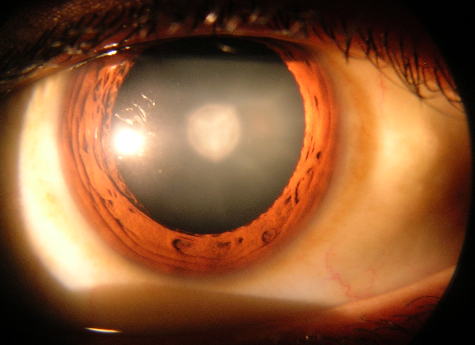
A catarata de início precoce e não sindrômica é uma condição ocular genética e rara. Ela é um problema de desenvolvimento do olho, mas não faz parte de uma síndrome maior (ou seja, não está ligada a outros problemas de saúde gerais). Ela pode se manifestar de muitas formas diferentes, tanto clinicamente quanto geneticamente. Geralmente, ela se apresenta como cataratas que afetam os dois olhos de maneira igual, não progridem (não pioram com o tempo) e aparecem ao nascimento ou na primeira infância. Outros problemas nos olhos podem ocorrer junto, como por exemplo, alterações no desenvolvimento da parte da frente do olho, colobomas, nistagmo (movimento involuntário dos olhos), córnea pequena, olho pequeno e miopia. No entanto, outros órgãos ou sistemas do corpo geralmente não são afetados.
Introdução
O que você precisa saber de cara
A catarata de início precoce e não sindrômica é uma condição ocular genética e rara. Ela é um problema de desenvolvimento do olho, mas não faz parte de uma síndrome maior (ou seja, não está ligada a outros problemas de saúde gerais). Ela pode se manifestar de muitas formas diferentes, tanto clinicamente quanto geneticamente. Geralmente, ela se apresenta como cataratas que afetam os dois olhos de maneira igual, não progridem (não pioram com o tempo) e aparecem ao nascimento ou na primeira infância. Outros problemas nos olhos podem ocorrer junto, como por exemplo, alterações no desenvolvimento da parte da frente do olho, colobomas, nistagmo (movimento involuntário dos olhos), córnea pequena, olho pequeno e miopia. No entanto, outros órgãos ou sistemas do corpo geralmente não são afetados.
Encontrou um erro ou informação desatualizada? Sugira uma correção →
Entender a doença
Do básico ao detalhe, leia no seu ritmo
Preparando trilha educativa...
Sinais e sintomas
O que aparece no corpo e com que frequência cada sintoma acontece
Partes do corpo afetadas
+ 9 sintomas em outras categorias
Características mais comuns
Os sintomas variam de pessoa para pessoa. Abaixo estão as 46 características clínicas mais associadas, ordenadas por frequência.
Linha do tempo da pesquisa
Encontrou um erro ou informação desatualizada? Sugira uma correção →
Genética e causas
O que está alterado no DNA e como passa nas famílias
Genes associados
34 genes identificados com associação a esta condição.
Heat-shock transcription factor that specifically binds heat shock promoter elements (HSE) (PubMed:22587838, PubMed:23507146). Required for denucleation and organelle rupture and degradation that occur during eye lens terminal differentiation, when fiber cells that compose the lens degrade all membrane-bound organelles in order to provide lens with transparency to allow the passage of light (By similarity). In this process, may regulate denucleation of lens fiber cells in part by activating DNAS
Nucleus
Cataract 5, multiple types
An opacification of the crystalline lens of the eye that frequently results in visual impairment or blindness. Opacities vary in morphology, are often confined to a portion of the lens, and may be static or progressive. CTRCT5 includes infantile, lamellar, zonular, nuclear, anterior polar, stellate, and Marner-type cataracts, among others. Finger malformation is observed in some kindreds.
Branching enzyme that converts linear into branched poly-N-acetyllactosaminoglycans. Introduces the blood group I antigen during embryonic development. It is closely associated with the development and maturation of erythroid cells Determines the expression of the blood group I antigen in erythrocytes
Golgi apparatus membrane
Cataract 13, with adult i phenotype
An opacification of the crystalline lens of the eye that frequently results in visual impairment or blindness. Opacities vary in morphology, are often confined to a portion of the lens, and may be static or progressive. In general, the more posteriorly located and dense an opacity, the greater the impact on visual function. CTRCT13 is associated with the rare adult i phenotype, in which adult red blood cells are rich in i antigen and contain low levels of I antigen.
Required for the correct formation of lens intermediate filaments as part of a complex composed of BFSP1, BFSP2 and CRYAA (PubMed:28935373). Plays a role in maintenance of retinal lens optical clarity (By similarity)
Cell membraneCytoplasmCytoplasm, cytoskeletonCytoplasm, cell cortex
Cataract 12, multiple types
An opacification of the crystalline lens of the eye that frequently results in visual impairment or blindness. Opacities vary in morphology, are often confined to a portion of the lens, and may be static or progressive. In general, the more posteriorly located and dense an opacity, the greater the impact on visual function. The opacities can be nuclear, sutural, stellate cortical, lamellar, cortical, nuclear embryonic, Y-sutural, punctate cortical, congenital or with juvenile- and adult-onset.
Crystallins are the dominant structural components of the vertebrate eye lens
Cataract 42
An opacification of the crystalline lens of the eye that frequently results in visual impairment or blindness. Opacities vary in morphology, are often confined to a portion of the lens, and may be static or progressive. In general, the more posteriorly located and dense an opacity, the greater the impact on visual function.
Vimentins are class-III intermediate filaments found in various non-epithelial cells, especially mesenchymal cells. Vimentin is attached to the nucleus, endoplasmic reticulum, and mitochondria, either laterally or terminally. Plays a role in cell directional movement, orientation, cell sheet organization and Golgi complex polarization at the cell migration front (By similarity). Protects SCRIB from proteasomal degradation and facilitates its localization to intermediate filaments in a cell conta
CytoplasmCytoplasm, cytoskeletonNucleus matrixCell membraneCell projection, axon
Cataract 30, multiple types
An opacification of the crystalline lens of the eye that frequently results in visual impairment or blindness. Opacities vary in morphology, are often confined to a portion of the lens, and may be static or progressive. In general, the more posteriorly located and dense an opacity, the greater the impact on visual function.
May contribute to the transparency and refractive index of the lens. Has chaperone-like activity, preventing aggregation of various proteins under a wide range of stress conditions. In lens epithelial cells, stabilizes the ATP6V1A protein, preventing its degradation by the proteasome (By similarity)
CytoplasmNucleusSecretedLysosome
Myopathy, myofibrillar, 2A, adult-onset
A form of myofibrillar myopathy, a group of chronic neuromuscular disorders characterized at ultrastructural level by disintegration of the sarcomeric Z disk and myofibrils, and replacement of the normal myofibrillar markings by small dense granules, or larger hyaline masses, or amorphous material. MFM2A is an autosomal dominant form characterized by weakness of the proximal and distal limb muscles, weakness of the neck, velopharynx and trunk muscles, respiratory insufficiency, hypertrophic cardiomyopathy, and cataract.
Crystallins are the dominant structural components of the vertebrate eye lens
Cataract 39, multiple types
An opacification of the crystalline lens of the eye that frequently results in visual impairment or blindness. Opacities vary in morphology, are often confined to a portion of the lens, and may be static or progressive. In general, the more posteriorly located and dense an opacity, the greater the impact on visual function. CTRCT39 includes lamellar, anterior polar, and complete cataracts.
Lipid kinase that can phosphorylate both monoacylglycerol and diacylglycerol to form lysophosphatidic acid (LPA) and phosphatidic acid (PA), respectively (PubMed:15939762). Does not phosphorylate sphingosine (PubMed:15939762). Phosphorylates ceramide (By similarity). Phosphorylates 1,2-dioleoylglycerol more rapidly than 2,3-dioleoylglycerol (By similarity). Independently of its lipid kinase activity, acts as a component of the TIM22 complex (PubMed:28712724, PubMed:28712726). The TIM22 complex m
Mitochondrion inner membraneMitochondrion intermembrane space
Mitochondrial DNA depletion syndrome 10
An autosomal recessive mitochondrial disorder characterized by congenital cataracts, hypertrophic cardiomyopathy, skeletal myopathy, exercise intolerance, and lactic acidosis. Mental development is normal, but affected individuals may die early from cardiomyopathy.
Crystallins are the dominant structural components of the vertebrate eye lens
Cataract 3, multiple types
An opacification of the crystalline lens of the eye that frequently results in visual impairment or blindness. Opacities vary in morphology, are often confined to a portion of the lens, and may be static or progressive. CTRCT3 includes congenital cerulean and sutural cataract with punctate and cerulean opacities, among others. Cerulean cataract is characterized by peripheral bluish and white opacifications organized in concentric layers with occasional central lesions arranged radially. The opacities are observed in the superficial layers of the fetal nucleus as well as the adult nucleus of the lens. Involvement is usually bilateral. Visual acuity is only mildly reduced in childhood. In adulthood, the opacifications may progress, making lens extraction necessary. Histologically the lesions are described as fusiform cavities between lens fibers which contain a deeply staining granular material. Although the lesions may take on various colors, a dull blue is the most common appearance and is responsible for the designation cerulean cataract. Sutural cataract with punctate and cerulean opacities is characterized by white opacification around the anterior and posterior Y sutures, and grayish and bluish, spindle shaped, oval punctate and cerulean opacities of various sizes arranged in lamellar form. The spots are more concentrated towards the peripheral layers and do not delineate the embryonal or fetal nucleus. Phenotypic variation with respect to the size and density of the sutural opacities as well as the number and position of punctate and cerulean spots is observed among affected subjects.
Required for the correct formation of lens intermediate filaments as part of a complex composed of BFSP1, BFSP2 and CRYAA (PubMed:28935373). Involved in altering the calcium regulation of MIP water permeability (PubMed:30790544)
Cell membraneCytoplasmCytoplasm, cytoskeletonCytoplasm, cell cortex
Cataract 33, multiple types
An opacification of the crystalline lens of the eye that frequently results in visual impairment or blindness. Opacities vary in morphology, are often confined to a portion of the lens, and may be static or progressive. In general, the more posteriorly located and dense an opacity, the greater the impact on visual function. CTRCT33 has juvenile-onset and the opacities are restricted to the cortex of the lens, not involving the nucleus.
Plays a critical role as a guanine nucleotide exchange factor (GEF) for CDC42 in several intracellular processes associated with the actin and microtubule cytoskeleton. Regulates the structure of apical junctions through F-actin organization in epithelial cells (PubMed:17015620, PubMed:19767742). Participates in the normal lumenogenesis of epithelial cell cysts by regulating spindle orientation (PubMed:20479467). Plays a role in ciliogenesis (By similarity). May play a role in membrane trafficki
CytoplasmGolgi apparatus, Golgi stackCytoplasm, cytoskeletonSynapseCell junction
Cataract 48
A form of cataract, an opacification of the crystalline lens of the eye that frequently results in visual impairment or blindness. Opacities vary in morphology, are often confined to a portion of the lens, and may be static or progressive. In general, the more posteriorly located and dense an opacity, the greater the impact on visual function. CTRCT48 is an autosomal recessive form characterized by infantile or early-childhood onset.
Transcriptional regulator which is important for the differentiation and maintenance of meso-diencephalic dopaminergic (mdDA) neurons during development. In addition to its importance during development, it also has roles in the long-term survival and maintenance of the mdDA neurons. Activates NR4A2/NURR1-mediated transcription of genes such as SLC6A3, SLC18A2, TH and DRD2 which are essential for development of mdDA neurons. Acts by decreasing the interaction of NR4A2/NURR1 with the corepressor
Nucleus
Anterior segment dysgenesis 1
A form of anterior segment dysgenesis, a group of defects affecting anterior structures of the eye including cornea, iris, lens, trabecular meshwork, and Schlemm canal. Anterior segment dysgeneses result from abnormal migration or differentiation of the neural crest derived mesenchymal cells that give rise to components of the anterior chamber during eye development. Different anterior segment anomalies may exist alone or in combination, including iris hypoplasia, enlarged or reduced corneal diameter, corneal vascularization and opacity, posterior embryotoxon, corectopia, polycoria, abnormal iridocorneal angle, ectopia lentis, and anterior synechiae between the iris and posterior corneal surface. Clinical conditions falling within the phenotypic spectrum of anterior segment dysgeneses include aniridia, Axenfeld anomaly, Reiger anomaly/syndrome, Peters anomaly, and iridogoniodysgenesis.
Present in the thicker 16-17 nm junctions of mammalian lens fiber cells, where it may contribute to cell junctional organization. Acts as a receptor for calmodulin. May play an important role in both lens development and cataractogenesis
Membrane
Cataract, multiple types 19
An opacification of the crystalline lens of the eye that frequently results in visual impairment or blindness. Opacities vary in morphology, are often confined to a portion of the lens, and may be static or progressive. In general, the more posteriorly located and dense an opacity, the greater the impact on visual function.
Crystallins are the dominant structural components of the vertebrate eye lens
Cataract 20, multiple types
An opacification of the crystalline lens of the eye that frequently results in visual impairment or blindness. Opacities vary in morphology, are often confined to a portion of the lens, and may be static or progressive. In general, the more posteriorly located and dense an opacity, the greater the impact on visual function. CTRCT20 includes progressive polymorphic anterior, posterior, or peripheral cortical.
Probable core component of the endosomal sorting required for transport complex III (ESCRT-III) which is involved in multivesicular bodies (MVBs) formation and sorting of endosomal cargo proteins into MVBs. MVBs contain intraluminal vesicles (ILVs) that are generated by invagination and scission from the limiting membrane of the endosome and mostly are delivered to lysosomes enabling degradation of membrane proteins, such as stimulated growth factor receptors, lysosomal enzymes and lipids. The M
Cytoplasm, cytosolLate endosome membraneMidbodyNucleus envelope
Cataract 31, multiple types
An opacification of the crystalline lens of the eye that frequently results in visual impairment or blindness. Opacities vary in morphology, are often confined to a portion of the lens, and may be static or progressive. In general, the more posteriorly located and dense an opacity, the greater the impact on visual function. CTRCT31 includes posterior polar, progressive posterior subcapsular, nuclear, and anterior subcapsular cataracts.
May function in cell morphology by maintaining the integrity of the circumferential actin ring and controlling lamellipod formation. Involved in the regulation eye, tooth, brain and craniofacial development
Apical cell membraneCell projection, lamellipodiumCell junction, tight junctionCell junction, focal adhesionCytoplasm
Nance-Horan syndrome
Rare X-linked disorder characterized by congenital cataracts, dental anomalies, dysmorphic features, and, in some cases, intellectual disability. Distinctive dental anomalies are seen in affected males, including supernumerary incisors and crown shaped permanent teeth. Characteristic facial features are anteverted pinnae, long face, and prominent nasal bridge and nose. Carrier females display milder variable symptoms of disease with lens opacities often involving the posterior Y sutures, and on occasion dental anomalies and the characteristic facial features described.
Contributes to the transparency and refractive index of the lens (PubMed:18302245). In its oxidized form (absence of intramolecular disulfide bond), acts as a chaperone, preventing aggregation of various proteins under a wide range of stress conditions (PubMed:18199971, PubMed:19595763, PubMed:22120592, PubMed:31792453). Required for the correct formation of lens intermediate filaments as part of a complex composed of BFSP1, BFSP2 and CRYAA (PubMed:28935373)
CytoplasmNucleus
Crystallins are the dominant structural components of the vertebrate eye lens
Cataract 17, multiple types
An opacification of the crystalline lens of the eye that frequently results in visual impairment or blindness. Opacities vary in morphology, are often confined to a portion of the lens, and may be static or progressive. In general, the more posteriorly located and dense an opacity, the greater the impact on visual function. CTRCT17 includes nuclear and pulverulent cataracts, among others. Nuclear cataracts affect the central nucleus of the eye, are often not highly visually significant. The density of the opacities varies greatly from fine dots to a dense, white and chalk-like, central cataract. The condition is usually bilateral. Nuclear cataracts are often combined with opacified cortical fibers encircling the nuclear opacity, which are referred to as cortical riders. Pulverulent cataracts are characterized by a dust-like, 'pulverised' appearance of the opacities which can be found in any part of the lens.
Plays a critical role in epithelial cell morphogenesis, polarity, adhesion and cytoskeletal organization in the lens (PubMed:26231217)
Apical cell membrane
Component of a progesterone-binding protein complex (PubMed:28396637). Binds progesterone (PubMed:25675345). Has many reported cellular functions (heme homeostasis, interaction with CYPs). Required for the maintenance of uterine histoarchitecture and normal female reproductive lifespan (By similarity). Intracellular heme chaperone. Regulates heme synthesis via interactions with FECH and acts as a heme donor for at least some hemoproteins (PubMed:27599036). Forms a ternary complex with TMEM97 rec
Microsome membraneSmooth endoplasmic reticulum membraneMitochondrion outer membraneSecreted
Participates in the regulation of cellular Ca(2+) homeostasis, at least partly, by modulating the filling state of the endoplasmic reticulum Ca(2+) store (PubMed:16989814). Negatively regulates the ER stress response and positively regulates the stability of V-ATPase subunits ATP6V1A and ATP1B1 by preventing their degradation through an unknown proteasome-independent mechanism (PubMed:23035048)
Endoplasmic reticulum membraneCytoplasmic vesicle, secretory vesicle
Wolfram syndrome 1
A rare disorder characterized by juvenile-onset insulin-dependent diabetes mellitus with optic atrophy. Other manifestations include diabetes insipidus, sensorineural deafness, dementia, psychiatric illnesses.
Structural component of lens fiber gap junctions (PubMed:30044662). Gap junctions are dodecameric channels that connect the cytoplasm of adjoining cells (By similarity). They are formed by the docking of two hexameric hemichannels, one from each cell membrane. Small molecules and ions diffuse from one cell to a neighboring cell via the central pore (PubMed:30044662)
Cell membraneCell junction, gap junction
Cataract 14, multiple types
An opacification of the crystalline lens of the eye that frequently results in visual impairment or blindness. Opacities vary in morphology, are often confined to a portion of the lens, and may be static or progressive. CTRCT14 includes zonular pulverulent cataract, among others. Zonular or lamellar cataracts are opacities, broad or narrow, usually consisting of powdery white dots affecting only certain layers or zones between the cortex and nucleus of an otherwise clear lens. The opacity may be so dense as to render the entire central region of the lens completely opaque, or so translucent that vision is hardly if at all impeded. Usually sharply separated from a clear cortex outside them, they may have projections from their outer edges known as riders or spokes.
Acts as a co-chaperone for HSP90 and is required for proper folding of the myosin motor domain. Plays a role in sarcomere formation during muscle cell development. Is necessary for normal early lens development
Cytoplasm, myofibril, sarcomere, Z lineCytoplasm, myofibril, sarcomere, A bandCytoplasm, perinuclear regionCytoplasm, cytosol
Cataract 43
An opacification of the crystalline lens of the eye that frequently results in visual impairment or blindness. Opacities vary in morphology, are often confined to a portion of the lens, and may be static or progressive. In general, the more posteriorly located and dense an opacity, the greater the impact on visual function.
Structural component of eye lens gap junctions (PubMed:18006672, PubMed:19756179). Gap junctions are dodecameric channels that connect the cytoplasm of adjoining cells. They are formed by the docking of two hexameric hemichannels, one from each cell membrane (By similarity). Small molecules and ions diffuse from one cell to a neighboring cell via the central pore (PubMed:18006672, PubMed:19756179)
Cell membraneCell junction, gap junction
Cataract 1, multiple types
An opacification of the crystalline lens of the eye that frequently results in visual impairment or blindness. Opacities vary in morphology, are often confined to a portion of the lens, and may be static or progressive. CTRCT1 includes congenital, zonular pulverulent, nuclear progressive, nuclear pulverulent, nuclear total, total, and posterior subcapsular types of cataract. Zonular or lamellar cataracts are opacities, broad or narrow, usually consisting of powdery white dots affecting only certain layers or zones between the cortex and nucleus of an otherwise clear lens. The opacity may be so dense as to render the entire central region of the lens completely opaque, or so translucent that vision is hardly if at all impeded. Zonular cataracts generally do not involve the embryonic nucleus, though sometimes they involve the fetal nucleus. Usually sharply separated from a clear cortex outside them, they may have projections from their outer edges known as riders or spokes. In some cases cataract is associated with microcornea without any other systemic anomaly or dysmorphism. Microcornea is defined by a corneal diameter inferior to 10 mm in both meridians in an otherwise normal eye.
Nuclear lamina-associated inner nuclear membrane protein that is involved in nuclear structure organization, maintenance of nuclear envelope (NE) integrity and NE reformation after mitosis (PubMed:16339967, PubMed:17097643, PubMed:28242692, PubMed:32494070). Plays a role as transmembrane adapter for the endosomal sorting complexes required for transport (ESCRT), and is thereby involved in ESCRT-mediated NE reformation (PubMed:28242692, PubMed:32494070). Promotes ESCRT-mediated NE closure by recr
Nucleus inner membraneNucleus envelopeCytoplasm, cytoskeleton, spindle
Cataract 46, juvenile-onset, with or without arrhythmic cardiomyopathy
A form of cataract, an opacification of the crystalline lens of the eye that frequently results in visual impairment or blindness. Opacities vary in morphology, are often confined to a portion of the lens, and may be static or progressive. In general, the more posteriorly located and dense an opacity, the greater the impact on visual function. CTRCT46 can be associated with variable onset of a severe form of arrhythmic cardiomyopathy resulting in sudden cardiac death.
Crystallins are the dominant structural components of the vertebrate eye lens
Cataract 4, multiple types
An opacification of the crystalline lens of the eye that frequently results in visual impairment or blindness. Opacities vary in morphology, are often confined to a portion of the lens, and may be static or progressive. CTRCT4 includes crystalline aculeiform, congenital cerulean and non-nuclear polymorphic cataracts, among others. Crystalline aculeiform cataract is characterized by fiberglass-like or needle-like crystals projecting in different directions, through or close to the axial region of the lens. Non-nuclear polymorphic cataract is a partial opacity with variable location between the fetal nucleus of the lens and the equator. The fetal nucleus is normal. The opacities are irregular and look similar to a bunch of grapes and may be present simultaneously in different lens layers. Congenital cerulean cataract is characterized by peripheral bluish and white opacifications organized in concentric layers with occasional central lesions arranged radially. The opacities are observed in the superficial layers of the fetal nucleus as well as the adult nucleus of the lens. Involvement is usually bilateral. Visual acuity is only mildly reduced in childhood. In adulthood, the opacifications may progress, making lens extraction necessary. Histologically the lesions are described as fusiform cavities between lens fibers which contain a deeply staining granular material. Although the lesions may take on various colors, a dull blue is the most common appearance and is responsible for the designation cerulean cataract.
Acts as a transcriptional activator or repressor. Involved in embryonic lens fiber cell development. Recruits the transcriptional coactivators CREBBP and/or EP300 to crystallin promoters leading to up-regulation of crystallin gene during lens fiber cell differentiation. Activates the expression of IL4 in T helper 2 (Th2) cells. Increases T-cell susceptibility to apoptosis by interacting with MYB and decreasing BCL2 expression. Together with PAX6, transactivates strongly the glucagon gene promote
Nucleus
Crystallins are the dominant structural components of the vertebrate eye lens
Cataract 2, multiple types
An opacification of the crystalline lens of the eye that frequently results in visual impairment or blindness. Opacities vary in morphology, are often confined to a portion of the lens, and may be static or progressive. CTRCT2 includes Coppock-like cataract, among others. Coppock-like cataract is a congenital pulverulent disk-like opacity involving the embryonic nucleus with many tiny white dots in the lamellar portion of the lens. It is usually bilateral and dominantly inherited. In some cases, CTRCT2 is associated with microcornea without any other systemic anomaly or dysmorphism. Microcornea is defined by a corneal diameter inferior to 10 mm in both meridians in an otherwise normal eye.
Receptor tyrosine kinase which binds promiscuously membrane-bound ephrin-A family ligands residing on adjacent cells, leading to contact-dependent bidirectional signaling into neighboring cells. The signaling pathway downstream of the receptor is referred to as forward signaling while the signaling pathway downstream of the ephrin ligand is referred to as reverse signaling. Activated by the ligand ephrin-A1/EFNA1 regulates migration, integrin-mediated adhesion, proliferation and differentiation
Cell membraneCell projection, ruffle membraneCell projection, lamellipodium membraneCell junction, focal adhesion
Cataract 6, multiple types
An opacification of the crystalline lens of the eye that frequently results in visual impairment or blindness. Opacities vary in morphology, are often confined to a portion of the lens, and may be static or progressive. CTRCT6 includes posterior polar and age-related cortical cataracts, among others. Posterior polar cataract is a subcapsular opacity, usually disk-shaped, located at the back of the lens. Age-related cortical cataract is a developmental punctate opacity restricted to the cortex. The cataract is white or cerulean, increases in number with age, but rarely affects vision.
Crystallins are the dominant structural components of the vertebrate eye lens
Cataract 10, multiple types
An opacification of the crystalline lens of the eye that frequently results in visual impairment or blindness. Opacities vary in morphology, are often confined to a portion of the lens, and may be static or progressive. CTRCT10 includes congenital zonular with sutural opacities, among others. This is a form of zonular cataract with an erect Y-shaped anterior and an inverted Y-shaped posterior sutural opacities. Zonular or lamellar cataracts are opacities, broad or narrow, usually consisting of powdery white dots affecting only certain layers or zones between the cortex and nucleus of an otherwise clear lens. The opacity may be so dense as to render the entire central region of the lens completely opaque, or so translucent that vision is hardly if at all impeded. Zonular cataracts generally do not involve the embryonic nucleus, though sometimes they involve the fetal nucleus. Usually sharply separated from a clear cortex outside them, they may have projections from their outer edges known as riders or spokes.
Aquaporins form homotetrameric transmembrane channels, with each monomer independently mediating water transport across the plasma membrane along its osmotic gradient (PubMed:11001937, PubMed:24120416). Specifically expressed in lens fiber cells, this aquaporin is crucial for maintaining lens water homeostasis and transparency. Beyond water permeability, it also acts as a cell-to-cell adhesion molecule, forming thin junctions between lens fiber cells that are essential for maintaining the ordere
Cell membraneCell junction
Cataract 15, multiple types
An opacification of the crystalline lens of the eye that frequently results in visual impairment or blindness. Opacities vary in morphology, are often confined to a portion of the lens, and may be static or progressive. CTRCT15 includes polymorphic, progressive punctate lamellar, cortical, anterior and posterior polar, nonprogressive lamellar with sutural opacities, embryonic nuclear, and pulverulent cortical, among others.
Crystallins are the dominant structural components of the vertebrate eye lens
Cataract 23, multiple types
An opacification of the crystalline lens of the eye that frequently results in visual impairment or blindness. Opacities vary in morphology, are often confined to a portion of the lens, and may be static or progressive. In general, the more posteriorly located and dense an opacity, the greater the impact on visual function. CTRCT23 is a zonular cataract. Zonular or lamellar cataracts are opacities, broad or narrow, usually consisting of powdery white dots affecting only certain layers or zones between the cortex and nucleus of an otherwise clear lens. The opacity may be so dense as to render the entire central region of the lens completely opaque, or so translucent that vision is hardly if at all impeded. Zonular cataracts generally do not involve the embryonic nucleus, though sometimes they involve the fetal nucleus. Usually sharply separated from a clear cortex outside them, they may have projections from their outer edges known as riders or spokes.
Key enzyme in the cholesterol biosynthesis pathway. Catalyzes the cyclization of (S)-2,3 oxidosqualene to lanosterol, a reaction that forms the sterol nucleus (PubMed:14766201, PubMed:26200341, PubMed:7639730). Through the production of lanosterol may regulate lens protein aggregation and increase transparency (PubMed:26200341)
Endoplasmic reticulum membrane
Cataract 44
An opacification of the crystalline lens of the eye that frequently results in visual impairment or blindness. Opacities vary in morphology, are often confined to a portion of the lens, and may be static or progressive. In general, the more posteriorly located and dense an opacity, the greater the impact on visual function.
Crystallins are the dominant structural components of the vertebrate eye lens
Cataract 22, multiple types
An opacification of the crystalline lens of the eye that frequently results in visual impairment or blindness. Opacities vary in morphology, are often confined to a portion of the lens, and may be static or progressive. In general, the more posteriorly located and dense an opacity, the greater the impact on visual function. CTRCT22 includes nuclear cataract among others. Nuclear cataracts affect the central nucleus of the eye, and are often not highly visually significant. The density of the opacities varies greatly from fine dots to a dense, white and chalk-like, central cataract. The condition is usually bilateral. Nuclear cataracts are often combined with opacified cortical fibers encircling the nuclear opacity, which are referred to as cortical riders.
Medicamentos aprovados (FDA)
1 medicamento encontrado nos registros da FDA americana.
Variantes genéticas (ClinVar)
138 variantes patogênicas registradas no ClinVar.
Classificação de variantes (ClinVar)
Distribuição de 7 variantes classificadas pelo ClinVar.
Vias biológicas (Reactome)
48 vias biológicas associadas aos genes desta condição.
Diagnóstico
Os sinais que médicos procuram e os exames que confirmam
Tratamento e manejo
Remédios, cuidados de apoio e o que precisa acompanhar
Onde tratar no SUS
Hospitais de referência no Brasil e o protocolo oficial do SUS (PCDT)
🇧🇷 Atendimento SUS — Catarata não-sindrômica de início precoce
Selecione um estado ou use sua localização para ver resultados.
Dados de DATASUS/CNES, SBGM, ABNeuro e Ministério da Saúde. Sempre confirme a disponibilidade diretamente com o estabelecimento.
Pesquisa ativa
Ensaios clínicos abertos e novidades científicas recentes
Ensaios em destaque
🟢 Recrutando agora
1 pesquisa recrutando participantes. Converse com seu médico sobre a possibilidade de participar.
Outros ensaios clínicos
Publicações mais relevantes
Case Series on Autosomal Recessive Non-Syndromic Retinitis Pigmentosa Caused by POMGNT1 Mutations with a Report of a New Variant.
Recessive Protein O-linked-mannose beta-1,2-N-acetylglucosaminyltransferase 1 (POMGNT1) mutations can cause early onset muscle-eye-brain disease but have also more recently been associated with non-syndromic Retinitis Pigmentosa. In this case series, we describe three sisters affected by non-syndromic autosomal recessive POMGNT1 retinopathy with a report of a new variant. The three patients received care at West Virginia University Eye Institute, including full ophthalmic examination with additional fundus imaging, optical coherence tomography (OCT), electroretinogram (ERG), and visual field testing. Diagnostic panel testing of 330 genes was also obtained. The proband was seen for cataract evaluation at age 42, and her fundus examination was suggestive of retinitis pigmentosa. Her oldest sister had been treated for acute anterior uveitis with retinal vasculitis. Another sister was diagnosed with multiple sclerosis (MS) and peripheral retinal degeneration. Posterior subcapsular cataracts were diagnosed between age 42 and 55 in all three sisters, each with constricted fields with preserved central vision. We identified one pathogenic POMGNT1 variant (c.751 + 1G > A) and one likely pathogenic variant (c.1010T > C p.Ile337Thr) in all three sisters. A thorough family history and examination of the siblings with genotyping might have led to an earlier diagnosis of retinal inherited disease and avoidance of immunomodulatory treatment in the oldest sibling.
A novel CRYBB2 mutation causes autosomal dominant cataract: A report from a Chinese family.
This study aimed to examine pathogenic mutation within one Chinese family of five-generations suffering from autosomal dominant cataract. Next-generation sequencing and Sanger sequencing were used to find the pathogenic variants. A rare mutation, c.563G > A, in CRYBB2 gene was found in the proband that showed symptom of non-syndromic congenital autosomal dominant cataract. This mutation had been found in all affected individuals and in one healthy infant, but it did not exist between two individuals who did not develop such disease in that family, as well as in 100 healthy subjects who showed no relation with that family. Cataracts in this family varied with different severity of lens opacities and elongation of axial length. One missense mutation c.563G > A is reported in the CRYBB2 gene among one Chinese family suffering from early-onset cataract, and associated novel phenotypes are the elongation of axial length and the types of cataract. Our results expand the spectrum of associated phenotypes of CRYBB2 mutation.
Dominant ARL3-related retinitis pigmentosa.
To clinically and genetically characterise a second family with dominant ARL3-related retinitis pigmentosa due to a specific ARL3 missense variant, p.(Tyr90Cys). Clinical examination included optical coherence tomography, electroretinography, and ultra-wide field retinal imaging with autofluorescence. Retrospective data were collected from the registry of inherited retinal diseases at Oslo university hospital. DNA was analysed by whole-exome sequencing and Sanger sequencing. The ARL3 missense variant was visualized in a 3D-protein structure. The phenotype was non-syndromic retinitis pigmentosa with cataract associated with early onset of decreased central vision and central retinal thinning. Sanger sequencing confirmed the presence of a de novo ARL3 missense variant p.(Tyr90Cys) in the index patient and his affected son. We did not find any other cases with rare ARL3 variants in a cohort of 431 patients with retinitis pigmentosa-like disease. By visualizing Tyr90 in the 3D protein structure, it seems to play an important role in packing of the α/β structure of ADP-ribosylation factor-like 3 (ARL3). When changing Tyr90 to cysteine, we observe a loss of interactions in the core of the α/β structure that is likely to affect folding and stability of ARL3. Our study confirms that the ARL3 missense variant p.(Tyr90Cys) causes retinitis pigmentosa. In 2016, Strom et al. reported the exact same variant in a mother and two children with RP, labelled ?RP83 in the OMIM database. Now the questionmark can be removed, and ARL3 should be added to the list of genes that may cause non-syndromic dominant retinitis pigmentosa.
Publicações recentes
A novel CRYBB2 mutation causes autosomal dominant cataract: A report from a Chinese family.
Recessive mutations in LEPREL1 underlie a recognizable lens subluxation phenotype.
Homozygous loss-of-function mutation of the LEPREL1 gene causes severe non-syndromic high myopia with early-onset cataract.
Non-syndromic posterior lenticonus a cause of childhood cataract: evidence for X-linked inheritance.
📚 EuropePMCmostrando 3
Case Series on Autosomal Recessive Non-Syndromic Retinitis Pigmentosa Caused by POMGNT1 Mutations with a Report of a New Variant.
Journal of clinical medicineA novel CRYBB2 mutation causes autosomal dominant cataract: A report from a Chinese family.
European journal of ophthalmologyDominant ARL3-related retinitis pigmentosa.
Ophthalmic geneticsAssociações
Organizações que acompanham esta doença — pra ter apoio e orientação
Ainda não temos associações cadastradas para Catarata não-sindrômica de início precoce.
É de uma associação que acompanha esta doença? Fale com a gente →
Comunidades
Grupos ativos de quem convive com esta doença aqui no Raras
Ainda não existe comunidade no Raras para Catarata não-sindrômica de início precoce
Pacientes, familiares e cuidadores se organizam em comunidades pra compartilhar experiências, fazer perguntas e se apoiar. Você pode ser o primeiro.
Tire suas dúvidas
Perguntas, dicas e experiências compartilhadas aqui na página
Participe da discussão
Faça login para postar dúvidas, compartilhar experiências e interagir com especialistas.
Fazer loginDoenças relacionadas
Doenças com sintomas parecidos — ajudam quem ainda está buscando diagnóstico
Referências e fontes
Bases de dados externas citadas neste artigo
Publicações científicas
Artigos indexados no PubMed ligados a esta doença no grafo RarasNet — título, periódico e PMID direto da fonte, sem intermediação de IA.
- Case Series on Autosomal Recessive Non-Syndromic Retinitis Pigmentosa Caused by POMGNT1 Mutations with a Report of a New Variant.
- A novel CRYBB2 mutation causes autosomal dominant cataract: A report from a Chinese family.
- Dominant ARL3-related retinitis pigmentosa.
- Recessive mutations in LEPREL1 underlie a recognizable lens subluxation phenotype.
- Homozygous loss-of-function mutation of the LEPREL1 gene causes severe non-syndromic high myopia with early-onset cataract.
- Non-syndromic posterior lenticonus a cause of childhood cataract: evidence for X-linked inheritance.
Bases de dados e fontes oficiais
Identificadores e referências canônicas usadas para montar este verbete.
- ORPHA:91492(Orphanet)
- OMIM OMIM:601371(OMIM)
- MONDO:0011060(MONDO)
- GARD:16801(GARD (NIH))
- Variantes catalogadas(ClinVar)
- Busca completa no PubMed(PubMed)
- Q18387306(Wikidata)
Dados compilados pelo RarasNet a partir de fontes abertas (Orphanet, OMIM, MONDO, PubMed/EuropePMC, ClinicalTrials.gov, DATASUS, PCDT/MS). Este conteúdo é informativo e não substitui avaliação médica.
Conteúdo mantido por Agente Raras · Médicos e pesquisadores podem colaborar
Catarata não-sindrômica de início precoce
📋 Origem dos dados
Esta página agrega dados de fontes públicas e oficiais. Dados sobre cobertura no SUS (PCDT, CEAF) são verificados ativamente por agente proativo (ver badge no infobox). Demais dados têm atribuição de fonte + data da última sincronização — clique para abrir o original.
- Doença rara (ontologia)
- fonte: Orphanet
- Identificador unificado
- fonte: MONDO
- Genética mendeliana
- fonte: OMIM
- Codificação WHO/SUS
- fonte: WHO ICD-10 / DATASUS
- CID-11 (futuro)
- fonte: WHO ICD-11
- NIH/GARD
- fonte: GARD (NIH)
- Dado público estruturado
- fonte: Wikidata
- Medicamentos aprovados FDA
- fonte: FDA OpenFDA