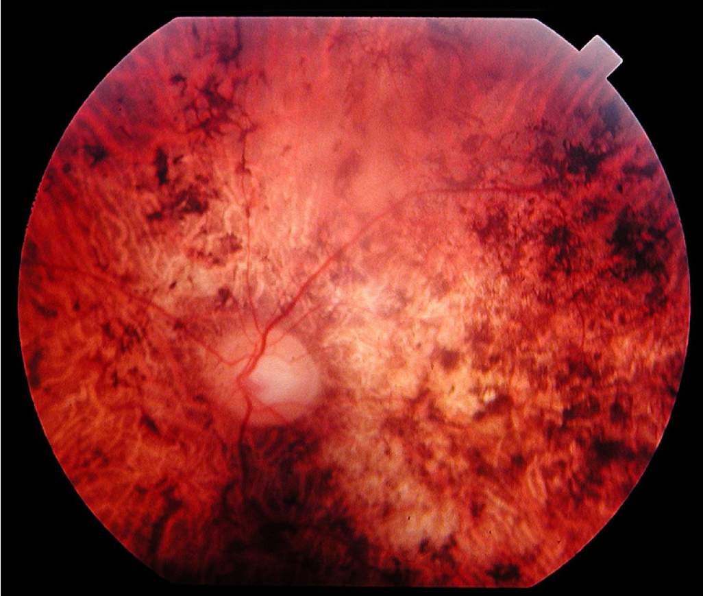
Problemas de visão genéticos que afetam a retina e são classificados como retinopatias pigmentares.
Introdução
O que você precisa saber de cara
Problemas de visão genéticos que afetam a retina e são classificados como retinopatias pigmentares.
Escala de raridade
<1/50kMuito rara
1/20kRara
1/10kPouco freq.
1/5kIncomum
1/2k
Encontrou um erro ou informação desatualizada? Sugira uma correção →
Entender a doença
Do básico ao detalhe, leia no seu ritmo
Preparando trilha educativa...
Sinais e sintomas
O que aparece no corpo e com que frequência cada sintoma acontece
Partes do corpo afetadas
+ 21 sintomas em outras categorias
Características mais comuns
Os sintomas variam de pessoa para pessoa. Abaixo estão as 83 características clínicas mais associadas, ordenadas por frequência.
Linha do tempo da pesquisa
Encontrou um erro ou informação desatualizada? Sugira uma correção →
Genética e causas
O que está alterado no DNA e como passa nas famílias
Genes associados
33 genes identificados com associação a esta condição. Padrão de herança: Autosomal dominant, Autosomal recessive, X-linked recessive.
Curadoria gene-doença
fontes oficiaisInvolved in ceramide synthesis
Golgi apparatus membraneEndoplasmic reticulum membrane
Cone-rod dystrophy 22
An autosomal recessive form of cone-rod dystrophy, an inherited retinal dystrophy characterized by retinal pigment deposits visible on fundus examination, predominantly in the macular region, and initial loss of cone photoreceptors followed by rod degeneration. This leads to decreased visual acuity and sensitivity in the central visual field, followed by loss of peripheral vision. Severe loss of vision occurs earlier than in retinitis pigmentosa, due to cone photoreceptors degenerating at a higher rate than rod photoreceptors.
Soluble retinoid carrier essential the proper function of both rod and cone photoreceptors. Participates in the regeneration of active 11-cis-retinol and 11-cis-retinaldehyde, from the inactive 11-trans products of the rhodopsin photocycle and in the de novo synthesis of these retinoids from 11-trans metabolic precursors. The cycling of retinoids between photoreceptor and adjacent pigment epithelium cells is known as the 'visual cycle'
Cytoplasm
Bothnia retinal dystrophy
A type of retinitis punctata albescens. Affected individuals show night blindness from early childhood with features consistent with retinitis punctata albescens and macular degeneration.
The small GTPases Rab are key regulators of intracellular membrane trafficking, from the formation of transport vesicles to their fusion with membranes (PubMed:8647132). Rabs cycle between an inactive GDP-bound form and an active GTP-bound form that is able to recruit to membranes different sets of downstream effectors directly responsible for vesicle formation, movement, tethering and fusion (PubMed:8647132). RAB28 is required for shedding and phagocytosis of cone cell outer segments (OS) discs
Cell membraneCytoplasm, cytoskeleton, cilium basal bodyCytoplasmNucleus
Cone-rod dystrophy 18
A form of cone-rod dystrophy, an inherited retinal dystrophy characterized by retinal pigment deposits visible on fundus examination, predominantly in the macular region, and initial loss of cone photoreceptors followed by rod degeneration. This leads to decreased visual acuity and sensitivity in the central visual field, followed by loss of peripheral vision. Severe loss of vision occurs earlier than in retinitis pigmentosa, due to cone photoreceptors degenerating at a higher rate than rod photoreceptors.
Essential for retina photoreceptor outer segment disk morphogenesis, may also play a role with ROM1 in the maintenance of outer segment disk structure (By similarity). Required for the maintenance of retinal outer nuclear layer thickness (By similarity). Required for the correct development and organization of the photoreceptor inner segment (By similarity)
MembraneCell projection, cilium, photoreceptor outer segmentPhotoreceptor inner segment
Retinitis pigmentosa 7
A retinal dystrophy belonging to the group of pigmentary retinopathies. Retinitis pigmentosa is characterized by retinal pigment deposits visible on fundus examination and primary loss of rod photoreceptor cells followed by secondary loss of cone photoreceptors. Patients typically have night vision blindness and loss of midperipheral visual field. As their condition progresses, they lose their far peripheral visual field and eventually central vision as well.
Outward-rectifying chloride channel involved in endolysosomal chloride homeostasis, membrane fusion and function. Conducts chloride currents up to hundreds of picoamperes. Regulates lysosomal calcium content by reducing the lysosomal membrane potential, thereby activating TRPML1 channel and further release of lysosomal calcium ions. Regulates the pH in endolysosomal compartments and may contribute to progressive acidification from endosome to lysosome. Permeable to other halides such as iodide a
Endosome membraneLysosome membrane
Ceroid lipofuscinosis, neuronal, 7
A form of neuronal ceroid lipofuscinosis with onset in early childhood. Neuronal ceroid lipofuscinoses are progressive neurodegenerative, lysosomal storage diseases characterized by intracellular accumulation of autofluorescent liposomal material, and clinically by seizures, dementia, visual loss, and/or cerebral atrophy. The lipopigment patterns observed most often in neuronal ceroid lipofuscinosis type 7 comprise mixed combinations of granular, curvilinear, fingerprint, and rectilinear profiles.
Cell surface receptor for PLXNB1, PLXNB2, PLXNB3 and PLXND1 that plays an important role in cell-cell signaling (By similarity). Regulates glutamatergic and GABAergic synapse development (By similarity). Promotes the development of inhibitory synapses in a PLXNB1-dependent manner and promotes the development of excitatory synapses in a PLXNB2-dependent manner (By similarity). Plays a role in priming antigen-specific T-cells, promotes differentiation of Th1 T-helper cells, and thereby contributes
Cell membrane
Retinitis pigmentosa 35
A retinal dystrophy belonging to the group of pigmentary retinopathies. Retinitis pigmentosa is characterized by retinal pigment deposits visible on fundus examination and primary loss of rod photoreceptor cells followed by secondary loss of cone photoreceptors. Patients typically have night vision blindness and loss of midperipheral visual field. As their condition progresses, they lose their far peripheral visual field and eventually central vision as well.
Involved in synaptic functions in photoreceptor cells, the signal transduction in immune cells as a Src family kinase activator, endosome recycling, the uptake of bacteria and endocytosis, protein trafficking in sensory neurons and as lipid-binding chaperone with specificity for a diverse subset of myristoylated proteins. Specifically binds the myristoyl moiety of a subset of N-terminally myristoylated proteins and is required for their localization. Binds myristoylated GNAT1 and is required for
Cytoplasm, cytoskeleton, microtubule organizing center, centrosomeCytoplasm, cytoskeleton, spindle poleCytoplasm, cytoskeleton, spindle
Immunodeficiency 13
A rare and heterogeneous syndrome defined by a reproducible reduction in the CD4 T-lymphocyte count (less than 300 cells per microliter or less than 20% of total T-cells) in the absence of HIV infection or other known causes of immunodeficiency. IMD13 predisposes to infections and malignancy.
Stimulates retinal guanylyl cyclase when free calcium ions concentration is low and inhibits guanylyl cyclase when free calcium ions concentration is elevated (PubMed:18706439, PubMed:19459154, PubMed:30184081, PubMed:30622141). This Ca(2+)-sensitive regulation of retinal guanylyl cyclase is a key event in recovery of the dark state of rod photoreceptors following light exposure (By similarity). May be involved in cone photoreceptor light response and recovery of response in bright light (By sim
MembranePhotoreceptor inner segmentCell projection, cilium, photoreceptor outer segment
Cone dystrophy 3
An autosomal dominant cone dystrophy. Cone dystrophies are retinal dystrophies characterized by progressive degeneration of the cone photoreceptors with preservation of rod function, as indicated by electroretinogram. However, some rod involvement may be present in some cone dystrophies, particularly at late stage. Affected individuals suffer from photophobia, loss of visual acuity, color vision and central visual field. Another sign is the absence of macular lesions for many years. Cone dystrophies are distinguished from the cone-rod dystrophies in which some loss of peripheral vision also occurs.
May be important in protein trafficking and/or protein folding and stabilization
CytoplasmNucleus
Leber congenital amaurosis 4
A severe dystrophy of the retina, typically becoming evident in the first years of life. Visual function is usually poor and often accompanied by nystagmus, sluggish or near-absent pupillary responses, photophobia, high hyperopia and keratoconus.
May function as scaffolding protein. Required for normal location of RPGR at the connecting cilium of photoreceptor cells. Required for normal disk morphogenesis and disk organization in the outer segment of photoreceptor cells and for survival of photoreceptor cells
Cell projection, cilium
Leber congenital amaurosis 6
A severe dystrophy of the retina, typically becoming evident in the first years of life. Visual function is usually poor and often accompanied by nystagmus, sluggish or near-absent pupillary responses, photophobia, high hyperopia and keratoconus.
Visual pigments are the light-absorbing molecules that mediate vision. They consist of an apoprotein, opsin, covalently linked to cis-retinal
Cell membrane
Colorblindness, partial, deutan series
An X-linked color vision defect characterized by a dichromasy in which red and green are confused, without loss of luminance or shift or shortening of the spectrum. Dichromasy is due to the use of only two types of photoreceptors, blue plus red in deuteranopia and blue plus green in protanopia.
May be involved in photoreceptor outer segment disk morphogenesis (By similarity)
CytoplasmPhotoreceptor inner segment
Cone-rod dystrophy 16
An inherited retinal dystrophy characterized by retinal pigment deposits visible on fundus examination, predominantly in the macular region, and initial loss of cone photoreceptors followed by rod degeneration. This leads to decreased visual acuity and sensitivity in the central visual field, followed by loss of peripheral vision. Severe loss of vision occurs earlier than in retinitis pigmentosa, due to cone photoreceptors degenerating at a higher rate than rod photoreceptors.
May play a role in cell differentiation, proliferation and apoptosis (PubMed:24556617). Binds cholesterol in cholesterol-containing plasma membrane microdomains and may play a role in the organization of the apical plasma membrane in epithelial cells. During early retinal development acts as a key regulator of disk morphogenesis. Involved in regulation of MAPK and Akt signaling pathways. In neuroblastoma cells suppresses cell differentiation such as neurite outgrowth in a RET-dependent manner (P
Apical cell membraneCell projection, microvillus membraneCell projection, cilium, photoreceptor outer segmentEndoplasmic reticulumEndoplasmic reticulum-Golgi intermediate compartment
Retinitis pigmentosa 41
A retinal dystrophy belonging to the group of pigmentary retinopathies. Retinitis pigmentosa is characterized by retinal pigment deposits visible on fundus examination and primary loss of rod photoreceptor cells followed by secondary loss of cone photoreceptors. Patients typically have night vision blindness and loss of midperipheral visual field. As their condition progresses, they lose their far peripheral visual field and eventually central vision as well.
Voltage-sensitive calcium channels (VSCC) mediate the entry of calcium ions into excitable cells and are also involved in a variety of calcium-dependent processes, including muscle contraction, hormone or neurotransmitter release, gene expression, cell motility, cell division and cell death. The isoform alpha-1F gives rise to L-type calcium currents. Long-lasting (L-type) calcium channels belong to the 'high-voltage activated' (HVA) group. They are blocked by dihydropyridines (DHP), phenylalkyla
Membrane
Night blindness, congenital stationary, 2A
A non-progressive retinal disorder characterized by impaired night vision, often associated with nystagmus and myopia.
Metalloprotease that cleaves and releases a number of molecules with important roles in tumorigenesis and angiogenesis, such as TEK, KDR, EPHB4, CD40, VCAM1 and CDH5. May mediate cell-cell, cell-matrix interactions and regulate the motility of cells via interactions with integrins May act as alpha-secretase for amyloid precursor protein (APP)
Cell membraneSecreted
Cone-rod dystrophy 9
An inherited retinal dystrophy characterized by retinal pigment deposits visible on fundus examination, predominantly in the macular region, and initial loss of cone photoreceptors followed by rod degeneration. This leads to decreased visual acuity and sensitivity in the central visual field, followed by loss of peripheral vision. Severe loss of vision occurs earlier than in retinitis pigmentosa, due to cone photoreceptors degenerating at a higher rate than rod photoreceptors.
Catalyzes the formation of NAD(+) from nicotinamide mononucleotide (NMN) and ATP (PubMed:17402747). Can also use the deamidated form; nicotinic acid mononucleotide (NaMN) as substrate with the same efficiency (PubMed:17402747). Can use triazofurin monophosphate (TrMP) as substrate (PubMed:17402747). Also catalyzes the reverse reaction, i.e. the pyrophosphorolytic cleavage of NAD(+) (PubMed:17402747). For the pyrophosphorolytic activity, prefers NAD(+) and NaAD as substrates and degrades NADH, ni
Nucleus
Leber congenital amaurosis 9
A severe dystrophy of the retina, typically becoming evident in the first years of life. Visual function is usually poor and often accompanied by nystagmus, sluggish or near-absent pupillary responses, photophobia, high hyperopia and keratoconus.
Catalyzes the transfer of phosphatidylinositol and phosphatidylcholine between membranes (in vitro) (By similarity). Binds calcium ions
Endomembrane system
Cone-rod dystrophy 5
An inherited retinal dystrophy characterized by retinal pigment deposits visible on fundus examination, predominantly in the macular region, and initial loss of cone photoreceptors followed by rod degeneration. This leads to decreased visual acuity and sensitivity in the central visual field, followed by loss of peripheral vision. Severe loss of vision occurs earlier than in retinitis pigmentosa, due to cone photoreceptors degenerating at a higher rate than rod photoreceptors.
Visual pigments are the light-absorbing molecules that mediate vision. They consist of an apoprotein, opsin, covalently linked to cis-retinal
Membrane
Colorblindness, partial, protan series
An X-linked color vision defect characterized by a dichromasy in which red and green are confused, with loss of luminance and shift of brightness and hue curves toward the short wave end of the spectrum. Dichromasy is due to the use of only two types of photoreceptors, blue plus red in deuteranopia and blue plus green in protanopia.
Pore-forming subunit of the cone cyclic nucleotide-gated channel. Mediates cone photoresponses at bright light converting transient changes in intracellular cGMP levels into electrical signals. In the dark, cGMP levels are high and keep the channel open enabling a steady inward current carried by Na(+) and Ca(2+) ions that leads to membrane depolarization and neurotransmitter release from synaptic terminals. Upon photon absorption cGMP levels decline leading to channel closure and membrane hyper
Cell membrane
Achromatopsia 2
An autosomal recessive, ocular stationary disorder due to the absence of functioning cone photoreceptors in the retina. It is characterized by total colorblindness, low visual acuity, photophobia and nystagmus.
Acts as a guanine-nucleotide releasing factor (GEF) for RAB8A and RAB37 by promoting the conversion of inactive RAB-GDP to the active form RAB-GTP (PubMed:20631154). GEF activity towards RAB8A may facilitate ciliary trafficking by modulating ciliary intracellular localization of RAB8A (PubMed:20631154). GEF activity towards RAB37 maintains autophagic homeostasis and retinal function (By similarity). Involved in photoreceptor integrity (By similarity). May control cilia formation by regulating ac
Cytoplasm, cytoskeleton, flagellum axonemeGolgi apparatusCell projection, ciliumCytoplasm, cytoskeleton, microtubule organizing center, centrosomeCytoplasm, cytoskeleton, cilium basal bodyCytoplasm, cytoskeleton, cilium axoneme
Retinitis pigmentosa 3
An X-linked retinal dystrophy belonging to the group of pigmentary retinopathies. Retinitis pigmentosa is characterized by retinal pigment deposits visible on fundus examination and primary loss of rod photoreceptor cells followed by secondary loss of cone photoreceptors. Patients typically have night vision blindness and loss of midperipheral visual field. As their condition progresses, they lose their far peripheral visual field and eventually central vision as well. In RP3, affected males have a severe phenotype, and carrier females show a wide spectrum of clinical features ranging from completely asymptomatic to severe retinitis pigmentosa. Heterozygous women can manifest a form of choroidoretinal degeneration which is distinguished from other types by the absence of visual defects in the presence of a brilliant, scintillating, golden-hued, patchy appearance most striking around the macula, called a tapetal-like retinal reflex.
Plays a role in the initiation of autophagy. In the retina, might be involved in the process of photoreceptor cells renewal and recycling to preserve visual function. Induces apoptotic cell death when coexpressed with DRAM1
Lysosome membranePhotoreceptor inner segmentApical cell membrane
Cone-rod dystrophy 21
A form of cone-rod dystrophy, an inherited retinal dystrophy characterized by retinal pigment deposits visible on fundus examination, predominantly in the macular region, and initial loss of cone photoreceptors followed by rod degeneration. This leads to decreased visual acuity and sensitivity in the central visual field, followed by loss of peripheral vision. Severe loss of vision occurs earlier than in retinitis pigmentosa, due to cone photoreceptors degenerating at a higher rate than rod photoreceptors.
Catalyzes the synthesis of cyclic GMP (cGMP) in rods and cones of photoreceptors. Plays an essential role in phototransduction, by mediating cGMP replenishment (PubMed:15123990, PubMed:21928830, PubMed:26100624, PubMed:30319355, PubMed:9600905). May also participate in the trafficking of membrane-associated proteins to the photoreceptor outer segment membrane (By similarity)
Photoreceptor outer segment membraneEndoplasmic reticulum membrane
Leber congenital amaurosis 1
A severe dystrophy of the retina, typically becoming evident in the first years of life. Visual function is usually poor and often accompanied by nystagmus, sluggish or near-absent pupillary responses, photophobia, high hyperopia and keratoconus.
Rab effector involved in exocytosis (By similarity). May act as scaffold protein that regulates neurotransmitter release at the active zone. Essential for maintaining normal probability of neurotransmitter release and for regulating release during short-term synaptic plasticity (By similarity). Plays a role in dendrite formation by melanocytes (PubMed:23999003)
Cell membraneSynapsePresynaptic cell membrane
May be involved in modulating the expression of photoreceptor specific genes. Binds to the Ret-1 and Bat-1 element within the rhodopsin promoter
Nucleus
Macular degeneration, age-related, 6
A form of age-related macular degeneration, a multifactorial eye disease and the most common cause of irreversible vision loss in the developed world. In most patients, the disease is manifest as ophthalmoscopically visible yellowish accumulations of protein and lipid that lie beneath the retinal pigment epithelium and within an elastin-containing structure known as Bruch membrane.
The alpha-2/delta subunit of voltage-dependent calcium channels regulates calcium current density and activation/inactivation kinetics of the calcium channel
Membrane
Retinal cone dystrophy 4
Characterized by minimal symptoms except for slowly progressive reduction in visual acuity.
Transcription factor that binds and transactivates the sequence 5'-TAATC[CA]-3' which is found upstream of several photoreceptor-specific genes, including the opsin genes. Acts synergistically with other transcription factors, such as NRL, RORB and RAX, to regulate photoreceptor cell-specific gene transcription. Essential for the maintenance of mammalian photoreceptors
Nucleus
Leber congenital amaurosis 7
A severe dystrophy of the retina, typically becoming evident in the first years of life. Visual function is usually poor and often accompanied by nystagmus, sluggish or near-absent pupillary responses, photophobia, high hyperopia and keratoconus.
Plays a role in cilia formation and/or maintenance (By similarity). Plays a role in the regulation of cell morphology and cytoskeletal organization (PubMed:21834987). Involved in DNA damage repair (PubMed:26290490)
MitochondrionCytoplasm, cytoskeleton, cilium basal bodyCell projection, cilium, photoreceptor outer segmentCytoplasm
Retinal dystrophy with or without macular staphyloma
An ocular disorder characterized by decreased vision which worsen over time, and dystrophic changes in the retina, such as retinal pigment epithelium mottling and vessel narrowing. Macular staphyloma, without high myopia, is present in some patients.
Flippase that catalyzes in an ATP-dependent manner the transport of retinal-phosphatidylethanolamine conjugates like 11-cis and all-trans isomers of N-retinylidene-phosphatidylethanolamine (N-Ret-PE) from the lumen to the cytoplasmic leaflet of photoreceptor outer segment disk membranes, where 11-cis-retinylidene-phosphatidylethanolamine is then isomerized to its all-trans isomer and reduced by RDH8 to produce all-trans-retinol. This transport activity ensures that all-trans-retinal generated fr
MembraneEndoplasmic reticulumCytoplasmic vesicleCell projection, cilium, photoreceptor outer segment
Stargardt disease 1
An autosomal recessive form of Stargardt disease, a retinal degenerative disease characterized by macular dystrophy, progressive bilateral atrophy of the foveal retinal pigment epithelium, and accumulation of fluorescent flecks around the macula and/or in the central and near-peripheral areas of the retina. STGD1 patients typically lose central vision in their first or second decade of life.
Precursor of the transcription factor form (Processed cyclic AMP-dependent transcription factor ATF-6 alpha), which is embedded in the endoplasmic reticulum membrane (PubMed:10564271, PubMed:11158310, PubMed:11779464). Endoplasmic reticulum stress promotes processing of this form, releasing the transcription factor form that translocates into the nucleus, where it activates transcription of genes involved in the unfolded protein response (UPR) (PubMed:10564271, PubMed:11158310, PubMed:11779464)
Endoplasmic reticulum membraneGolgi apparatus membraneNucleus
Achromatopsia 7
A form of achromatopsia, an ocular stationary disorder due to the absence of functioning cone photoreceptors in the retina. It is characterized by total colorblindness, low visual acuity, photophobia and nystagmus.
Plays an important role in centriole assembly and/or stability and ciliogenesis (PubMed:20008567, PubMed:32060285). Involved in early steps of centriole duplication, as well as in the later steps of centriole length control (PubMed:19109428). Acts in concert with POC1A to ensure centriole integrity and proper mitotic spindle formation (PubMed:32060285). Required for primary cilia formation, ciliary length and also cell proliferation (PubMed:23015594). Required for retinal integrity (PubMed:25044
Cytoplasm, cytoskeleton, microtubule organizing center, centrosomeCytoplasm, cytoskeleton, microtubule organizing center, centrosome, centrioleCytoplasm, cytoskeleton, cilium basal bodyCytoplasm, cytoskeleton, spindle pole
Cone-rod dystrophy 20
A form of cone-rod dystrophy, an inherited retinal dystrophy characterized by retinal pigment deposits visible on fundus examination, predominantly in the macular region, and initial loss of cone photoreceptors followed by rod degeneration. This leads to decreased visual acuity and sensitivity in the central visual field, followed by loss of peripheral vision. Severe loss of vision occurs earlier than in retinitis pigmentosa, due to cone photoreceptors degenerating at a higher rate than rod photoreceptors.
Polyglutamylase which modifies tubulin, generating polyglutamate side chains on the gamma-carboxyl group of specific glutamate residues within the C-terminal tail of tubulin. Preferentially mediates ATP-dependent initiation step of the polyglutamylation reaction over the elongation step. Preferentially modifies the alpha-tubulin tail over a beta-tail (By similarity). Required for CCSAP localization to both polyglutamylated spindle and cilia microtubules (PubMed:22493317). Increases the effects o
Cell projection, ciliumCytoplasm, cytoskeleton, cilium basal bodyNucleusCytoplasm
Cone-rod dystrophy 19
A form of cone-rod dystrophy, an inherited retinal dystrophy characterized by retinal pigment deposits visible on fundus examination, predominantly in the macular region, and initial loss of cone photoreceptors followed by rod degeneration. This leads to decreased visual acuity and sensitivity in the central visual field, followed by loss of peripheral vision. Severe loss of vision occurs earlier than in retinitis pigmentosa, due to cone photoreceptors degenerating at a higher rate than rod photoreceptors.
Potential calcium-dependent cell-adhesion protein. May be required for the structural integrity of the outer segment (OS) of photoreceptor cells (By similarity)
Cell membrane
Cone-rod dystrophy 15
An autosomal recessive retinal dystrophy characterized by retinal pigment deposits visible on fundus examination, predominantly in the macular region, and initial loss of cone photoreceptors followed by rod degeneration. This leads to decreased visual acuity and sensitivity in the central visual field, followed by loss of peripheral vision. Severe loss of vision occurs earlier than in retinitis pigmentosa, due to cone photoreceptors degenerating at a higher rate than rod photoreceptors.
Variantes genéticas (ClinVar)
479 variantes patogênicas registradas no ClinVar.
Classificação de variantes (ClinVar)
Distribuição de 4.696 variantes classificadas pelo ClinVar.
Vias biológicas (Reactome)
30 vias biológicas associadas aos genes desta condição.
Diagnóstico
Os sinais que médicos procuram e os exames que confirmam
Tratamento e manejo
Remédios, cuidados de apoio e o que precisa acompanhar
Onde tratar no SUS
Hospitais de referência no Brasil e o protocolo oficial do SUS (PCDT)
🇧🇷 Atendimento SUS — Distrofia dos cones e bastonetes
Selecione um estado ou use sua localização para ver resultados.
Dados de DATASUS/CNES, SBGM, ABNeuro e Ministério da Saúde. Sempre confirme a disponibilidade diretamente com o estabelecimento.
Pesquisa ativa
Ensaios clínicos abertos e novidades científicas recentes
Ensaios em destaque
🟢 Recrutando agora
6 pesquisas recrutando participantes. Converse com seu médico sobre a possibilidade de participar.
Outros ensaios clínicos
40 ensaios clínicos encontrados, 7 ativos.
Publicações mais relevantes
RGR-mediated photopic visual cycle and oxidative stress: potential mechanisms for cone vision impairment and retinal degeneration in retinitis pigmentosa linked to D1080N-IRBP.
Interphotoreceptor retinoid-binding protein (IRBP), an extracellular glycoprotein, supports cone-mediated vision through unclear molecular mechanisms. Over 30 different mutations in the IRBP gene have been found in patients with retinitis pigmentosa, childhood-onset retinal dystrophy with high myopia, and cone-rod dystrophy, yet their pathogenicity and underlying pathogenic mechanisms have not been studied in animal models. Here, we found that extracellular IRBP significantly increased the quantities of the extracellular, but not intracellular, 11-cis-retinol and 11-cis-retinal synthesized by retinal G protein-coupled receptor (RGR) in coordination with retinol dehydrogenases and green light stimuli. Retinoid trafficking in the retina and recovery of S-cone and rod maximum photoresponses were substantially delayed in male and female mice of a new retinitis pigmentosa model linked to the human D1080N-IRBP. The mutant IRBP was unstable, not secreted, and retained in the endoplasmic reticulum of photoreceptors due to formation of insoluble high molecular complexes via disulfide bonds. Young mutant mice exhibited profound reduction in photoresponses to ultraviolet stimuli without a significant S-opsin reduction and S-cone structural degeneration. In contrast, M-cones exhibited early and progressive degeneration, accompanied by mislocalization of M-opsin to the soma and synaptic region of M-cones. Rods also underwent early and progressive degeneration. Oxidative and inflammatory stresses as well as pro-apoptotic proteins such as activated caspase-3, BAX, and apoptosis-inducing factor were markedly increased in the mutant mouse retina. These findings identify both a role of IRBP in the RGR-mediated photopic visual cycle supporting daytime color vision and the molecular mechanism by which D1080N-IRBP causes vision impairment and photoreceptor degeneration.Significance Statement IRBP, an interphotoreceptor matrix protein, supports cone-mediated color vision via unclear mechanism. Mutations in IRBP are associated with blinding diseases with poorly understood pathogenic mechanism. Here, we found that IRBP plays an important role in the RGR-mediated photopic visual cycle essential cone-mediated vision. Consistent with this, recovery of S-cone photoresponse was significantly delayed in a newly developed mouse model carrying a human disease-causing IRBP mutation. This mutation abolished IRBP secretion and led to progressive degeneration of rods and M-cones, accompanied by elevation of oxidative stress, inflammatory cytokines, and proapoptotic proteins in the retina. These results identify both a functional mechanism of IRBP in promoting cone-mediated vision and a pathogenic mechanism by which IRBP mutations cause vision loss and photoreceptor degeneration.
Advanced therapeutic approaches for inherited retinal diseases: an umbrella review.
To evaluate the efficacy and safety of advanced therapeutic approaches for inherited retinal disease (IRD) using evidence from systematic reviews and meta-analyses. Umbrella review. We searched for Epistemonikos, PubMed, Scopus, PsycInfo, Google Scholar, Joanna Briggs Institute Evidence Synthesis, the Cochrane Database of Systematic Reviews and Database of Abstracts of Reviews of Effects from inception to November 2024. This included English-language systematic review and meta-analysis assessing advanced therapies in patients with IRD (including congenital retinal dystrophies, retinal dystrophies, retinitis pigmentosa (RP), Stargardt disease, X linked RP, achromatopsia, cone-rod dystrophy, choroideraemia and X linked retinoschisis). Reviews that did not meet the methodological quality threshold were excluded. Two reviewers independently screened and extracted the data, with disagreements resolved by consensus. Findings were synthesised narratively due to the substantial overlap of primary studies. Six systematic reviews and meta-analyses published from 2020 onwards were included, comprising between 6 and 21 primary studies per review. The therapies evaluated included gene therapy, cell-based therapy and stem cell-based interventions. Reported effect estimates showed modest to clinically meaningful improvements in best-corrected visual acuity and retinal structural outcomes in selected IRD subtypes, although effect sizes varied widely across interventions and conditions. The GRADE certainty of evidence ranged from moderate to low, reflecting bias, imprecision and heterogeneity risks. Substantial overlap of primary studies was observed (corrected covered area = 28.9%), precluding quantitative pooling across reviews. The findings suggest notable improvements in visual acuity, retinal structure and other critical outcomes, with therapies such as cell therapy, gene therapy and stem cell therapy showing promising results in enhancing treatment efficacy. Although there are examples of successes with supportive evidence, the overall evidence is not sufficiently strong to make general recommendations, as studies still need to be evaluated on a case-by-case basis. Further high-quality, large-scale randomised controlled trials are needed to better confirm their efficacy and safety.
Novel Genotype-Phenotype Correlations in CRB1-Retinopathies: Insights from Isoforms and Protein Domains Linked to Disease Severity.
This study evaluates genotype-phenotype correlations in CRB1-retinopathies using standardized phenotypic classification and comprehensive analysis of Crumbs homolog 1 (CRB1)-A and CRB1-B involvement alongside in silico protein modeling analysis. Retrospective multicenter cohort study. A total of 389 patients with biallelic disease-causing CRB1 variants from 50 international cohorts, including 73 patients from Moorfields Eye Hospital. Phenotypes were reclassified using standardized diagnostic criteria. Genotype-phenotype correlations were assessed based on CRB1 isoform involvement and protein domain localization of variants, supported by in silico structural modeling. Associations between CRB1 variant location, isoform involvement, and clinical phenotypes including Leber congenital amaurosis/early onset severe retinal dystrophy (LCA/EOSRD), retinitis pigmentosa (RP), cone-rod dystrophy, and macular dystrophy (MD). All patients had variants affecting CRB1-A, with none exclusively affecting CRB1-B. Mutations specific to CRB1-A, sparing CRB1-B were associated with MD. Mutations in exons 6, 7, and 9 were associated to LCA/EOSRD and RP phenotypes, whereas exon 2 variants were linked to MD. Genotype-phenotype correlations included c.1841G>T p.(Gly614Val) linked to LCA/EOSRD and variants exclusively involving exon 11 and 12. Similarly, the variants c.2506C>A p.(Pro836Thr) and c.498_506del p.(Ile167_Gly169del) were linked to MD. Crumbs homolog 1-A must be affected for disease manifestation, while sparing of CRB1-B leads to milder phenotypes. Novel genotype-phenotype correlations were found using standardized phenotypic classification. Understanding protein structure and isoform involvement is crucial for accurate diagnosis, prognosis, and the development of targeted therapies. The authors have no proprietary or commercial interest in any materials discussed in this article.
Saliva Proteomics Shows Immune Activation and Metabolic Shifts in Female Jalili Syndrome Patients.
Jalili syndrome (JS) is an autosomal recessive disorder with cone-rod dystrophy and amelogenesis imperfecta caused by CNNM4 variants. This study describes salivary proteome patterns observed in a small female JS cohort to characterize the oral molecular environment. Unstimulated saliva was collected from three related female JS patients carrying CNNM4 c.1475G>A (p.Gly492Asp) and six age-matched female unaffected controls. Tandem mass tag (TMT)-based quantitative proteomics was performed. Eighty-seven uniquely quantified salivary proteins were identified. Thirty-three proteins showed higher abundance in JS saliva (log2FC > 0.6; adjusted p < 0.05), including neutrophil/innate immune proteins (MPO, CTSG, SERPINB1) and carbohydrate-metabolism enzymes (ENO1/ENO2, GAPDH, TKT, TALDO1). LDHA showed a group-specific detection pattern, being detected in all JS samples but not detected in controls under the current workflow. Functional annotation and interaction analyses highlighted themes related to innate immunity and carbohydrate metabolism. In this small female-only cohort, the salivary proteome profile observed in JS was characterized by increased abundance of proteins annotated to innate immune defense and carbohydrate-associated metabolic processes. These findings are descriptive and should be interpreted in the context of oral clinical status.
Clinical Validation of a CRX Variant Leading to a Cone-Rod Dystrophy.
Patients with cone-rod dystrophy (CORD) due to CRX mutations have progressive visual impairment characterized by central vision loss, photophobia, and color vision defects. On ophthalmic examination, patients with CRX-associated CORD may have macular abnormalities, changes in the retinal pigment epithelium, and progressive macular degeneration, affecting central visual function. Variants in the CRX gene located on chromosome 19q13 lead to photoreceptor dysfunction and retinal degeneration. Variant classification in CRX presents unique challenges, as approximately half of heterozygous missense variants may be benign, requiring careful phenotype-genotype correlation for accurate pathogenicity determination. Our patient had progressive vision loss, bilateral macular abnormalities, and visual symptoms compatible with CORD. The patient's clinical findings included central visual field defects, reduced multifocal electroretinography responses, and preserved full-field electroretinography consistent with macular-restricted disease. Next-generation sequencing showed a heterozygous, likely pathogenic variant c.166G>A (p.Ala56Thr) located within the homeodomain at residue 56. Comprehensive ophthalmic examination, electrophysiological testing, and genetic studies may all help reach a CORD diagnosis. This case highlights the importance of phenotype-driven variant interpretation for reclassifying CRX variants from "likely pathogenic" to "pathogenic" and raises clinical awareness for the critical role of genotype-phenotype concordance in inherited retinal dystrophies.
Publicações recentes
Expanding the Clinical and Genetic Spectrum of TTLL5-Associated Retinal Dystrophy: A Single-Center Cohort Study.
Early-onset hydroxychloroquine maculopathy: on the importance of genetic work-up.
Isolated bull's eye maculopathy in two siblings with biallelic TULP1 variants.
Saliva Proteomics Shows Immune Activation and Metabolic Shifts in Female Jalili Syndrome Patients.
Clinical Validation of a CRX Variant Leading to a Cone-Rod Dystrophy.
📚 EuropePMC290 artigos no totalmostrando 200
Saliva Proteomics Shows Immune Activation and Metabolic Shifts in Female Jalili Syndrome Patients.
Oral diseasesClinical Validation of a CRX Variant Leading to a Cone-Rod Dystrophy.
CureusRGR-mediated photopic visual cycle and oxidative stress: potential mechanisms for cone vision impairment and retinal degeneration in retinitis pigmentosa linked to D1080N-IRBP.
The Journal of neuroscience : the official journal of the Society for NeuroscienceAdvanced therapeutic approaches for inherited retinal diseases: an umbrella review.
BMJ openCDHR1 variants in a Japanese family with inherited retinal dystrophy and intrafamilial phenotypic variability.
Frontiers in ophthalmologyRPGRorf15 nanopore long-read sequencing improves retinitis pigmentosa molecular diagnosis for men and women.
Human geneticsCdhr1a and pcdh15b link photoreceptor outer segments with inner segment calyceal processes revealing a potential mechanism for cone-rod dystrophy.
bioRxiv : the preprint server for biologyNovel Genotype-Phenotype Correlations in CRB1-Retinopathies: Insights from Isoforms and Protein Domains Linked to Disease Severity.
Ophthalmology scienceCDHR1-Associated Retinal Dystrophies: Expanding the Clinical and Genetic Spectrum with a Hungarian Cohort.
GenesNasal Retinal Degeneration Is a Feature of a Subset of CRX-Associated Retinopathies.
GenesDelayed Clinical Diagnosis of Alström Syndrome in a Resource-Limited Setting: A Case Report From Rural Pakistan.
CureusGenotypic and phenotypic landscape of novel RPGR variants in patients from Western Canada.
Canadian journal of ophthalmology. Journal canadien d'ophtalmologieAlström syndrome: a cross-sectional and follow-up study of 127 patients in China, highlighting genetic variant spectrum and cardiac features.
Orphanet journal of rare diseasesIdentification of a Novel Splice-Site Variant in CACNA1F With Variable Phenotypic Expression in a Chinese Family.
Molecular genetics & genomic medicineThe Specific Pathogenicity Pattern of the Different CRB1 Isoforms Conditions Clinical Severity in Inherited Retinal Dystrophies.
International journal of molecular sciencesShort stature, optic atrophy, and Pelger-Huët anomaly (SOPH) syndrome: report of a case lacking neutrophil morphologic changes and review of literature.
Ophthalmic geneticsDiscrete Wavelet Transform Analysis of PERG Signal Energies for Differentiating Retinal Pathologies.
Annual International Conference of the IEEE Engineering in Medicine and Biology Society. IEEE Engineering in Medicine and Biology Society. Annual International ConferenceTwo genes, one culprit - a functional candidate validation of a SPATA7 deletion in dogs with day blindness/retinal degeneration.
PLoS geneticsPatient-Reported Social Impact of Molecularly Confirmed Macular Dystrophies and Cone-Rod Dystrophies.
Journal of clinical medicineClinical and Molecular Findings in PROM1-Associated Inherited Retinal Dystrophies.
GenesPOC1B-associated cone-rod dystrophy with bilateral optic disc swelling: A novel clinical observation.
American journal of ophthalmology case reportsNovel mutation in CNNM4 gene in a Chinese family with Jalili syndrome and literature review.
International journal of ophthalmologyUnveiling Inflammation-Like Retinal Remodeling in CRB1-Associated Inherited Retinal Dystrophies: Insights From a Multicenter Study.
American journal of ophthalmology[Ophthalmological care of patients with Bardet-Biedl syndrome].
Die OphthalmologieCentral Retinal Sensitivity Decline in RPGR-Related Retinal Phenotypes.
American journal of ophthalmologyAAV-Mediated Human Prominin-1 Gene Therapy Rescues Photoreceptor Degeneration in Prominin-1 Knockout Model.
FASEB journal : official publication of the Federation of American Societies for Experimental BiologyDysfunction of Unc119, a Transducin-Binding Protein, Leads to Cone-Rod Dystrophy through Activating JAK-Stat and NF-κB Inflammatory Pathways in the Mouse Retina.
The Journal of neuroscience : the official journal of the Society for NeuroscienceA Rare Case of Bardet-Biedl Syndrome Caused by a Heterozygous Point Variant in BBS7 and a CNV Involved BBS7.
Molecular syndromologyA novel missense TUBB4B variant outside of the canonical hotspot is associated with cone-rod dystrophy and sensorineural hearing loss.
Ophthalmic geneticsA 15 bp non-canonical splice-site deletion in ABCA4 linked with retinitis pigmentosa and myopia: insights into splicing defects and phenotypic variability.
Molecular biology reportsDeciphering the impact of ABCA4 genetic variants of unknown significance in inherited retinal disease through computational and functional approaches.
Advances in protein chemistry and structural biologyThe Undiagnosed Diseases Network (UDN) Solves Ocular Syndromic Diagnostic Dilemmas.
American journal of ophthalmologyCiliopathy: Alström Syndrome.
Advances in experimental medicine and biologyProgressive Cone Dystrophy and Cone-Rod Dystrophy.
Advances in experimental medicine and biologyA Case of Siblings with End-Stage Kidney Disease and Retinal Degeneration Suggestive of Partial Alström Syndrome.
NephronBardet-Biedl syndrome: a multisystem disorder with rare dermatological manifestations.
JPMA. The Journal of the Pakistan Medical AssociationDe novo variant in GUCY2D gene causing atypical cone-rod dystrophy in a consanguineous family and literature review.
International journal of ophthalmologyIdentification and functional characterization of ABCA4 gene variants in three patients with Stargardt disease or retinitis pigmentosa.
Frontiers in geneticsTapetal-like reflex in X-linked RPGR-associated retinopathy.
Ophthalmic geneticsCharacterisation and prevalence of inherited retinal diseases in the Finnish population reveals enrichment of population-specific phenotypes and causative variants.
The British journal of ophthalmologyAtp1b2Atp1b1 Knock-In Mice Exhibit a Cone-Rod Dystrophy-Like Phenotype.
CellsGenetic landscape of inherited retinal dystrophies in a Welsh tertiary referral centre.
The British journal of ophthalmologyFemale Simplex Carriers of X-Linked Retinal Dystrophies: A Case Series.
Case reports in ophthalmologyDark Adaptometry as a Diagnostic Tool in Retinal Diseases: Mechanisms and Clinical Utility.
Journal of clinical medicineLongitudinal study in autosomal recessive PROM1 inherited retinal disease.
Ophthalmic geneticsA case with bilateral C-shaped autofluorescence in retinal degeneration.
American journal of ophthalmology case reportsThe pathogenicity of a novel frame-shift variant c.2321delC of PROM1 in an autosomal recessive cone-rod dystrophy pedigree may be associated with augment of autophagy.
Experimental eye researchElevated Visual Crowding in CRB1-Associated Retinopathies: Understanding Functional Visual Deficits Using Child-Friendly Computerized Testing.
Investigative ophthalmology & visual scienceBilateral macular colobomata: expanded phenotype of PCARE/C2ORF71.
Ophthalmic geneticsA New Phenotypic Expression in a Patient With a Mutation in the CACNA1F Gene.
CureusClinical Applications of the Cone Contrast Test in Ophthalmology and Neurology.
Journal of clinical medicineEffectiveness of the Dual GIP/GLP1-Agonist Tirzepatide in 2 Cases of Alström Syndrome, a Rare Obesity Syndrome.
The Journal of clinical endocrinology and metabolismClinical Exome-Based Redefinition and Reclassification of Retinitis Pigmentosa.
Journal of Korean medical scienceVariants in CFAP410 cause a range of retinal and skeletal phenotypes.
NPJ genomic medicineExpanding the Clinical Spectrum of CRB1-Retinopathies: A Novel Genotype-Phenotype Correlation with Macular Dystrophy and Elevated Intraocular Pressure.
International journal of molecular sciencesA novel mutation in CNNM4 is associated with a case of Jalili syndrome in Egypt.
Documenta ophthalmologica. Advances in ophthalmologyAutosomal Dominant RP1 c.2613dupA (p.Arg872Thrfs*2) Variant Retinitis Pigmentosa Shows Linear Loss of the Ellipsoid Zone over Time with Highly Variable Phenotype.
Ophthalmologica. Journal international d'ophtalmologie. International journal of ophthalmology. Zeitschrift fur AugenheilkundeAssessment of CRB1-Associated Retinopathies Using the S-MAIA Fast Protocol and Spectral-Domain Optical Coherence Tomography.
BiomedicinesSystematic review of genotype-phenotype associations in CRX-associated retinal dystrophies.
BMJ open ophthalmologyDeterminants of diagnostic yield in a multi-ethnic Asian inherited retinal disease cohort.
European journal of human genetics : EJHGInherited retinal degeneration in Malay and Indian populations of Singapore and Malaysia: a prospective multicentre study.
Ophthalmic geneticsNovel compound heterozygous variants in SIX6 cause a PAX2 like Dysplastic Optic Disc with macular abnormalities without coexistent microphthalmia or cataract.
Ophthalmic geneticsCone Rod Homeobox (CRX): literature review and new insights.
Ophthalmic geneticsA novel recurrent ARL3 variant c.209G > A p.(Gly70Glu) causes variable non-syndromic dominant retinal dystrophy with defective lipidated protein transport in human retinal stem cell models.
Human molecular geneticsRetinal degeneration in spinocerebellar ataxia type 7: an overview of the current knowledge.
Arquivos brasileiros de oftalmologiaThe BXD32 Mouse: A High-Fidelity Model of Chronic Retinal Inflammation and Photoreceptor Degeneration.
Advances in experimental medicine and biologyA Novel Compound Heterozygous Variant in the ABHD12 Gene Cause PHARC Syndrome in a Chinese Family: The Proband Presenting New Genotype and Phenotype.
Molecular genetics & genomic medicineIdentification of Novel Variants in the NHS in Four Turkish Patients With Nance-Horan Syndrome.
American journal of medical genetics. Part AExpansion of genotypic and phenotypic findings in ADAMTS18-related ocular pathology.
American journal of ophthalmology case reportsA Novel Variant in TUBB4B Causes Progressive Cone-Rod Dystrophy and Early Onset Sensorineural Hearing Loss.
Molecular genetics & genomic medicineHardy-Rand-Rittler colour vision testing in cone and cone-rod dystrophies: correlation with structural and functional outcome measures.
Eye (London, England)Neuronal ceroid lipofuscinosis 11 (CLN11) presenting with early-onset cone-rod dystrophy and learning difficulties.
NeurogeneticsLongitudinal Assessment of Structural and Functional Changes in Rod-cone Dystrophy: A 10-year Follow-up Study.
Ophthalmology scienceUnraveling the genetic spectrum of inherited deaf-blindness in Portugal.
Orphanet journal of rare diseasesPhosphoribosyl pyrophosphate synthetase 1 (PRPS1) associated retinal degeneration: an international study.
Ophthalmic geneticsRORA-neurodevelopmental disorder: A unique triad of developmental disabilities, cerebellar anomalies, and myoclonic seizures.
Genetics in medicine : official journal of the American College of Medical GeneticsAssessing Contrast Sensitivity Function in CRB1-Retinopathies: Exploring Child-Friendly Measures of Visual Function.
Translational vision science & technologyFunctional and pathogenic insights into CNNM4 variants in Jalili syndrome.
Scientific reportsMyriad of congenital excavated optic disc anomalies in achondroplasia.
BMJ case reportsA novel homozygous missense variant in POC1B causes cone dystrophy in a consanguineous Pakistani family.
Ophthalmic geneticsCone-Rod Dystrophy and Progressive Visual Loss as the First Manifestation of Neuronal Ceroid Lipofuscinosis Type 7: A Case Report.
Clinical case reportsClinical and Histopathologic Findings in Jalili Syndrome.
Ophthalmology. RetinaLack of retinal degeneration in a Dram2 knockout mouse model.
Vision researchPhenotypic variability of RP1-related inherited retinal dystrophy associated with the c.5797 C > T (p.Arg1933*) variant in the Japanese population.
Scientific reportsDelayed-onset cord1 progressive retinal atrophy in English Springer Spaniels genetically affected with the RPGRIP1 variant.
Veterinary ophthalmologyAlström syndrome-wide clinical variability within the same variant: a case report and literature review.
Frontiers in pediatricsAcute Angle Closure Glaucoma in a Patient With Jalili-Smith Syndrome.
Cureusprominin-1-null Xenopus laevis develop subretinal drusenoid-like deposits, cone-rod dystrophy and RPE atrophy.
Journal of cell scienceBiallelic Loss-of-Function Variants in UBAP1L and Nonsyndromic Retinal Dystrophies.
JAMA ophthalmologyExpanding the genotypic and phenotypic spectra with a novel variant in the ciliopathy gene, CFAP410, associated with selective cone degeneration.
Ophthalmic geneticsA Novel CEP78 Variant Presenting as Cone Dystrophy and Hearing Loss.
Ophthalmic surgery, lasers & imaging retinaClinical, Genetic, and Histopathological Characteristics of CRX-associated Retinal Dystrophies.
Ophthalmology. RetinaA GUCY2D variant associated cone-rod dystrophy with electronegative ERG: A case report and review.
American journal of ophthalmology case reportsRod-sparing in a bardet-biedl syndrome patient with mutations in the ARL6 gene.
Documenta ophthalmologica. Advances in ophthalmologyNovel Variants in ABCA4-Related Retinopathies with Structural Re-Assessment of Variants of Uncertain Significance.
Ophthalmologica. Journal international d'ophtalmologie. International journal of ophthalmology. Zeitschrift fur AugenheilkundeRetinal cells derived from patients with DRAM2-dependent CORD21 dystrophy exhibit key lysosomal enzyme deficiency and lysosomal content accumulation.
Stem cell reportsDetailed phenotype and long-term follow-up of RAB28-associated cone-rod dystrophy.
Ophthalmic geneticsClinical exome analysis and targeted gene repair of the c.1354dupT variant in iPSC lines from patients with PROM1-related retinopathies exhibiting diverse phenotypes.
Stem cell research & therapyClinical characterizations and molecular genetic study of two co-segregating variants in PDZD7 and PDE6C genes leading simultaneously to non-syndromic hearing loss and achromatopsia.
BMC medical genomicsThe Clinical and Mutational Spectrum of Bardet-Biedl Syndrome in Saudi Arabia.
GenesClinical and Genetic Findings in a Cohort of Patients with PRPF31-Associated Retinal Dystrophy.
American journal of ophthalmologyProminin-1 null Xenopus laevis develop subretinal drusenoid-like deposits, cone-rod dystrophy, and RPE atrophy.
bioRxiv : the preprint server for biologyAlström Syndrome: A Review Focusing on Its Diverse Clinical Manifestations and Their Etiology as a Ciliopathy.
Yonago acta medicaIn Silico CRISPR-Cas-Mediated Base Editing Strategies for Early-Onset, Severe Cone-Rod Retinal Degeneration in Three Crumbs homolog 1 Patients, including the Novel Variant c.2833G>A.
GenesDe novo variation in ARID1B gene causes Coffin-Siris syndrome 1 in a Chinese family with excessive early-onset high myopia.
BMC medical genomicsExploring the diverse clinical and variant spectrum of CEP78-associated syndrome: Novel pathogenic variants identified in a case series.
American journal of medical genetics. Part AClinical, Ophthalmic, and Genetic Characterization of RPGRIP1-Associated Leber Congenital Amaurosis/Early-Onset Severe Retinal Dystrophy.
American journal of ophthalmologyNationwide Prevalence of Inherited Retinal Diseases in the Israeli Population.
JAMA ophthalmologyABCA4-related retinopathies in Lebanon.
HeliyonMetabolomics facilitates differential diagnosis in common inherited retinal degenerations by exploring their profiles of serum metabolites.
Nature communicationsA CASE OF ALSTRÖM SYNDROME WITH A NOVEL VARIANT IN ALMS1 GENE PRESENTING WITH CONE ROD DYSTROPHY AS FIRST FINDING.
Retinal cases & brief reportsNationwide epidemiologic survey on incidence of macular dystrophy in Japan.
Japanese journal of ophthalmologyIQCB1 (NPHP5)-Retinopathy: Clinical and Genetic Characterization and Natural History.
American journal of ophthalmologyRescue of cone and rod photoreceptor function in a CDHR1-model of age-related retinal degeneration.
Molecular therapy : the journal of the American Society of Gene TherapyDisease-specific variant interpretation highlighted the genetic findings in 2325 Japanese patients with retinitis pigmentosa and allied diseases.
Journal of medical geneticsFrequency and Distribution of Ophthalmic Surgical Procedures among Patients with Inherited Retinal Diseases.
Ophthalmology. RetinaAland Island Eye Disease with Retinoschisis in the Clinical Spectrum of CACNA1F-Associated Retinopathy-A Case Report.
International journal of molecular sciencesPRPH2-Related Retinal Dystrophies: Mutational Spectrum in 103 Families from a Spanish Cohort.
International journal of molecular sciencesMutational Profile and Retinal Phenotypes of PCARE-Related Cone-Rod Dystrophies in a Mexican Cohort.
Journal of ophthalmologyStructural and functional characterization of an individual with the M285R KCNV2 hypomorphic allele.
Ophthalmic geneticsATXN7-Related Cone-Rod Dystrophy: The Integrated Functional Evaluation of the Cerebellum (CERMOI) Study.
JAMA ophthalmologyLoss-of-function variants in UBAP1L cause autosomal recessive retinal degeneration.
Genetics in medicine : official journal of the American College of Medical GeneticsNovel hemizygous single-nucleotide duplication in RPGR in a patient with retinal dystrophy and sensorineural hearing loss.
Molecular genetics & genomic medicineA Retrospective Longitudinal Study of 460 Patients with ABCA4-Associated Retinal Disease.
OphthalmologyExcessive tubulin glutamylation leads to progressive cone-rod dystrophy and loss of outer segment integrity.
Human molecular geneticsVariants in UBAP1L lead to autosomal recessive rod-cone and cone-rod dystrophy.
Genetics in medicine : official journal of the American College of Medical GeneticsAdult-onset neuronal intranuclear inclusion disease related retinal degeneration: a Chinese case series.
Frontiers in medicinePeripapillary vessel density in eyes with cone-rod dystrophy.
PloS oneProtein modeling and in silico analysis to assess pathogenicity of ABCA4 variants in patients with inherited retinal disease.
Molecular visionGenetics of Retinitis Pigmentosa and Other Hereditary Retinal Disorders in Western Switzerland.
Ophthalmic researchUnique phenotypic-genotypic correlation in Saudi patients with ALMS1 mutations.
Saudi journal of ophthalmology : official journal of the Saudi Ophthalmological SocietyDouble Hyperautofluorescence Rings as a Sign of CFAP410-related Retinopathy.
Investigative ophthalmology & visual scienceDentofacial manifestations in a child with Jalili syndrome.
Special care in dentistry : official publication of the American Association of Hospital Dentists, the Academy of Dentistry for the Handicapped, and the American Society for Geriatric DentistryHIGH MYOPIA IS COMMON IN PATIENTS WITH X-LINKED RETINOPATHIES: Myopic Maculopathy Analysis.
Retina (Philadelphia, Pa.)Phenotypic and Genetic Alterations in Adult-Onset Cone and Cone-Rod Dystrophy.
Ophthalmic researchSubluxated cataractous lens and high myopia: An uncommon association in an achondroplasia child.
Oman journal of ophthalmologyFour different gene-related cone-rod dystrophy: clinical and genetic findings in six Chinese families with diverse modes of inheritance.
Frontiers in geneticsAlström's Syndrome, Leber's Hereditary Optic Neuropathy, or Retinitis Pigmentosa? A Case of Misdiagnosis.
Case reports in ophthalmological medicineRecombinant protein delivery enables modulation of the phototransduction cascade in mouse retina.
Cellular and molecular life sciences : CMLSHeterogeneity in the progression of retinal pathologies in mice harboring patient mimicking Impg2 mutations.
Human molecular geneticsApplication of Electrophysiology in Non-Macular Inherited Retinal Dystrophies.
Journal of clinical medicineWhole genome sequencing for inherited retinal diseases in the Korean National Project of Bio Big Data.
Graefe's archive for clinical and experimental ophthalmology = Albrecht von Graefes Archiv fur klinische und experimentelle OphthalmologieSstr2 Defines the Cone Differentiation-Competent Late-Stage Retinal Progenitor Cells in the Developing Mouse Retina.
Stem cells translational medicineClinical, Genotypic, and Imaging Characterization of the Spectrum of ABCA4 Retinopathies.
Ophthalmology. RetinaPathogenicity and functional analysis of CFAP410 mutations causing cone-rod dystrophy with macular staphyloma.
Frontiers in medicineThe New Era of Therapeutic Strategies for the Treatment of Retinitis Pigmentosa: A Narrative Review of Pathomolecular Mechanisms for the Development of Cell-Based Therapies.
BiomedicinesPrevalence of inherited retinal diseases in a large Egyptian cohort.
BMC ophthalmologyExome sequencing in retinal dystrophy patients reveals a novel candidate gene ER membrane protein complex subunit 3.
HeliyonABCA4 c.6480-35A>G, a novel branchpoint variant associated with Stargardt disease.
Frontiers in geneticsA natural history study of autosomal dominant GUCY2D-associated cone-rod dystrophy.
Documenta ophthalmologica. Advances in ophthalmologyProgression of Rare Inherited Retinal Dystrophies May Be Monitored by Adaptive Optics Imaging.
Life (Basel, Switzerland)Foveal Hypoplasia in CRB1-Related Retinopathies.
International journal of molecular sciencesHypomorphic CDHR1 variants may result in retinitis pigmentosa with relative preservation of cone function.
Ophthalmic geneticsRPGR: Deep Phenotyping and Genetic Characterization With Findings Specific to the 3'-end of ORF15.
Investigative ophthalmology & visual scienceExacerbated response to oxidative stress in the Retinitis Pigmentosa CerklKD/KO mouse model triggers retinal degeneration pathways upon acute light stress.
Redox biologyMolecular characterization of MAP9 in the photoreceptor sensory cilia as a modifier in canine RPGRIP1-associated cone-rod dystrophy.
Frontiers in cellular neuroscienceDanon Disease: Entire LAMP2 Gene Deletion with Unusual Clinical Presentation-Case Report and Review of the Literature.
GenesCharacteristics of Rare Inherited Retinal Dystrophies in Adaptive Optics-A Study on 53 Eyes.
Diagnostics (Basel, Switzerland)Prevalence and optical coherence tomography analyses of outer retinal tubulations in Chinese population with inherited retinal diseases.
Eye (London, England)Coats-like Vasculopathy in Inherited Retinal Disease: Prevalence, Characteristics, Genetics, and Management.
OphthalmologyProgressive Cone-Rod Dystrophy and RPE Dysfunction in Mitfmi/+ Mice.
GenesThe origins of the full-field flash electroretinogram b-wave.
Frontiers in molecular neuroscienceClinical and Molecular Aspects of C2orf71/PCARE in Retinal Diseases.
International journal of molecular sciencesGene Augmentation for Autosomal Dominant CRX-Associated Retinopathies.
Advances in experimental medicine and biologyPresumed Unindicated Implantation of Posterior Chamber Phakic Intraocular Lens.
The American journal of case reportsDevelopment of an AAV-CRISPR-Cas9-based treatment for dominant cone-rod dystrophy 6.
Molecular therapy. Methods & clinical developmentComprehensive Genotyping and Phenotyping Analysis of GUCY2D-Associated Rod- and Cone-Dominated Dystrophies.
American journal of ophthalmologyGene therapy for RAB28: What can we learn from zebrafish?
Vision researchGenetic and Clinical Profile of Retinopathies Due to Disease-Causing Variants in Leber Congenital Amaurosis (LCA)-Associated Genes in a Large German Cohort.
International journal of molecular sciencesGenotypic Profile and Clinical Characteristics of CRX-Associated Retinopathy in Koreans.
GenesNationwide genetic analysis of more than 600 families with inherited eye diseases in Argentina.
NPJ genomic medicineRPGR-Related Retinopathy: Clinical Features, Molecular Genetics, and Gene Replacement Therapy.
Cold Spring Harbor perspectives in medicineMouse all-cone retina models of Cav1.4 synaptopathy.
Frontiers in molecular neuroscienceClinical Heterogeneity in Two Siblings Harbouring a Heterozygous PRPH2 Pathogenic Variant.
Klinische Monatsblatter fur AugenheilkundeStructural and Pathogenic Impacts of ABCA4 Variants in Retinal Degenerations-An In-Silico Study.
International journal of molecular sciencesHomozygous frameshift variant in POC1B causes male infertility with oligoasthenoteratozoospermia in human and mice.
Human molecular geneticsPENTOSAN POLYSULFATE SODIUM (ELMIRON) MACULOPATHY: A Genetic Perspective.
Retina (Philadelphia, Pa.)Choroidal structure investigated by choroidal vascularity index in patients with inherited retinal diseases.
International journal of retina and vitreousNatural disease history of a canine model of oligogenic RPGRIP1-cone-rod dystrophy establishes variable effects of previously and newly mapped modifier loci.
Human molecular geneticsA Rare Case of Achondroplasia With Bilateral Developmental Cataract.
CureusCDHR1-Related Cone-Rod Dystrophy: Clinical Characteristics, Imaging Findings, and Genetic Test Results-A Case Report.
Medicina (Kaunas, Lithuania)Genetic Characteristics and Long-Term Follow-Up of Slovenian Patients with RPGR Retinal Dystrophy.
International journal of molecular sciencesCellular and Molecular Mechanisms of Pathogenesis Underlying Inherited Retinal Dystrophies.
BiomoleculesBiallelic Variants in TULP1 Are Associated with Heterogeneous Phenotypes of Retinal Dystrophy.
International journal of molecular sciencesLongitudinal Structure-Function Evaluation in a Patient with CDHR1-Associated Retinal Dystrophy: Progressive Visual Function Loss with Retinal Remodeling.
Diagnostics (Basel, Switzerland)Eyes Shut Homolog-Associated Retinal Degeneration: Natural History, Genetic Landscape, and Phenotypic Spectrum.
Ophthalmology. RetinaRPGRIP1-related retinal disease presenting as isolated cone dysfunction.
Ophthalmic geneticsAbsence of CEP78 causes photoreceptor and sperm flagella impairments in mice and a human individual.
eLifeRPGRIP1 variant associated with pigmented paravenous chorioretinal atrophy.
European journal of ophthalmologyA mouse model of cone photoreceptor function loss (cpfl9) with degeneration due to a mutation in Gucy2e.
Frontiers in molecular neurosciencePhenotypic variability in PRPH2 as demonstrated by a family with incomplete penetrance of autosomal dominant cone-rod dystrophy.
Documenta ophthalmologica. Advances in ophthalmologyPrescribing patterns of low vision devices in patients with cone-related dystrophies.
Indian journal of ophthalmologyPRPH2-Associated Retinopathy: Novel Variants and Genotype-Phenotype Correlations.
Ophthalmology. RetinaInvestigation of Structural Alterations in Inherited Retinal Diseases: A Quantitative SD-OCT-Analysis of Retinal Layer Thicknesses in Light of Underlying Genetic Mutations.
International journal of molecular sciencesPCYT1A Missense Variant in Vizslas with Disproportionate Dwarfism.
GenesROSAH syndrome mimicking chronic uveitis.
Clinical geneticsVarious phenotypes of autosomal dominant cone-rod dystrophy with cone-rod homeobox mutation in two Chinese families.
International journal of ophthalmologyClinical and Molecular Features of a Chinese Cohort With Syndromic and Nonsyndromic Retinal Dystrophies Related to the CEP290 Gene.
American journal of ophthalmologyThe impact of modifier genes on cone-rod dystrophy heterogeneity: An explorative familial pilot study and a hypothesis on neurotransmission impairment.
PloS oneUse of the Medmont Dark-Adapted Chromatic Perimeter for Assessing Rod Function in Retinitis Pigmentosa.
Methods in molecular biology (Clifton, N.J.)Associações
Organizações que acompanham esta doença — pra ter apoio e orientação
Ainda não temos associações cadastradas para Distrofia dos cones e bastonetes.
É de uma associação que acompanha esta doença? Fale com a gente →
Comunidades
Grupos ativos de quem convive com esta doença aqui no Raras
Ainda não existe comunidade no Raras para Distrofia dos cones e bastonetes
Pacientes, familiares e cuidadores se organizam em comunidades pra compartilhar experiências, fazer perguntas e se apoiar. Você pode ser o primeiro.
Tire suas dúvidas
Perguntas, dicas e experiências compartilhadas aqui na página
Participe da discussão
Faça login para postar dúvidas, compartilhar experiências e interagir com especialistas.
Fazer loginDoenças relacionadas
Doenças com sintomas parecidos — ajudam quem ainda está buscando diagnóstico
Referências e fontes
Bases de dados externas citadas neste artigo
Publicações científicas
Artigos indexados no PubMed ligados a esta doença no grafo RarasNet — título, periódico e PMID direto da fonte, sem intermediação de IA.
- RGR-mediated photopic visual cycle and oxidative stress: potential mechanisms for cone vision impairment and retinal degeneration in retinitis pigmentosa linked to D1080N-IRBP.The Journal of neuroscience : the official journal of the Society for Neuroscience· 2026· PMID 41775632mais citado
- Advanced therapeutic approaches for inherited retinal diseases: an umbrella review.
- Novel Genotype-Phenotype Correlations in CRB1-Retinopathies: Insights from Isoforms and Protein Domains Linked to Disease Severity.
- Saliva Proteomics Shows Immune Activation and Metabolic Shifts in Female Jalili Syndrome Patients.
- Clinical Validation of a CRX Variant Leading to a Cone-Rod Dystrophy.
- Expanding the Clinical and Genetic Spectrum of TTLL5-Associated Retinal Dystrophy: A Single-Center Cohort Study.
- Early-onset hydroxychloroquine maculopathy: on the importance of genetic work-up.
- Isolated bull's eye maculopathy in two siblings with biallelic TULP1 variants.
Bases de dados e fontes oficiais
Identificadores e referências canônicas usadas para montar este verbete.
- ORPHA:1872(Orphanet)
- MONDO:0015993(MONDO)
- GARD:10790(GARD (NIH))
- Variantes catalogadas(ClinVar)
- Busca completa no PubMed(PubMed)
- Q18553315(Wikidata)
Dados compilados pelo RarasNet a partir de fontes abertas (Orphanet, OMIM, MONDO, PubMed/EuropePMC, ClinicalTrials.gov, DATASUS, PCDT/MS). Este conteúdo é informativo e não substitui avaliação médica.
Conteúdo mantido por Agente Raras · Médicos e pesquisadores podem colaborar
Distrofia dos cones e bastonetes
📋 Origem dos dados
Esta página agrega dados de fontes públicas e oficiais. Dados sobre cobertura no SUS (PCDT, CEAF) são verificados ativamente por agente proativo (ver badge no infobox). Demais dados têm atribuição de fonte + data da última sincronização — clique para abrir o original.
- Doença rara (ontologia)
- fonte: Orphanet
- Identificador unificado
- fonte: MONDO
- Codificação WHO/SUS
- fonte: WHO ICD-10 / DATASUS
- CID-11 (futuro)
- fonte: WHO ICD-11
- NIH/GARD
- fonte: GARD (NIH)
- Indexação biomédica
- fonte: MeSH (NLM)
- Dado público estruturado
- fonte: Wikidata
- Ensaios clínicos
- fonte: ClinicalTrials.gov