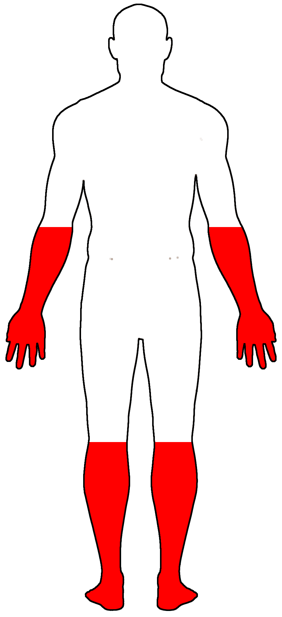
Uma miopatia distal (doença muscular que afeta as extremidades, como pés e mãos), caracterizada por fraqueza na parte de trás da perna, na região da panturrilha (especialmente nos músculos gastrocnêmio e sóleo), e associada a dificuldades para ficar na ponta dos pés.
Introdução
O que você precisa saber de cara
Uma miopatia distal (doença muscular que afeta as extremidades, como pés e mãos), caracterizada por fraqueza na parte de trás da perna, na região da panturrilha (especialmente nos músculos gastrocnêmio e sóleo), e associada a dificuldades para ficar na ponta dos pés.
Escala de raridade
<1/50kMuito rara
1/20kRara
1/10kPouco freq.
1/5kIncomum
1/2k
Encontrou um erro ou informação desatualizada? Sugira uma correção →
Entender a doença
Do básico ao detalhe, leia no seu ritmo
Preparando trilha educativa...
Sinais e sintomas
O que aparece no corpo e com que frequência cada sintoma acontece
Partes do corpo afetadas
+ 12 sintomas em outras categorias
Características mais comuns
Os sintomas variam de pessoa para pessoa. Abaixo estão as 44 características clínicas mais associadas, ordenadas por frequência.
Linha do tempo da pesquisa
Encontrou um erro ou informação desatualizada? Sugira uma correção →
Genética e causas
O que está alterado no DNA e como passa nas famílias
Genes associados
2 genes identificados com associação a esta condição. Padrão de herança: Autosomal recessive.
Key calcium ion sensor involved in the Ca(2+)-triggered synaptic vesicle-plasma membrane fusion. Plays a role in the sarcolemma repair mechanism of both skeletal muscle and cardiomyocytes that permits rapid resealing of membranes disrupted by mechanical stress (By similarity)
Cell membrane, sarcolemmaCytoplasmic vesicle membraneCell membraneLate endosome membrane
Muscular dystrophy, limb-girdle, autosomal recessive 2
An autosomal recessive degenerative myopathy characterized by weakness and atrophy starting in the proximal pelvifemoral muscles, with onset in the late teens or later, massive elevation of serum creatine kinase levels and slow progression. Scapular muscle involvement is minor and not present at onset. Upper limb girdle involvement follows some years after the onset in lower limbs.
Plays a role in plasma membrane repair in a process involving annexins (PubMed:33496727). Does not exhibit calcium-activated chloride channel (CaCC) activity
Endoplasmic reticulum membraneCell membrane
Gnathodiaphyseal dysplasia
Rare skeletal syndrome characterized by bone fragility, sclerosis of tubular bones, and cemento-osseous lesions of the jawbone. Patients experience frequent bone fractures caused by trivial accidents in childhood; however the fractures heal normally without bone deformity. The jaw lesions replace the tooth-bearing segments of the maxilla and mandible with fibrous connective tissues, including various amounts of cementum-like calcified mass, sometimes causing facial deformities. Patients also have a propensity for jaw infection and often suffer from purulent osteomyelitis-like symptoms, such as swelling of and pus discharge from the gums, mobility of the teeth, insufficient healing after tooth extraction and exposure of the lesions into the oral cavity.
Variantes genéticas (ClinVar)
1.475 variantes patogênicas registradas no ClinVar.
Classificação de variantes (ClinVar)
Distribuição de 3 variantes classificadas pelo ClinVar.
Vias biológicas (Reactome)
3 vias biológicas associadas aos genes desta condição.
Diagnóstico
Os sinais que médicos procuram e os exames que confirmam
Tratamento e manejo
Remédios, cuidados de apoio e o que precisa acompanhar
Onde tratar no SUS
Hospitais de referência no Brasil e o protocolo oficial do SUS (PCDT)
🇧🇷 Atendimento SUS — Miopatia de Miyoshi
Selecione um estado ou use sua localização para ver resultados.
Dados de DATASUS/CNES, SBGM, ABNeuro e Ministério da Saúde. Sempre confirme a disponibilidade diretamente com o estabelecimento.
Pesquisa ativa
Ensaios clínicos abertos e novidades científicas recentes
Ensaios em destaque
🟢 Recrutando agora
1 pesquisa recrutando participantes. Converse com seu médico sobre a possibilidade de participar.
Outros ensaios clínicos
6 ensaios clínicos encontrados, 1 ativos.
Publicações mais relevantes
DYSF gene variant spectrum in Arab populations across eight countries: A systematic review.
Dysferlinopathies are a subset of autosomal recessive muscular dystrophies resulting from pathogenic variants in the dysferlin (DYSF) gene. The prevalence of dysferlinopathies remains inadequately defined. This review aims to elucidate the mutational spectrum of DYSF in Arab populations. A systematic search was conducted in PubMed, ScienceDirect, Scopus, and Web of Science up to September 15, 2025. We identified 48 unique DYSF variants documented in the literature across eight Arab countries, resulting in 49 country-entries due to one variant being reported in two countries. The distribution of reported variants was as follows: Saudi Arabia 32.7% (16/49), Algeria 20.4% (10/49), Egypt 20.4% (10/49), Tunisia 10.2% (5/49), Morocco 6.1% (3/49), Libya 4.1% (2/49), Lebanon 4.1% (2/49), and Oman 2.0% (1/49). Clinical presentations were categorized based on phenotype assignments across variants, totaling 52 assignments as some variants were associated with multiple phenotypes: limb-girdle muscular dystrophy, recessive type 2 (LGMDR2) 50% (26/52), proximodistal 15% (8/52), Miyoshi myopathy 8% (4/52), distal myopathy with anterior tibial onset (DMAT) 4% (2/52), and asymptomatic hyperCKemia 4% (2/52). In terms of molecular consequences (denominator = 48 unique variants), frameshift variants constituted 36% (17/48), missense variants 29% (14/48), nonsense variants 15% (7/48), splice donor variants 6% (3/48), splice acceptor variants 4% (2/48), intronic variants 2% (1/48), and synonymous variants 2% (1/48). Documenting these variants across populations facilitates diagnosis and informs future public health strategies. Notably, no cohort study based in Morocco has focused on the genetics of dysferlinopathy; existing Moroccan data are limited to isolated case reports. Dysferlinopathy includes a spectrum of muscle disease characterized by two major phenotypes: Miyoshi muscular dystrophy (MMD) and limb-girdle muscular dystrophy type 2B (LGMD2B); and two minor phenotypes: asymptomatic hyperCKemia and distal myopathy with anterior tibial onset (DMAT). MMD (median age of onset 19 years) is characterized by muscle weakness and atrophy, most marked in the distal parts of the legs, especially the gastrocnemius and soleus muscles. Over a period of years, the weakness and atrophy spread to the thighs and gluteal muscles. The forearms may become mildly atrophic with decrease in grip strength; the small muscles of the hands are spared. LGMD2B is characterized by early weakness and atrophy of the pelvic and shoulder girdle muscles in adolescence or young adulthood, with slow progression. Other phenotypes in this spectrum are scapuloperoneal syndrome and congenital muscular dystrophy. Asymptomatic hyperCKemia is characterized by marked elevation of serum CK concentration only. DMAT is characterized by early and predominant distal muscle weakness, particularly of the muscles of the anterior compartment of the legs. The diagnosis of dysferlinopathy is established in a proband with suggestive findings and biallelic pathogenic variants in DYSF identified by molecular genetic testing. Treatment of manifestations: There is no approved therapy for dysferlinopathy. Treatment is symptomatic only. Management should be tailored to the individual and the specific subtype. Individualized management may include physical therapy, use of mechanical aids, surgical intervention for orthopedic complications, respiratory aids, and social and emotional support. Surveillance: Annual monitoring of muscle strength, physical function, activities of daily living, joint range of motion, balance, and respiratory function, and for evidence of cardiomyopathy for individuals with cardiac involvement. Agents/circumstances to avoid: Weight control to avoid obesity. Dysferlinopathy is inherited in an autosomal recessive manner. If both parents are known to be heterozygous for a DYSF pathogenic variant, each sib of an affected individual has at conception a 25% chance of being affected, a 50% chance of being an asymptomatic carrier, and a 25% chance of being unaffected and not a carrier. Once the DYSF pathogenic variants have been identified in an affected family member, carrier testing for at-risk relatives, prenatal testing for a pregnancy at increased risk, and preimplantation genetic testing are possible.
In Vitro Correction of Point Mutations in the DYSF Gene Using Prime Editing.
Dysferlinopathy is caused by over 500 mutations in the gene encoding dysferlin, including close to 300 point mutations. One option to cure the disease is to use a gene therapy to correct these mutations at the root. Prime editing is a technique which can replace the mutated nucleotide with the wild-type nucleotide. In this article, prime editing is used to correct several point mutations in the DYSF gene responsible for dysferlinopathy. In vitro editing of HEK293T cells reaches up to 31%. Notably, editing was more efficient in myoblasts than in patient-derived fibroblasts. The prime editing technique was also used to create a new myoblast clone containing a patient mutation from a healthy myoblast cell line.
Dysferlinopathy as cause of long-term hyperCKemia with preserved strength.
Dysferlin (DYSF) has a crucial role in sarcolemmal repair. While DYSF mutations commonly manifest as limb-girdle muscular dystrophy (LGMDR2) or distal Miyoshi myopathy, atypical manifestations, such as asymptomatic hyperCKemia and pseudometabolic myopathy, are rarely reported. We describe clinical, serologic, radiologic, genetic, and muscle pathology findings of three patients with rare dysferlinopathy phenotypes and long-term follow up in one of them. We also review the literature pertinent to uncommon forms of dysferlinopathy presenting with hyperCKemia and pseudometabolic phenotype. Patient 1 is a 51-year-old female with exercise-induced myalgia predominantly affecting calf muscles for 7 years. She had a 22-year history of asymptomatic hyperCKemia (CK 812-2,223 U/L). Neurologic exam showed mild calf enlargement without weakness. CT of the lower limb revealed fatty infiltration of distal peroneal and calf muscles. Genetic testing showed two DYSF variants, c.2163-2A > G (pathogenic) and c.866C > G, p.Ser289Cys (VUS), unknown if heteroallelic. Muscle biopsy demonstrated nuclei internalization and absent dysferlin immunoreactivity. Patient 2 is a 20-year-old male, football player, with an episode of exercise-induced myalgia followed by asymptomatic persistent hyperCKemia (729-2,645 U/L). He had normal strength but mild calf muscle atrophy. Muscle MRI demonstrated subtle T2 hyperintensity in the posterior leg compartment musculature. He has two heteroallelic DYSF variants, c.6008G > A, p.Gly2003Asp (pathogenic) and c.854C > T, p.Thr285Met (VUS). Muscle biopsy showed no myopathic changes but reduced dysferlin immunoreactivity. Patient 3 is a 58-year-old female with incidentally detected asymptomatic hyperCKemia (CK: 249-2,096 U/L) for 2 years. She had normal strength and normal lower limb muscle MRI. She carries two heteroallelic DYSF variants, c.2517del, p.Met840Trpfs*108 (pathogenic) and c.6058C > T, p.Arg2020Cys (VUS). Muscle biopsy showed minimal myopathic changes and attenuated dysferlin immunoreactivity. Reduced dysferlin expression was confirmed by western blot in patients 2 and 3. Needle EMG was normal in all patients. Dysferlinopathy should be considered in the differential diagnosis of metabolic myopathies and asymptomatic hyperCKemia. Patient 1's long history of hyperCKemia without weakness over two decades suggests that CK elevation in dysferlinopathy does not necessarily predict development of weakness. Additionally, the lack of dystrophic changes on muscle biopsy of patients with asymptomatic or minimally symptomatic hyperCKemia should not discourage the search for dysferlin deficiency in muscle, particularly in the setting of DYSF variants.
An interesting report of POPDC3 limb girdle muscular dystrophy R26 from India.
Popeye domain containing 3 (POPDC3) gene encodes a protein involved in membrane trafficking and is highly expressed in skeletal muscles. POPDC3 pathogenic variants are associated with LGMDR26. Only a few reports of POPDC3 LGMD exist worldwide and none from India. Herein, we describe the first case of POPDC3 LGMD26. This is a case report from a neurology referral center in India. All the clinical, laboratory and electrophysiological data were collected from the medical records. A 34-year-old man born to non-consanguineous parents presented with progressive proximal weakness of lower limbs from 22 years of age. He developed calf muscle pain and recurrent falls on walking for 7 years. He had atrophy of calves (medial gastrocnemius more than lateral) along with weakness of hip extensor, adductors and knee flexors and normal upper limb power, resembling Miyoshi myopathy. Serum creatine kinase ranged from 3524 to 6531 U/L. Muscle MRI showed selective atrophy of gluteus maximus, quadriceps femoris, semimembranosus and gastrocnemius with sparing of rectus femoris, gracilis and sartorius. Muscle biopsy done elsewhere and reported to show dystrophic features and immunohistochemistry showed positive staining for dystrophins and sarcoglycans. Clinically the possibility of LGMDR2/Dysferlinopathy, was considered and whole exome sequencing was done which revealed a novel homozygous pathogenic nonsense premature termination codon (PTC) variant (NM_022361.5) c.316C > T (NP_079130.2:) p.Arg106Ter) in exon 2 of POPDC3 gene. This is the first report of POPDC3- LGMDR26 from India detected among a large cohort (461 genetically confirmed cases). POPDC3 gene variations should be considered in distal onset LGMDs with markedly elevated serum creatine kinase levels.
A Late-Onset Presentation of Miyoshi Myopathy: A Case Report.
Miyoshi myopathy is a muscular dystrophy disease characterized by muscle weakness and atrophy generally in distal muscle groups, such as in the legs and arms. Miyoshi myopathy is thought to occur due to genetic mutations in the DYSF gene, which codes for the dysferlin protein, which is critical for muscle cell membrane integrity and muscle fiber adhesiveness. The first symptoms begin in early adulthood and include weakness and atrophy in the calves, gait abnormalities, pain and discomfort in affected muscles, and difficulty jumping or walking on tiptoes. Patients generally are diagnosed by a combination of physical exam findings, genetic testing, muscle biopsy, and elevated creatinine kinase (CK) levels. Management of the disease progression includes physical therapy to strengthen the muscles, nutritional support, occupational therapy, and assisted device education. While not life-threatening, Miyoshi myopathy outlook is generally considered moderate to poor due to significant muscle weakness and eventual loss of mobility usually in 10-20 years after onset. We present a unique case of a 66-year-old male patient complaining of pain in his bilateral calves after having had a series of back surgeries 10 years prior. A diagnosis of Miyoshi myopathy, a rare occurrence in this age group, was made based on CK levels. In this report, we will discuss the pathophysiology, disease progression, and management of Miyoshi myopathy.
Publicações recentes
Dysferlinopathies: phenotypic study of a Moroccan series of 28 cases.
DYSF gene variant spectrum in Arab populations across eight countries: A systematic review.
An interesting report of POPDC3 limb girdle muscular dystrophy R26 from India.
A Late-Onset Presentation of Miyoshi Myopathy: A Case Report.
📚 EuropePMC53 artigos no totalmostrando 89
DYSF gene variant spectrum in Arab populations across eight countries: A systematic review.
Biomolecules & biomedicineAn interesting report of POPDC3 limb girdle muscular dystrophy R26 from India.
Journal of neuromuscular diseasesA Late-Onset Presentation of Miyoshi Myopathy: A Case Report.
CureusWhole Exome Sequencing Identified a Stop-Gained Mutation in DYSF Gene Associated With Dysferlinopathy in an Iranian Family.
International journal of genomicsTips to Design Effective Splice-Switching Antisense Oligonucleotides for Exon Skipping and Exon Inclusion.
Methods in molecular biology (Clifton, N.J.)An Overview of Recent Advances and Clinical Applications of Exon Skipping and Splice Modulation for Muscular Dystrophy and Various Genetic Diseases.
Methods in molecular biology (Clifton, N.J.)In Vitro Correction of Point Mutations in the DYSF Gene Using Prime Editing.
International journal of molecular sciencesDysferlinopathy as cause of long-term hyperCKemia with preserved strength.
Orphanet journal of rare diseasesClinical Presentation, Diagnosis, and Genetic Insights of Miyoshi Myopathy: A Case Report and Literature Review.
CureusPerformance of upper limb entry item to predict forced vital capacity in dysferlin-deficient limb girdle muscular dystrophy.
Neuromuscular disorders : NMDBrain of miyoshi myopathy/dysferlinopathy patients presents with structural and metabolic anomalies.
Scientific reportsLimb Girdle Muscular Dystrophy Type 2B (LGMD2B): Diagnosis and Therapeutic Possibilities.
International journal of molecular sciencesThe Dysferlinopathies Conundrum: Clinical Spectra, Disease Mechanism and Genetic Approaches for Treatments.
BiomoleculesPhenotypic and genotypic analysis of a patient with Miyoshi myopathy caused by truncated protein.
GeneMiyoshi myopathy associated with spine rigidity and multiple contractures: a case report.
BMC musculoskeletal disordersHigh Prevalence of a c.5979dupA Variant in the Dysferlin Gene (DYSF) in Individuals from a Semiarid Region of Brazil.
Current genomicsPortrait of Dysferlinopathy: Diagnosis and Development of Therapy.
Journal of clinical medicineDysferlinopathy in Tunisia: clinical spectrum, genetic background and prognostic profile.
Neuromuscular disorders : NMDTetraspanin CD82 Associates with Trafficking Vesicle in Muscle Cells and Binds to Dysferlin and Myoferlin.
Advanced biologyThe Dysferlin C2A Domain Binds PI(4,5)P2 and Penetrates Membranes.
Journal of molecular biologyUtilization of Targeted RNA-Seq for the Resolution of Variant Pathogenicity and Enhancement of Diagnostic Yield in Dysferlinopathy.
Journal of personalized medicineMyostatin and follistatin as monitoring and prognostic biomarkers in dysferlinopathy.
Neuromuscular disorders : NMDMiyoshi Muscular Dystrophy Type 1 with Mutated DYSF Gene Misdiagnosed as Becker Muscular Dystrophy: A Case Report and Literature Review.
GenesGenotype-phenotype correlation and natural history study of dysferlinopathy: a single-centre experience from India.
NeurogeneticsMorpholino-Mediated Exons 28-29 Skipping of Dysferlin and Characterization of Multiexon-skipped Dysferlin using RT-PCR, Immunoblotting, and Membrane Wounding Assay.
Methods in molecular biology (Clifton, N.J.)Anoctamin-5 Muscular Dystrophy: Report of Two Cases with Different Phenotypes and Genotypes from the Indian Subcontinent.
Neurology IndiaThe clinical, myopathological, and molecular characteristics of 26 Chinese patients with dysferlinopathy: a high proportion of misdiagnosis and novel variants.
BMC neurologyAnoctamin 5 (ANO5) Muscle Disorders: A Narrative Review.
GenesRecurrent, non-traumatic, non-exertional rhabdomyolysis after immunologic stimuli in a healthy adolescent female: a case report.
BMC pediatricsDysferlinopathies: Clinical and genetic variability.
Clinical geneticsA Novel Homozygous Variant in DYSF Gene Is Associated with Autosomal Recessive Limb Girdle Muscular Dystrophy R2/2B.
International journal of molecular sciencesMS1/MMD1 homologues in the moss Physcomitrium patens are required for male and female gametogenesis.
The New phytologistProgression to Loss of Ambulation Among Patients with Autosomal Recessive Limb-girdle Muscular Dystrophy: A Systematic Review.
Journal of neuromuscular diseasesClinical, Neurophysiological, Radiological, Pathological, and Genetic Features of Dysferlinopathy in Saudi Arabia.
Frontiers in neuroscienceCalpainopathy (Leyden-Mobius Limb-Girdle Muscular Dystrophy Type 2A Phenotype) and Dysferlinopathy (Miyoshi Distal Myopathy Limb-Girdle Muscular Dystrophy Type 2B Phenotype) of Preadolescent Onset: Case Reports of Two Male Filipinos.
CureusCardiac and pulmonary findings in dysferlinopathy: A 3-year, longitudinal study.
Muscle & nerveThe C2 domains of dysferlin: roles in membrane localization, Ca2+ signalling and sarcolemmal repair.
The Journal of physiologyMagnetic resonance imaging pattern variability in dysferlinopathy.
Acta myologica : myopathies and cardiomyopathies : official journal of the Mediterranean Society of MyologyMutation at a new allele of the dysferlin gene causes Miyoshi myopathy: A case report.
Journal of musculoskeletal & neuronal interactionsA new dysferlin gene mutation in a Portuguese family with Miyoshi myopathy.
BMJ case reportsEfficient ssODN-Mediated Targeting by Avoiding Cellular Inhibitory RNAs through Precomplexed CRISPR-Cas9/sgRNA Ribonucleoprotein.
Stem cell reportsMiyoshi myopathy and limb girdle muscular dystrophy R2 are the same disease.
Neuromuscular disorders : NMDSubclinical Cardiomyopathy in Miyoshi Myopathy Detected by Late Gadolinium Enhancement Cardiac Magnetic Resonance Imaging.
International heart journalIntensive Teenage Activity Is Associated With Greater Muscle Hyperintensity on T1W Magnetic Resonance Imaging in Adults With Dysferlinopathy.
Frontiers in neurologyA novel dysferlin gene mutation in a Filipino male with Miyoshi myopathy.
Clinical neurology and neurosurgeryNull variants in DYSF result in earlier symptom onset.
Clinical geneticsPhenotypic Spectrum of Myopathies with Recessive Anoctamin-5 Mutations.
Journal of neuromuscular diseasesFunctions of Vertebrate Ferlins.
CellsFunctional muscle hypertrophy by increased insulin-like growth factor 1 does not require dysferlin.
Muscle & nervePhenotypic Drug Screening for Dysferlinopathy Using Patient-Derived Induced Pluripotent Stem Cells.
Stem cells translational medicineIdentification of Novel Antisense-Mediated Exon Skipping Targets in DYSF for Therapeutic Treatment of Dysferlinopathy.
Molecular therapy. Nucleic acidsDiltiazem improves contractile properties of skeletal muscle in dysferlin-deficient BLAJ mice, but does not reduce contraction-induced muscle damage.
Physiological reportsDevelopment of muscular dystrophy in a CRISPR-engineered mutant rabbit model with frame-disrupting ANO5 mutations.
Cell death & diseaseA novel ANO5 splicing variant in a LGMD2L patient leads to production of a truncated aggregation-prone Ano5 peptide.
The journal of pathology. Clinical researchMuscle Cells Fix Breaches by Orchestrating a Membrane Repair Ballet.
Journal of neuromuscular diseasesA Case of Obsessive-Compulsive Disorder Comorbid with Miyoshi Myopathy.
Indian journal of psychological medicineNovel duplication mutation of the DYSF gene in a Pakistani family with Miyoshi Myopathy.
Saudi medical journalIncreased nonHDL cholesterol levels cause muscle wasting and ambulatory dysfunction in the mouse model of LGMD2B.
Journal of lipid researchNovel, de novo dysferlin gene mutations in a patient with Miyoshi myopathy.
Neuroscience lettersDesigning Effective Antisense Oligonucleotides for Exon Skipping.
Methods in molecular biology (Clifton, N.J.)Coupling of excitation to Ca2+ release is modulated by dysferlin.
The Journal of physiologyLimb-Girdle Muscular Dystrophy 2B and Miyoshi Presentations of Dysferlinopathy.
The American journal of the medical sciencesCorrigendum: Twenty-Year Clinical Progression of Dysferlinopathy in Patients from Dagestan.
Frontiers in neurologyTwenty-Year Clinical Progression of Dysferlinopathy in Patients from Dagestan.
Frontiers in neurologyHyperckemia and myalgia are common presentations of anoctamin-5-related myopathy in French patients.
Muscle & nerveDiscordant manifestation in brothers with Miyoshi myopathy.
Journal of the neurological sciencesDysferlin mediates membrane tubulation and links T-tubule biogenesis to muscular dystrophy.
Journal of cell sciencePolymyositis without Beneficial Response to Steroid Therapy: Should Miyoshi Myopathy be a Differential Diagnosis?
The American journal of case reportsAtypical Miyoshi distal myopathy: A case report.
Experimental and therapeutic medicineDysferlin mutations and mitochondrial dysfunction.
Neuromuscular disorders : NMDProgress and challenges in diagnosis of dysferlinopathy.
Muscle & nerveThe Clinical Outcome Study for dysferlinopathy: An international multicenter study.
Neurology. GeneticsMembrane repair of human skeletal muscle cells requires Annexin-A5.
Biochimica et biophysica actaDysferlin Binds SNAREs (Soluble N-Ethylmaleimide-sensitive Factor (NSF) Attachment Protein Receptors) and Stimulates Membrane Fusion in a Calcium-sensitive Manner.
The Journal of biological chemistry[Modeling muscular diseases by patient-derived iPS cells].
Nihon yakurigaku zasshi. Folia pharmacologica JaponicaDefective membrane fusion and repair in Anoctamin5-deficient muscular dystrophy.
Human molecular geneticsPathogenic mutation R959W alters recognition dynamics of dysferlin inner DysF domain.
Molecular bioSystemsGenetic disruption of Ano5 in mice does not recapitulate human ANO5-deficient muscular dystrophy.
Skeletal muscleDysferlinopathy in Iran: Clinical and genetic report.
Journal of the neurological sciencesDysferlinopathy Fibroblasts Are Defective in Plasma Membrane Repair.
PLoS currentsDysferlinopathy in Switzerland: clinical phenotypes and potential founder effects.
BMC neurologyDysferlin deficiency blunts β-adrenergic-dependent lusitropic function of mouse heart.
The Journal of physiologyExon 32 Skipping of Dysferlin Rescues Membrane Repair in Patients' Cells.
Journal of neuromuscular diseasesCalf heads on a trophy sign: Miyoshi myopathy.
Journal of neurosciences in rural practiceRespiratory and cardiac function in japanese patients with dysferlinopathy.
Muscle & nerveDysferlin deficiency confers increased susceptibility to coxsackievirus-induced cardiomyopathy.
Cellular microbiologyCD4+ cells, macrophages, MHC-I and C5b-9 involve the pathogenesis of dysferlinopathy.
International journal of clinical and experimental pathologyClinical heterogeneity and a high proportion of novel mutations in a Chinese cohort of patients with dysferlinopathy.
Neurology IndiaCase report of an adolescent girl with limb-girdle muscular dystrophy type 2B - the usefulness of muscle protein immunostaining in the diagnosis of dysferlinopathies.
Folia neuropathologicaAssociações
Organizações que acompanham esta doença — pra ter apoio e orientação
Ainda não temos associações cadastradas para Miopatia de Miyoshi.
É de uma associação que acompanha esta doença? Fale com a gente →
Comunidades
Grupos ativos de quem convive com esta doença aqui no Raras
Ainda não existe comunidade no Raras para Miopatia de Miyoshi
Pacientes, familiares e cuidadores se organizam em comunidades pra compartilhar experiências, fazer perguntas e se apoiar. Você pode ser o primeiro.
Tire suas dúvidas
Perguntas, dicas e experiências compartilhadas aqui na página
Participe da discussão
Faça login para postar dúvidas, compartilhar experiências e interagir com especialistas.
Fazer loginDoenças relacionadas
Doenças com sintomas parecidos — ajudam quem ainda está buscando diagnóstico
Referências e fontes
Bases de dados externas citadas neste artigo
Publicações científicas
Artigos indexados no PubMed ligados a esta doença no grafo RarasNet — título, periódico e PMID direto da fonte, sem intermediação de IA.
- DYSF gene variant spectrum in Arab populations across eight countries: A systematic review.
- In Vitro Correction of Point Mutations in the DYSF Gene Using Prime Editing.
- Dysferlinopathy as cause of long-term hyperCKemia with preserved strength.
- An interesting report of POPDC3 limb girdle muscular dystrophy R26 from India.
- A Late-Onset Presentation of Miyoshi Myopathy: A Case Report.
- Dysferlinopathies: phenotypic study of a Moroccan series of 28 cases.
- Dysferlinopathy.
Bases de dados e fontes oficiais
Identificadores e referências canônicas usadas para montar este verbete.
- ORPHA:45448(Orphanet)
- MONDO:0009685(MONDO)
- GARD:9676(GARD (NIH))
- Variantes catalogadas(ClinVar)
- Busca completa no PubMed(PubMed)
- Q60195034(Wikidata)
Dados compilados pelo RarasNet a partir de fontes abertas (Orphanet, OMIM, MONDO, PubMed/EuropePMC, ClinicalTrials.gov, DATASUS, PCDT/MS). Este conteúdo é informativo e não substitui avaliação médica.
Conteúdo mantido por Agente Raras · Médicos e pesquisadores podem colaborar
Miopatia de Miyoshi
📋 Origem dos dados
Esta página agrega dados de fontes públicas e oficiais. Dados sobre cobertura no SUS (PCDT, CEAF) são verificados ativamente por agente proativo (ver badge no infobox). Demais dados têm atribuição de fonte + data da última sincronização — clique para abrir o original.
- Doença rara (ontologia)
- fonte: Orphanet
- Identificador unificado
- fonte: MONDO
- Codificação WHO/SUS
- fonte: WHO ICD-10 / DATASUS
- CID-11 (futuro)
- fonte: WHO ICD-11
- NIH/GARD
- fonte: GARD (NIH)
- Indexação biomédica
- fonte: MeSH (NLM)
- Dado público estruturado
- fonte: Wikidata
- Ensaios clínicos
- fonte: ClinicalTrials.gov