A ataxia espinocerebelar (AEC) é uma doença genética, progressiva e degenerativa com múltiplos tipos, cada um dos quais pode ser considerado uma condição neurológica por si só. Estima-se que 150.000 pessoas nos Estados Unidos tenham um diagnóstico de ataxia espinocerebelar a qualquer momento. A AEC é hereditária, progressiva e degenerativa. Não há tratamento eficaz ou cura conhecidos. A AEC pode afetar qualquer pessoa de qualquer idade. A doença é causada por um gene recessivo ou dominante.
Introdução
O que você precisa saber de cara
Visão geral
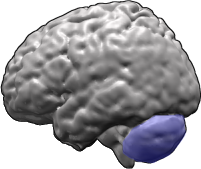
A ataxia cerebelosa degenerativa e progressiva autossômica recessiva é uma doença neurológica rara que afeta principalmente o cerebelo, a parte do cérebro responsável pelo equilíbrio e coordenação dos movimentos. O termo "autossômica recessiva" significa que a condição se manifesta quando a pessoa herda duas cópias alteradas do gene associado, uma de cada genitor. A doença é caracterizada por uma degeneração progressiva do cerebelo, levando a dificuldades crescentes de coordenação motora e equilíbrio.[1][3]
Sinais e sintomas
Os sinais e sintomas da ataxia cerebelosa degenerativa e progressiva autossômica recessiva são variados e podem incluir tremor intencional (tremor que ocorre durante movimentos voluntários), ataxia sensorial (falta de coordenação devido à perda de sensibilidade), neuropatia periférica (danos nos nervos que afetam a sensação e o movimento), fraqueza muscular dos membros inferiores e amiotrofia distal (perda de massa muscular nas extremidades). Outros sintomas relatados incluem movimentos involuntários, movimentos sacádicos em onda quadrada (movimentos oculares anormais), baixa estatura, metacarpo curto (ossos da mão encurtados), palma curta e tamanho testicular diminuído.[1][3]
Alguns pacientes podem apresentar encefalopatia epiléptica (convulsões associadas a alterações cerebrais), rabdomiólise aguda (destruição muscular rápida), atividade anormal da lactato desidrogenase (enzima muscular), atrofia ou degeneração do tronco cerebral, hiperintensidade do sinal T2 na ressonância magnética da medula espinhal, necrose avascular da epífise femoral capital (morte do tecido ósseo na cabeça do fêmur), morfologia anormal do bulbo raquidiano, degeneração axonal (dano às fibras nervosas), vacúolos com bordas (alterações nas células musculares), agrupamento de tipos de fibras musculares, reflexo patelar hiperativo e distrofia muscular. O início dos sintomas geralmente ocorre na infância.[1][3]
Causas genéticas
A ataxia cerebelosa degenerativa e progressiva autossômica recessiva é causada por alterações (mutações) em genes específicos. Os genes associados a esta condição incluem: FLVCR1 (que produz uma proteína transportadora de colina e etanolamina), CTDP1 (envolvido na regulação da transcrição do DNA), SIL1 (que auxilia no dobramento de proteínas), FXN (que produz a frataxina, importante para a função mitocondrial), TBCE (envolvido na formação dos microtúbulos celulares) e TWNK (que produz a helicase Twinkle, essencial para a replicação do DNA mitocondrial). Mutações em qualquer um desses genes podem interromper funções celulares críticas, levando à degeneração progressiva do cerebelo.[1][4]
Diagnóstico
O diagnóstico da ataxia cerebelosa degenerativa e progressiva autossômica recessiva é baseado na avaliação clínica dos sintomas, histórico familiar e exames complementares. Testes genéticos estão disponíveis para confirmar a presença de mutações nos genes associados. Atualmente, existem 10 testes genéticos disponíveis para esta condição, e 372 variantes genéticas foram registradas no ClinVar, um banco de dados público que relaciona variantes genéticas a fenótipos clínicos. O diagnóstico precoce pode ajudar no planejamento do acompanhamento e manejo dos sintomas.[1][4]
Tratamento e manejo
Atualmente, não há cura para a ataxia cerebelosa degenerativa e progressiva autossômica recessiva. O tratamento é focado no manejo dos sintomas e na melhora da qualidade de vida. Isso pode incluir fisioterapia para manter a mobilidade e força muscular, terapia ocupacional para auxiliar nas atividades diárias, e fonoaudiologia para dificuldades de fala e deglutição. O acompanhamento com neurologista é essencial para monitorar a progressão da doença e ajustar as intervenções conforme necessário. Não há medicamentos específicos aprovados para esta condição, e nenhum tratamento está disponível no Sistema Único de Saúde (SUS) para esta doença.[1]
Prognóstico e qualidade de vida
O prognóstico da ataxia cerebelosa degenerativa e progressiva autossômica recessiva varia de acordo com a gravidade dos sintomas e a idade de início. Por ser uma condição progressiva, os sintomas tendem a piorar ao longo do tempo, podendo levar a dificuldades significativas de locomoção e coordenação. O suporte multidisciplinar, incluindo fisioterapia, terapia ocupacional e acompanhamento psicológico, é fundamental para ajudar o paciente e sua família a lidar com os desafios da doença e manter a melhor qualidade de vida possível.[1][3]
Conteúdo informativo gerado e mantido automaticamente a partir de fontes oficiais (Orphanet, HPO, OMIM, SUS). Não substitui avaliação médica.
A ataxia espinocerebelar (AEC) é uma doença genética, progressiva e degenerativa com múltiplos tipos, cada um dos quais pode ser considerado uma condição neurológica por si só. Estima-se que 150.000 pessoas nos Estados Unidos tenham um diagnóstico de ataxia espinocerebelar a qualquer momento. A AEC é hereditária, progressiva e degenerativa. Não há tratamento eficaz ou cura conhecidos. A AEC pode afetar qualquer pessoa de qualquer idade. A doença é causada por um gene recessivo ou dominante.
Tem tratamento?
Encontrou um erro ou informação desatualizada? Sugira uma correção →
Entender a doença
Do básico ao detalhe, leia no seu ritmo
Preparando trilha educativa...
Sinais e sintomas
O que aparece no corpo e com que frequência cada sintoma acontece
Visão geral
A ataxia cerebelosa degenerativa e progressiva autossômica recessiva é uma doença neurológica rara que afeta principalmente o cerebelo, a parte do cérebro responsável pelo equilíbrio e coordenação dos movimentos. O termo "autossômica recessiva" significa que a condição se manifesta quando a pessoa herda duas cópias alteradas do gene associado, uma de cada genitor. A doença é caracterizada por uma degeneração progressiva do cerebelo, levando a dificuldades crescentes de coordenação motora e equilíbrio.[1][3]
Sinais e sintomas
Os sinais e sintomas da ataxia cerebelosa degenerativa e progressiva autossômica recessiva são variados e podem incluir tremor intencional (tremor que ocorre durante movimentos voluntários), ataxia sensorial (falta de coordenação devido à perda de sensibilidade), neuropatia periférica (danos nos nervos que afetam a sensação e o movimento), fraqueza muscular dos membros inferiores e amiotrofia distal (perda de massa muscular nas extremidades). Outros sintomas relatados incluem movimentos involuntários, movimentos sacádicos em onda quadrada (movimentos oculares anormais), baixa estatura, metacarpo curto (ossos da mão encurtados), palma curta e tamanho testicular diminuído.[1][3]
Alguns pacientes podem apresentar encefalopatia epiléptica (convulsões associadas a alterações cerebrais), rabdomiólise aguda (destruição muscular rápida), atividade anormal da lactato desidrogenase (enzima muscular), atrofia ou degeneração do tronco cerebral, hiperintensidade do sinal T2 na ressonância magnética da medula espinhal, necrose avascular da epífise femoral capital (morte do tecido ósseo na cabeça do fêmur), morfologia anormal do bulbo raquidiano, degeneração axonal (dano às fibras nervosas), vacúolos com bordas (alterações nas células musculares), agrupamento de tipos de fibras musculares, reflexo patelar hiperativo e distrofia muscular. O início dos sintomas geralmente ocorre na infância.[1][3]
Causas genéticas
A ataxia cerebelosa degenerativa e progressiva autossômica recessiva é causada por alterações (mutações) em genes específicos. Os genes associados a esta condição incluem: FLVCR1 (que produz uma proteína transportadora de colina e etanolamina), CTDP1 (envolvido na regulação da transcrição do DNA), SIL1 (que auxilia no dobramento de proteínas), FXN (que produz a frataxina, importante para a função mitocondrial), TBCE (envolvido na formação dos microtúbulos celulares) e TWNK (que produz a helicase Twinkle, essencial para a replicação do DNA mitocondrial). Mutações em qualquer um desses genes podem interromper funções celulares críticas, levando à degeneração progressiva do cerebelo.[1][4]
Diagnóstico
O diagnóstico da ataxia cerebelosa degenerativa e progressiva autossômica recessiva é baseado na avaliação clínica dos sintomas, histórico familiar e exames complementares. Testes genéticos estão disponíveis para confirmar a presença de mutações nos genes associados. Atualmente, existem 10 testes genéticos disponíveis para esta condição, e 372 variantes genéticas foram registradas no ClinVar, um banco de dados público que relaciona variantes genéticas a fenótipos clínicos. O diagnóstico precoce pode ajudar no planejamento do acompanhamento e manejo dos sintomas.[1][4]
Tratamento e manejo
Atualmente, não há cura para a ataxia cerebelosa degenerativa e progressiva autossômica recessiva. O tratamento é focado no manejo dos sintomas e na melhora da qualidade de vida. Isso pode incluir fisioterapia para manter a mobilidade e força muscular, terapia ocupacional para auxiliar nas atividades diárias, e fonoaudiologia para dificuldades de fala e deglutição. O acompanhamento com neurologista é essencial para monitorar a progressão da doença e ajustar as intervenções conforme necessário. Não há medicamentos específicos aprovados para esta condição, e nenhum tratamento está disponível no Sistema Único de Saúde (SUS) para esta doença.[1]
Prognóstico e qualidade de vida
O prognóstico da ataxia cerebelosa degenerativa e progressiva autossômica recessiva varia de acordo com a gravidade dos sintomas e a idade de início. Por ser uma condição progressiva, os sintomas tendem a piorar ao longo do tempo, podendo levar a dificuldades significativas de locomoção e coordenação. O suporte multidisciplinar, incluindo fisioterapia, terapia ocupacional e acompanhamento psicológico, é fundamental para ajudar o paciente e sua família a lidar com os desafios da doença e manter a melhor qualidade de vida possível.[1][3]
Conteúdo informativo gerado e mantido automaticamente a partir de fontes oficiais (Orphanet, HPO, OMIM, SUS). Não substitui avaliação médica.
Partes do corpo afetadas
+ 101 sintomas em outras categorias
Características mais comuns
Os sintomas variam de pessoa para pessoa. Abaixo estão as 246 características clínicas mais associadas, ordenadas por frequência.
Linha do tempo da pesquisa
Encontrou um erro ou informação desatualizada? Sugira uma correção →
Genética e causas
O que está alterado no DNA e como passa nas famílias
Genes associados
6 genes identificados com associação a esta condição.
Uniporter that mediates the transport of extracellular choline and ethanolamine into cells, thereby playing a key role in phospholipid biosynthesis (PubMed:37100056, PubMed:38693265, PubMed:38778100, PubMed:39306721). Choline and ethanolamine are the precursors of phosphatidylcholine and phosphatidylethanolamine, respectively, the two most abundant phospholipids (PubMed:38693265, PubMed:38778100). Transport is not coupled with proton transport and is exclusively driven by the choline (or ethanol
Cell membraneMitochondrion membrane
Retinopathy-sensory neuropathy syndrome
An autosomal recessive neurodegenerative syndrome beginning in infancy with areflexia and retinitis pigmentosa. Nyctalopia (night blindness) and peripheral visual field loss are usually evident during late childhood or teenage years, with subsequent progressive constriction of the visual fields and loss of central retinal function over time. A sensory ataxia caused by degeneration of the posterior columns of the spinal cord results in a loss of proprioceptive sensation that is clinically evident in the second decade of life and gradually progresses. Scoliosis, camptodactyly, achalasia, gastrointestinal dysmotility, and a sensory peripheral neuropathy are variable features of the disease. Affected individuals have no clinical or radiological evidence of cerebral or cerebellar involvement. Some patients have pain insensitivity and commonly manifest self-injury, ulcers and amputations.
Processively dephosphorylates 'Ser-2' and 'Ser-5' of the heptad repeats YSPTSPS in the C-terminal domain of the largest RNA polymerase II subunit. This promotes the activity of RNA polymerase II. Plays a role in the exit from mitosis by dephosphorylating crucial mitotic substrates (USP44, CDC20 and WEE1) that are required for M-phase-promoting factor (MPF)/CDK1 inactivation
NucleusCytoplasm, cytoskeleton, microtubule organizing center, centrosomeCytoplasm, cytoskeleton, spindle poleMidbody
Congenital cataracts, facial dysmorphism, and neuropathy
An autosomal recessive developmental disorder characterized by a complex clinical phenotype with seemingly unrelated features involving multiple organs and systems. Developmental abnormalities include congenital cataracts and microcorneae, hypomyelination of the peripheral nervous system, impaired physical growth, delayed early motor and intellectual development, facial dysmorphism and hypogonadism. Central nervous system involvement, with cerebral and spinal cord atrophy, may be the result of disrupted development with superimposed degenerative changes. Affected individuals are prone to severe rhabdomyolysis after viral infections and to serious complications related to general anesthesia (such as pulmonary edema and epileptic seizures).
Required for protein translocation and folding in the endoplasmic reticulum (ER). Functions as a nucleotide exchange factor for the ER lumenal chaperone HSPA5
Endoplasmic reticulum lumen
Marinesco-Sjoegren syndrome
Autosomal recessive multisystem disorder which is characterized by cerebellar ataxia due to cerebellar atrophy, with Purkinje and granule cell loss and myopathy featuring marked muscle replacement with fat and connective tissue. Other cardinal features include bilateral cataracts, hypergonadotrophic hypogonadism and mild to severe intellectual disability. Skeletal abnormalities, short stature, dysarthria, strabismus and nystagmus are also frequent findings. Mutational inactivation of this protein may result in ER stress-induced cell death signaling or malfunctioning chaperone machineries that mishandle client proteins which are critical for the organs targeted in MSS.
Functions as an activator of persulfide transfer to the scaffoding protein ISCU as component of the core iron-sulfur cluster (ISC) assembly complex and participates to the [2Fe-2S] cluster assembly (PubMed:12785837, PubMed:24971490). Accelerates sulfur transfer from NFS1 persulfide intermediate to ISCU and to small thiols such as L-cysteine and glutathione leading to persulfuration of these thiols and ultimately sulfide release (PubMed:24971490). Binds ferrous ion and is released from FXN upon t
MitochondrionCytoplasm, cytosol
Friedreich ataxia
Autosomal recessive, progressive degenerative disease characterized by neurodegeneration and cardiomyopathy it is the most common inherited ataxia. The disorder is usually manifest before adolescence and is generally characterized by incoordination of limb movements, dysarthria, nystagmus, diminished or absent tendon reflexes, Babinski sign, impairment of position and vibratory senses, scoliosis, pes cavus, and hammer toe. In most patients, FRDA is due to GAA triplet repeat expansions in the first intron of the frataxin gene. But in some cases the disease is due to mutations in the coding region.
Tubulin-folding protein; involved in the second step of the tubulin folding pathway and in the regulation of tubulin heterodimer dissociation. Required for correct organization of microtubule cytoskeleton and mitotic splindle, and maintenance of the neuronal microtubule network
CytoplasmCytoplasm, cytoskeleton
Hypoparathyroidism-retardation-dysmorphism syndrome
An autosomal recessive multisystem disorder characterized by hypoparathyroidism, intrauterine and postnatal growth retardation, psychomotor retardation, epilepsy, microcephaly, and facial dysmorphism.
Mitochondrial helicase involved in mtDNA replication and repair (PubMed:12975372, PubMed:15167897, PubMed:17324440, PubMed:18039713, PubMed:18971204, PubMed:25824949, PubMed:26887820, PubMed:27226550). Might have a role in mtDNA repair (PubMed:27226550). Has DNA strand separation activity needed to form a processive replication fork for leading strand synthesis which is catalyzed by the formation of a replisome complex with POLG and mtSDB (PubMed:12975372, PubMed:15167897, PubMed:18039713, PubMe
Mitochondrion matrix, mitochondrion nucleoidMitochondrion inner membrane
Progressive external ophthalmoplegia with mitochondrial DNA deletions, autosomal dominant, 3
A disorder characterized by progressive weakness of ocular muscles and levator muscle of the upper eyelid. In a minority of cases, it is associated with skeletal myopathy, which predominantly involves axial or proximal muscles and which causes abnormal fatigability and even permanent muscle weakness. Ragged-red fibers and atrophy are found on muscle biopsy. A large proportion of chronic ophthalmoplegias are associated with other symptoms, leading to a multisystemic pattern of this disease. Additional symptoms are variable, and may include cataracts, hearing loss, sensory axonal neuropathy, ataxia, depression, hypogonadism, and parkinsonism.
Medicamentos e terapias
Mecanismo: Nuclear factor erythroid 2-related factor 2 activator
Mecanismo: Quinone reductase 1 modulator
Mecanismo: Peroxisome proliferator-activated receptor gamma agonist
Mecanismo: Interferon gamma receptor agonist
Mecanismo: Neuronal acetylcholine receptor; alpha4/beta2 partial agonist
Mecanismo: Peroxisome proliferator-activated receptor gamma agonist
Mecanismo: Erythropoietin receptor agonist
Mecanismo: HMG-CoA reductase inhibitor
Mecanismo: Glucocorticoid receptor agonist
Variantes genéticas (ClinVar)
372 variantes patogênicas registradas no ClinVar.
Vias biológicas (Reactome)
24 vias biológicas associadas aos genes desta condição.
Diagnóstico
Os sinais que médicos procuram e os exames que confirmam
Tratamento e manejo
Remédios, cuidados de apoio e o que precisa acompanhar
Onde tratar no SUS
Hospitais de referência no Brasil e o protocolo oficial do SUS (PCDT)
🇧🇷 Atendimento SUS — Ataxia cerebelosa degenerativa e progressiva autossômica recessiva
Selecione um estado ou use sua localização para ver resultados.
Dados de DATASUS/CNES, SBGM, ABNeuro e Ministério da Saúde. Sempre confirme a disponibilidade diretamente com o estabelecimento.
Pesquisa ativa
Ensaios clínicos abertos e novidades científicas recentes
Ensaios em destaque
🟢 Recrutando agora
1 pesquisa recrutando participantes. Converse com seu médico sobre a possibilidade de participar.
Outros ensaios clínicos
Publicações mais relevantes
Longitudinal Analysis of Natural History Progression of Rare and Ultra-Rare Cerebellar Ataxias Using Item Response Theory.
Degenerative cerebellar ataxias comprise a heterogeneous group of rare and ultra-rare genetic diseases. While disease-modifying treatments are now on the horizon for many ataxias, robust trial designs and analysis methods are lacking. To better inform trial designs, we applied item response theory (IRT) modeling to evaluate the natural history progression of several ataxias, assessed with the widely used scale for assessment and rating of ataxia (SARA). A longitudinal IRT model was built utilizing real-world data from the large autosomal recessive cerebellar ataxia (ARCA) registry. Disease progression was evaluated for the overall cohort as well as for the 10 most common ARCA genotypes. Sample sizes were calculated for simulated trials with autosomal recessive spastic ataxia Charlevoix-Saguenay (ARSACS) and polymerase gamma (POLG) ataxia, as showcased, across multiple design and analysis scenarios. Longitudinal IRT models were able to describe the changes in the latent variable underlying SARA as a function of time since ataxia onset for both the overall ARCA cohort and the common genotypes. The typical progression rates varied across genotypes between relatively high in POLG (~ 0.98 SARA points/year at SARA = 20) and very low in COQ8A ataxia (~ 0.003 SARA points/year at SARA = 20). Smaller trial sizes were required in case of faster progression, longer trials (~ 75-90% less with 5 years vs. 2 years), and larger drug effects (~ 70-80% less with 100% vs. 50% inhibition). Simulating under the developed IRT model, the longitudinal IRT model had the highest power, with a well-controlled type I error, compared to total score models or end-of-treatment analyses. The established longitudinal IRT framework allows efficient utilization of natural history data and ultimately facilitates the design and analysis of treatment trials in rare and ultra-rare genetic ataxias.
Clinicopathological and pedigree investigation of a novel spinocerebellar neurological disease in juvenile Quarter Horses in North America.
In 2020, a novel neurologic disease was observed in juvenile Quarter Horses (QHs) in North America. It was unknown if this was an aberrant manifestation of another previously described neurological disorder in foals, such as equine neuroaxonal dystrophy/equine degenerative myeloencephalopathy (eNAD/EDM). To describe the clinical findings, outcomes, and postmortem changes with Equine Juvenile Spinocerebellar Ataxia (EJSCA), differentiate the disease from other similar neurological disorders, and determine a mode of inheritance. Twelve neurologically affected QH foals and the dams. Genomic DNA was isolated and pedigrees were manually constructed. All foals (n = 12/12) had a history of acute onset of neurological deficits with no history of trauma. Neurological deficits were characterized by asymmetrical spinal ataxia, with pelvic limbs more severely affected than thoracic limbs. Clinicopathological abnormalities included high serum activity of gamma-glutamyl transferase and hyperglycemia. All foals became recumbent (median, 3 days: [0-18 days]), which necessitated humane euthanasia (n = 11/12, 92%; the remaining case was found dead). Histological evaluation at postmortem revealed dilated myelin sheaths and digestion chambers within the spinal cord, most prominently in the dorsal spinocerebellar tracts. Pedigree analysis revealed a likely autosomal recessive mode of inheritance. EJSCA is a uniformly fatal, rapidly progressive, likely autosomal recessive neurological disease of QHs <1 month of age in North America that is etiologically distinct from other clinically similar neurological disorders. Once the causative variant for EJSCA is validated, carriers can be identified through genetic testing to inform breeding decisions.
A Rare Phenotype of Inherited Cerebellar Ataxia.
Ataxia is a syndrome of imbalance and incoordination, categorized as hereditary ataxias, degenerative ataxias (non-hereditary), and acquired ataxias. Hereditary ataxia is further classified based on its mode of inheritance. Here, we have reported a case of early-onset autosomal recessive cerebellar ataxia with retained reflexes in a young male with positive family history. A young male presented with ten years history of tremors in both hands and head, aggravated with work and relieved with rest, and imbalance while walking, which has now progressed to the level where the patient cannot walk without support. The patient's younger brother also had a similar history. Central nervous system examination revealed cerebellar ataxia with retained reflexes. After ruling out other causes of ataxia in this age group by investigations, we could make the diagnosis of early-onset cerebellar ataxia with retained tendon reflexes (autosomal recessive). Presenting as a disease of variable presentation, the important diagnostic cues are classification and localization of ataxia. The investigations should be focusing on those cases of ataxias that are treatable. Family history is important to identify hereditary ataxias, as well as in genetic counselling of the affected patients.
Case Report: Infantile Cerebellar-Retinal Degeneration With Compound Heterozygous Variants in ACO2 Gene-Long-Term Follow-Up of a Sibling.
Infantile cerebellar-retinal degeneration (ICRD) is an extremely rare, infantile-onset neuro-degenerative disease, characterized by autosomal recessive inherited, global developmental delay (GDD), progressive cerebellar and cortical atrophy, and retinal degeneration. In 2012, a biallelic pathogenic variant in ACO2 gene (NM_001098.3) was found to be causative of this disease. To date, approximately 44 variants displaying various clinical features have been reported. Here, we report a case of two siblings with compound heterozygous variants in the ACO2 gene. Two siblings without perinatal problems were born to healthy non-consanguineous Korean parents. They showed GDD and seizures since infancy. Their first brain magnetic resonance imaging (MRI), electroencephalography, and metabolic workup revealed no abnormal findings. As they grew, they developed symptoms including ataxia, dysmetria, poor sitting balance, and myopia. Follow-up brain MRI findings revealed atrophy of the cerebellum and optic nerve. Through exome sequencing of both siblings and their parents, we identified the following compound heterozygous variants in the ACO2: c.85C > T (p.Arg29Trp) and c.2303C > A (p.Ala768Asp). These two variants were categorized as likely pathogenic based on ACMG/AMP guidelines. In conclusion, this case help to broaden the genetic and clinical spectrum of the ACO2 variants associated with ICRD. We have also documented the long-term clinical course and serial brain MRI findings for two patients with this extremely rare disease.
Dysfunction of cerebellar microglia in Ataxia-telangiectasia.
Ataxia-telangiectasia (A-T) is a multisystem autosomal recessive disease caused by mutations in the ATM gene and characterized by cerebellar atrophy, progressive ataxia, immunodeficiency, male and female sterility, radiosensitivity, cancer predisposition, growth retardation, insulin-resistant diabetes, and premature aging. ATM phosphorylates more than 1500 target proteins, which are involved in cell cycle control, DNA repair, apoptosis, modulation of chromatin structure, and other cytoplasmic as well as mitochondrial processes. In our quest to better understand the mechanisms by which ATM deficiency causes cerebellar degeneration, we hypothesized that specific vulnerabilities of cerebellar microglia underlie the etiology of A-T. Our hypothesis is based on the recent finding that dysfunction of glial cells affect a variety of process leading to impaired neuronal functionality (Song et al., 2019). Whereas astrocytes and neurons descend from the neural tube, microglia originate from the hematopoietic system, invade the brain at early embryonic stage, and become the innate immune cells of the central nervous system and important participants in development of synaptic plasticity. Here we demonstrate that microglia derived from Atm-/- mouse cerebellum display accelerated cell migration and are severely impaired in phagocytosis, secretion of neurotrophic factors, and mitochondrial activity, suggestive of apoptotic processes. Interestingly, no microglial impairment was detected in Atm-deficient cerebral cortex, and Atm deficiency had less impact on astroglia than microglia. Collectively, our findings validate the roles of glial cells in cerebellar attrition in A-T.
Publicações recentes
Longitudinal Analysis of Natural History Progression of Rare and Ultra-Rare Cerebellar Ataxias Using Item Response Theory.
Clinicopathological and pedigree investigation of a novel spinocerebellar neurological disease in juvenile Quarter Horses in North America.
Dysfunction of cerebellar microglia in Ataxia-telangiectasia.
History of Ataxias and Paraplegias with an Annotation on the First Description of Striatonigral Degeneration.
Genetic and phenotypic features of patients with childhood ataxias diagnosed by next-generation sequencing gene panel.
📚 EuropePMCmostrando 12
Longitudinal Analysis of Natural History Progression of Rare and Ultra-Rare Cerebellar Ataxias Using Item Response Theory.
Clinical pharmacology and therapeuticsClinicopathological and pedigree investigation of a novel spinocerebellar neurological disease in juvenile Quarter Horses in North America.
Journal of veterinary internal medicineA Rare Phenotype of Inherited Cerebellar Ataxia.
CureusCase Report: Infantile Cerebellar-Retinal Degeneration With Compound Heterozygous Variants in ACO2 Gene-Long-Term Follow-Up of a Sibling.
Frontiers in geneticsDysfunction of cerebellar microglia in Ataxia-telangiectasia.
GliaHistory of Ataxias and Paraplegias with an Annotation on the First Description of Striatonigral Degeneration.
Cerebellum (London, England)Genetic and phenotypic features of patients with childhood ataxias diagnosed by next-generation sequencing gene panel.
Brain & developmentThe Role of Iron in Friedreich's Ataxia: Insights From Studies in Human Tissues and Cellular and Animal Models.
Frontiers in neuroscienceAutosomal Recessive Cerebellar Ataxias: Paving the Way toward Targeted Molecular Therapies.
NeuronGDAP2 mutations implicate susceptibility to cellular stress in a new form of cerebellar ataxia.
Brain : a journal of neurologyMutations in NKX6-2 Cause Progressive Spastic Ataxia and Hypomyelination.
American journal of human geneticsGenetic and pharmacological evidence implicates cathepsins in Niemann-Pick C cerebellar degeneration.
Human molecular geneticsAssociações
Organizações que acompanham esta doença — pra ter apoio e orientação
Ainda não temos associações cadastradas para Ataxia cerebelosa degenerativa e progressiva autossômica recessiva.
É de uma associação que acompanha esta doença? Fale com a gente →
Comunidades
Grupos ativos de quem convive com esta doença aqui no Raras
Ainda não existe comunidade no Raras para Ataxia cerebelosa degenerativa e progressiva autossômica recessiva
Pacientes, familiares e cuidadores se organizam em comunidades pra compartilhar experiências, fazer perguntas e se apoiar. Você pode ser o primeiro.
Tire suas dúvidas
Perguntas, dicas e experiências compartilhadas aqui na página
Participe da discussão
Faça login para postar dúvidas, compartilhar experiências e interagir com especialistas.
Fazer loginDoenças relacionadas
Doenças com sintomas parecidos — ajudam quem ainda está buscando diagnóstico
Referências e fontes
Bases de dados externas citadas neste artigo
Publicações científicas
Artigos indexados no PubMed ligados a esta doença no grafo RarasNet — título, periódico e PMID direto da fonte, sem intermediação de IA.
- Longitudinal Analysis of Natural History Progression of Rare and Ultra-Rare Cerebellar Ataxias Using Item Response Theory.
- Clinicopathological and pedigree investigation of a novel spinocerebellar neurological disease in juvenile Quarter Horses in North America.
- A Rare Phenotype of Inherited Cerebellar Ataxia.
- Case Report: Infantile Cerebellar-Retinal Degeneration With Compound Heterozygous Variants in ACO2 Gene-Long-Term Follow-Up of a Sibling.
- Dysfunction of cerebellar microglia in Ataxia-telangiectasia.
- History of Ataxias and Paraplegias with an Annotation on the First Description of Striatonigral Degeneration.
- Genetic and phenotypic features of patients with childhood ataxias diagnosed by next-generation sequencing gene panel.
Bases de dados e fontes oficiais
Identificadores e referências canônicas usadas para montar este verbete.
- ORPHA:98098(Orphanet)
- MONDO:0020046(MONDO)
- GARD:19415(GARD (NIH))
- Variantes catalogadas(ClinVar)
- Busca completa no PubMed(PubMed)
- Q55346094(Wikidata)
Dados compilados pelo RarasNet a partir de fontes abertas (Orphanet, OMIM, MONDO, PubMed/EuropePMC, ClinicalTrials.gov, DATASUS, PCDT/MS). Este conteúdo é informativo e não substitui avaliação médica.
Conteúdo mantido por Agente Raras · Médicos e pesquisadores podem colaborar
Ataxia cerebelosa degenerativa e progressiva autossômica recessiva
📋 Origem dos dados
Esta página agrega dados de fontes públicas e oficiais. Dados sobre cobertura no SUS (PCDT, CEAF) são verificados ativamente por agente proativo (ver badge no infobox). Demais dados têm atribuição de fonte + data da última sincronização — clique para abrir o original.
- Doença rara (ontologia)
- fonte: Orphanet
- Identificador unificado
- fonte: MONDO
- NIH/GARD
- fonte: GARD (NIH)
- Dado público estruturado
- fonte: Wikidata
- Medicamentos (literatura)
- fonte: Orphanet
- Ensaios clínicos
- fonte: ClinicalTrials.gov