A ataxia espinocerebelar tipo 1 (SCA1) é um subtipo de ataxia cerebelar autossômica dominante tipo I (ADCA tipo I) caracterizada por disartria, dificuldades de escrita, ataxia de membros e comumente nistagmo e anormalidades sacádicas.
Introdução
O que você precisa saber de cara
A ataxia espinocerebelar tipo 1 (SCA1) é um subtipo de ataxia cerebelar autossômica dominante tipo I (ADCA tipo I) caracterizada por disartria, dificuldades de escrita, ataxia de membros e comumente nistagmo e anormalidades sacádicas.
Tem tratamento?
Escala de raridade
<1/50kMuito rara
1/20kRara
1/10kPouco freq.
1/5kIncomum
1/2k
Encontrou um erro ou informação desatualizada? Sugira uma correção →
Entender a doença
Do básico ao detalhe, leia no seu ritmo
Preparando trilha educativa...
Sinais e sintomas
O que aparece no corpo e com que frequência cada sintoma acontece
Partes do corpo afetadas
+ 41 sintomas em outras categorias
Características mais comuns
Os sintomas variam de pessoa para pessoa. Abaixo estão as 76 características clínicas mais associadas, ordenadas por frequência.
Linha do tempo da pesquisa
Encontrou um erro ou informação desatualizada? Sugira uma correção →
Genética e causas
O que está alterado no DNA e como passa nas famílias
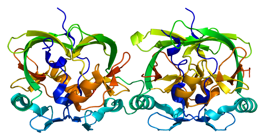
Genes associados
1 gene identificado com associação a esta condição. Padrão de herança: Autosomal dominant.
Curadoria gene-doença
fontes oficiaisChromatin-binding factor that repress Notch signaling in the absence of Notch intracellular domain by acting as a CBF1 corepressor. Binds to the HEY promoter and might assist, along with NCOR2, RBPJ-mediated repression. Binds RNA in vitro. May be involved in RNA metabolism (PubMed:21475249). In concert with CIC and ATXN1L, involved in brain development (By similarity)
CytoplasmNucleus
Spinocerebellar ataxia 1
Spinocerebellar ataxia is a clinically and genetically heterogeneous group of cerebellar disorders. Patients show progressive incoordination of gait and often poor coordination of hands, speech and eye movements, due to cerebellum degeneration with variable involvement of the brainstem and spinal cord. SCA1 belongs to the autosomal dominant cerebellar ataxias type I (ADCA I) which are characterized by cerebellar ataxia in combination with additional clinical features like optic atrophy, ophthalmoplegia, bulbar and extrapyramidal signs, peripheral neuropathy and dementia. SCA1 is caused by expansion of a CAG repeat in the coding region of ATXN1. Longer expansions result in earlier onset and more severe clinical manifestations of the disease.
Medicamentos e terapias
Mecanismo: Glycogen synthase kinase-3 inhibitor
Variantes genéticas (ClinVar)
28 variantes patogênicas registradas no ClinVar.
Classificação de variantes (ClinVar)
Distribuição de 17 variantes classificadas pelo ClinVar.
Diagnóstico
Os sinais que médicos procuram e os exames que confirmam
Tratamento e manejo
Remédios, cuidados de apoio e o que precisa acompanhar
Onde tratar no SUS
Hospitais de referência no Brasil e o protocolo oficial do SUS (PCDT)
🇧🇷 Atendimento SUS — Ataxia espinocerebelar tipo 1
Selecione um estado ou use sua localização para ver resultados.
Dados de DATASUS/CNES, SBGM, ABNeuro e Ministério da Saúde. Sempre confirme a disponibilidade diretamente com o estabelecimento.
Pesquisa ativa
Ensaios clínicos abertos e novidades científicas recentes
Ensaios em destaque
🟢 Recrutando agora
3 pesquisas recrutando participantes. Converse com seu médico sobre a possibilidade de participar.
Outros ensaios clínicos
17 ensaios clínicos encontrados, 7 ativos.
Publicações mais relevantes
Neural basis for mutant ATAXIN-1 induced respiratory dysfunction in mouse models of spinocerebellar ataxia type 1.
Spinocerebellar ataxia type 1 is a neurodegenerative disease characterized by motor dysfunction and premature death usually from compromised swallowing and respiration. Using plethysmography, we characterized respiration in the conditional f-ATXN1146Q/2Q SCA1 model. We found a progressive elevation of baseline respiration that impairs ability of f-ATXN1146Q/2Q mice to increase breathing during challenge. To delineate regions contributing to respiratory dysfunction, f-ATXN1146Q/2Q mice were crossed with Nestin-Cre and Acta1-Cre mice, respectively. Respiration improved by removing mATXN1 from neural lineages, but not from skeletal muscle demonstrating mATXN1 in the central nervous system is a key driver of respiratory dysfunction in SCA1 mouse models. Moreover, respiratory dysfunction in SCA1 mice involves two aspects: behavioral dysregulation exhibited as increased movement during plethysmography, and functional dysregulation of respiratory circuitry. As both of these aspects are rescued by deleting mATXN1 from neural cells, we further investigated the role of cerebellar Purkinje cells and chemosensing neurons in the brain stem in SCA1 respiratory phenotype. Our results indicate complex multiregional etiology of respiratory dysfunction. Mechanistically we found that in contrast to most other SCA1 symptoms, nuclear localization of mATXN1 does not play a key role in respiratory dysfunction.
Mutant ATXN1 impacts human and mouse microglia and contributes to cognitive, mood, and motor deficits in SCA1 mice.
Microglia, resident immune cells of the brain, are important players in neurodegeneration. While microglial activation is a hallmark of many neurodegenerative diseases, the specific role of microglia intrinsic factors in microglial activation and disease pathogenesis remains unknown. Spinocerebellar ataxia type-1 (SCA1) is an inherited autosomal dominant neurodegenerative disease characterized by severe neuronal loss and early microglial activation in the cerebellum. SCA1 is caused by CAG repeat expansion in the ubiquitously expressed ATAXIN1 (ATXN1) gene. Using human microglia differentiated from SCA1 patient derived iPSCs, we found that mutant ATXN1 is sufficient to alter morphology, gene and protein expression in human microglia in a cell-autonomous manner. Moreover, compared to controls, human SCA1 microglia exhibited increased phagocytosis and pro-inflammatory cytokine production, indicating an immune priming. To determine the extent to which mutant ATXN1 in microglia contributes to SCA1 pathogenesis and behavioral symptoms, we removed mutant ATXN1 from microglia and macrophages in a novel conditional SCA1 mouse model, f-ATXN1146Q/2Q mice. Microglial mutant ATXN1 reduction led to a marked correction in microglia phenotype, in particular in the transcriptomic signature of interferon type 1 mediated immune response, reduced microglial density and resulted in smaller microglia with reduced branching in the cerebellum. Pathology of Purkinje neurons and cerebellar astrogliosis were also ameliorated. Utilizing a battery of behavioral tests, we found that microglia and macrophage mutant ATXN1 reduction ameliorated cognitive, mood, and motor deficits in SCA1 mice. Together, these results indicate that mutant ATXN1 directly impacts microglial phenotype in SCA1, contributing to SCA1 pathology and behavioral deficits.
Impedance-based phenotypic profiling of metabotropic glutamate receptor ligand responses in SCA1 human iPSC-derived neuronal cultures.
Group 1 metabotropic glutamate receptors (mGluRs) are a family of G protein-coupled receptors (GPCRs) including mGluR1 and mGluR5. These receptors are expressed throughout the central nervous system and play an important role in synaptic plasticity. Dysfunction of mGluR1 in cerebellar Purkinje cells (PC) is observed in spinocerebellar ataxia type 1 (SCA1), an autosomal dominant neurodegenerative disorder caused by an expanded CAG repeat in the ATXN1 gene. MGluR1 dysfunction disrupts PC signaling and ultimately contributes to PC death and overall progressive cerebellar dysfunction observed in SCA1. To investigate mGluR1/5 pharmacology in the context of SCA1, mGluR1/5 function was assessed in SCA1 and control human induced pluripotent stem cell (hiPS)cell-derived neuronal cultures using impedance measurements in the xCELLigence real-time cell analyzer (RTCA) system. Culture conditions and protocols for evaluating glutamate receptor activity were first optimized. Subsequently, GRM1 and GRM5 expression levels were assessed and glutamatergic signaling function was characterized in SCA1 hiPS cell-derived neuronal cultures using both the non-selective endogenous ligand L-glutamate and the selective orthosteric agonist for mGluR1/5, (RS)-3,5-Dihydroxyphenylglycine. To specifically characterize mGluR1 function, mGluR1-specific positive- and negative allosteric modulators (Ro0711401 and JNJ16259685 respectively) were tested. Results showed reduced mGluR1 and decreased mGluR5 RNA expression levels and diminished mGluR1/5 responses in the SCA1 hiPS cell-derived neuronal cultures. These results underline the potential utility of impedance measurements for characterizing GPCR function and pharmacological testing in a high-throughput manner in patient-derived neuronal cultures.
Retinal morphology in spinocerebellar ataxia type 1 (SCA1) mice: A stereological analysis across different age groups.
Spinocerebellar ataxia type 1 (SCA1) affects not only the cerebellum but also the retina; however, retinal pathology remains poorly characterised in murine models of SCA1. To fill this gap, we performed a comprehensive stereological analysis of the retinal structure of SCA1154Q/2Q knock-in mice and their healthy SCA12Q/2Q littermates at 6 and 10 months of age. We compared animals across genotypes at each age and across ages within each genotype. Using unbiased stereology, we quantified the total retinal volume, volumes of individual retinal layers, total photoreceptor numbers, numbers of rods and cones, and total cell numbers in the inner nuclear and ganglion cell layers. Structural abnormalities, including disorganisation of photoreceptor outer segments and reduced volumes of both photoreceptor inner and outer segments, were evident in SCA1 mice as early as 6 months. By 10 months, these alterations had progressed, with a decrease in the number of ganglion cells and a reduced proportion of cones among the total photoreceptors. Wild-type mice also exhibited age-related changes, but the pattern and magnitude differed, suggesting distinct mechanisms of normal ageing versus SCA1-related neurodegeneration. Our findings demonstrate that retinal remodelling in SCA1 mice parallels changes observed in human patients, validating this model for investigating visual system involvement in SCA1. These results emphasize the need to consider retinal pathology when interpreting behavioural or motor deficits and in designing future preclinical interventions.
TMEM206 gene knockout improves balance performance in SCA1 transgenic mice.
TMEM206 was identified as a conserved chloride channel that underlies widely expressed, proton-activated, outwardly rectifying chloride currents. Spinocerebellar ataxia type 1 (SCA1) is one of polyglutamine diseases, and is characterized as a progressive and autosomal dominant genetic disease, which is caused by an increasing number of CAG repeats in the ataxin-1 gene. TMEM206 was confirmed to interact with ataxin-1. This study suggests that TMEM206-ataxin-1 interaction might involve in the pathological mechanisms of SCA1. To elucidate the mechanisms of SCA1 involved in proton-activated chloride channel gating, we bred TMEM206 knockout mice using SCA1 model mice. Motor coordination and balance in mice was evaluated using rotarod test and grip strength. These studies showed that genetic depletion of TMEM206 has slight impacts on the SCA1 mice weight, and partially improves motor incoordination in Atxn1154Q/2Q mice. No alteration in grip strength was found in Atxn1154Q/2Q mice with depletion of the TMEM206 gene. Our studies indicate that the TMEM206 knockout appears to emerge as a potential therapy method for SCA1 mice.
Publicações recentes
Long term administration of selective NMDA GluN2B receptor blocker Ro25-6981 attenuates neurodegeneration in mouse model of spinocerebellar ataxia type 1 (SCA1).
Early and Progressive Spinal Cord Atrophy in Spinocerebellar Ataxia Type 1.
Neural basis for mutant ATAXIN-1 induced respiratory dysfunction in mouse models of spinocerebellar ataxia type 1.
Mutant ATXN1 impacts human and mouse microglia and contributes to cognitive, mood, and motor deficits in SCA1 mice.
Impedance-based phenotypic profiling of metabotropic glutamate receptor ligand responses in SCA1 human iPSC-derived neuronal cultures.
📚 EuropePMC270 artigos no totalmostrando 194
Neural basis for mutant ATAXIN-1 induced respiratory dysfunction in mouse models of spinocerebellar ataxia type 1.
Neurobiology of diseaseMutant ATXN1 impacts human and mouse microglia and contributes to cognitive, mood, and motor deficits in SCA1 mice.
bioRxiv : the preprint server for biologyImpedance-based phenotypic profiling of metabotropic glutamate receptor ligand responses in SCA1 human iPSC-derived neuronal cultures.
Neurobiology of diseaseRetinal morphology in spinocerebellar ataxia type 1 (SCA1) mice: A stereological analysis across different age groups.
Experimental eye researchTranslational Relevance of SCA1 Models for the Development of Therapies for Spinocerebellar Ataxia Type 1.
BiomedicinesOral KDS2010, a Monoamine Oxidase-B (MAO-B) Inhibitor, Slows the Deterioration of Motor Coordination in Genetic SCA1 Models by Inhibiting Astrocytic MAO-B-Mediated Inflammation.
Journal of neurochemistryStriatal pathology in Spinocerebellar Ataxia Type 1 mice: A comparative study with Huntington's disease.
bioRxiv : the preprint server for biologyTMEM206 gene knockout improves balance performance in SCA1 transgenic mice.
IBRO neuroscience reportsPsychomotor and non-motor correlates of cognition in spinocerebellar ataxias Types 1, 2, 3, and 6.
Brain communicationsFunctional divergence of Capicua isoforms explains differential tissue vulnerability in neurological disease.
bioRxiv : the preprint server for biologyComparative Analysis of Two Autophagy-Enhancing Small Molecules (AUTEN-67 and -99) in a Drosophila Model of Spinocerebellar Ataxia Type 1.
International journal of molecular sciencesPredictive models for ataxia progression and conversion in spinocerebellar ataxia type 1 and 3.
Brain : a journal of neurologySpinocerebellar Ataxia Type 1 (SCA1) Cell Models Display Widespread Mitochondrial and Extra-Nuclear Alterations.
Journal of molecular neuroscience : MNThe predictive validity of pontine volume change in spinocerebellar ataxia type 1 (SCA1).
Neurobiology of diseaseSCA1 in Brazil: Local Cohort Profile and Scientific Contributions.
Cerebellum (London, England)Sex Differences in Spinocerebellar Ataxia Type 1: Clinical Presentation and Progression.
Cerebellum (London, England)Chemical Targeting of the ATXN1 aa99-163 Interaction Site Suppresses polyQ-Expanded Protein Dimerization.
ACS omegaIdentification of Splicing Regulatory Activity of ATXN1 and Its Associated Domains.
BiomoleculesFingolimod Prevents Neuroinflammation but Has a Limited Effect on the Development of Ataxia in a Mouse Model for SCA1.
International journal of molecular sciencesExploring mutation carriers' preferences regarding onset and progression of disease predictions for adult-onset genetic neurodegenerative diseases: a qualitative interview study.
Human geneticsAn expanded polyglutamine in ATAXIN1 results in a loss-of-function that exacerbates severity of Multiple Sclerosis in an EAE mouse model.
Research squareThe pattern and dynamics of white matter alterations in Spinocerebellar ataxia type 1: A diffusion-weighted magnetic resonance imaging study.
NeuroImage. ClinicalSex Differences in a Novel Mouse Model of Spinocerebellar Ataxia Type 1 (SCA1).
International journal of molecular sciencesmitoXplorer 3.0, A Web Tool for Exploring Mitochondrial Dynamics in Single-cell RNA-seq Data.
Journal of molecular biologyNatural compounds as therapeutic candidates for spinocerebellar ataxia type 1: a computational approach.
In silico pharmacologyPrediction of protein interactions with function in protein (de-)phosphorylation.
PloS oneRevisiting huntingtin activity and localization signals in the context of protein structure.
Journal of Huntington's diseaseGeneration of a human induced pluripotent stem cell line (UNIFEi001-A) from a patient with Spinocerebellar ataxia type 1 (SCA1).
Stem cell researchCRISPR-Cas9-directed gene therapy for spinocerebellar ataxia type 1.
Molecular therapy. Nucleic acidsEnhanced Age-Dependent Motor Impairment in Males of Drosophila melanogaster Modeling Spinocerebellar Ataxia Type 1 Is Linked to Dysregulation of a Matrix Metalloproteinase.
BiologyIncreased intrinsic membrane excitability is associated with olivary hypertrophy in spinocerebellar ataxia type 1.
Human molecular geneticsCas9 editing of ATXN1 in a spinocerebellar ataxia type 1 mice and human iPSC-derived neurons.
Molecular therapy. Nucleic acidsExpanded ATXN1 alters transcription and calcium signaling in SCA1 human motor neurons differentiated from induced pluripotent stem cells.
Neurobiology of diseaseEarly-onset phenotype in a patient with an intermediate allele and a large SCA1 expansion: a case report.
BMC neurologyA Neural Basis for Mutant ATAXIN-1 Induced Respiratory Dysfunction in Mouse Models of Spinocerebellar Ataxia Type 1.
bioRxiv : the preprint server for biologyMultimodal, Longitudinal Profiling of SCA1 Identifies Predictors of Disease Severity and Progression.
Annals of neurologyCerebellar contribution to cognitive deficits and prefrontal cortex dysfunction in Spinocerebellar Ataxia Type 1 (SCA1).
bioRxiv : the preprint server for biologyMolecular therapy for polyQ disorders: from bench to clinical trials.
Trends in molecular medicineCerebellar Heterogeneity and Selective vulnerability in Spinocerebellar Ataxia Type 1 (SCA1).
Neurobiology of diseaseDynamic molecular network analysis of iPSC-Purkinje cells differentiation delineates roles of ISG15 in SCA1 at the earliest stage.
Communications biologyPGT-M for spinocerebellar ataxia type 1: development of a STR panel and a report of two clinical cases.
Journal of assisted reproduction and geneticsMemantine suppresses the excitotoxicity but fails to rescue the ataxic phenotype in SCA1 model mice.
Biomedicine & pharmacotherapy = Biomedecine & pharmacotherapieCo-existence of Myelin Oligodendrocyte Glycoprotien Antibody-associated Disease (MOGAD) and Spinocerebellar Ataxia type 1 (SCA1): A case report.
Neurological sciences : official journal of the Italian Neurological Society and of the Italian Society of Clinical NeurophysiologyMapping SCA1 regional vulnerabilities reveals neural and skeletal muscle contributions to disease.
JCI insightQuantitative Evaluation of Stance as a Sensitive Biomarker of Postural Ataxia Development in Preclinical SCA1 Mutation Carriers.
Cerebellum (London, England)Probing cerebellar circuit dysfunction in rodent models of spinocerebellar ataxia by means of in vivo two-photon calcium imaging.
STAR protocolsCerebellar Volumetry in Ataxias: Relation to Ataxia Severity and Duration.
Cerebellum (London, England)Different Purkinje cell pathologies cause specific patterns of progressive gait ataxia in mice.
Neurobiology of diseaseAnesthetic Management of a Patient With Spinocerebellar Ataxia Type 1.
Anesthesia progressIntranuclear inclusions of polyQ-expanded ATXN1 sequester RNA molecules.
Frontiers in molecular neuroscienceLongitudinal single-cell transcriptional dynamics throughout neurodegeneration in SCA1.
NeuronIncreased intrinsic membrane excitability is associated with hypertrophic olivary degeneration in spinocerebellar ataxia type 1.
bioRxiv : the preprint server for biologyHD and SCA1: Tales from two 30-year journeys since gene discovery.
NeuronAntagonistic roles of canonical and Alternative-RPA in disease-associated tandem CAG repeat instability.
CellDysregulation of alternative splicing in spinocerebellar ataxia type 1.
Human molecular geneticsAltered calcium signaling in Bergmann glia contributes to spinocerebellar ataxia type-1 in a mouse model of SCA1.
Neurobiology of diseaseSpinocerebellar ataxia type 1: It's not just about Purkinje cells.
NeuronCAT Interruption as a Protective Factor in Chinese Patients with Spinocerebellar Ataxia Type 1.
Cerebellum (London, England)Experimental Treatment with Edaravone in a Mouse Model of Spinocerebellar Ataxia 1.
International journal of molecular sciencesRibosomal protein SA is a common component of neuronal intranuclear inclusions in polyglutamine diseases and Marinesco bodies.
Neuropathology : official journal of the Japanese Society of NeuropathologyEarly molecular layer interneuron hyperactivity triggers Purkinje neuron degeneration in SCA1.
NeuronSpinocerebellar Ataxia Type 1 Characteristics in Patient-Derived Fibroblast and iPSC-Derived Neuronal Cultures.
Movement disorders : official journal of the Movement Disorder SocietyComposite autonomic severity scoring in spinocerebellar ataxia type 1 and 2.
Journal of clinical neuroscience : official journal of the Neurosurgical Society of AustralasiaTherapeutic Strategies for Spinocerebellar Ataxia Type 1.
BiomoleculesThe potential value of disease-modifying therapy in patients with spinocerebellar ataxia type 1: an early health economic modeling study.
Journal of neurologySCAview: an Intuitive Visual Approach to the Integrative Analysis of Clinical Data in Spinocerebellar Ataxias.
Cerebellum (London, England)Alterations in oligodendrocyte transcriptional networks reveal region-specific vulnerabilities to neurological disease.
iScienceDisruption of the ATXN1-CIC complex reveals the role of additional nuclear ATXN1 interactors in spinocerebellar ataxia type 1.
NeuronMotor neuron involvement threatens survival in spinocerebellar ataxia type 1.
Neuropathology and applied neurobiologyOn the identification of potential novel therapeutic targets for spinocerebellar ataxia type 1 (SCA1) neurodegenerative disease using EvoPPI3.
Journal of integrative bioinformaticsDelineating regional vulnerability in the neurodegenerative disease SCA1 using a conditional mutant ATXN1 mouse.
bioRxiv : the preprint server for biologyBaseline Clinical and Blood Biomarkers in Patients With Preataxic and Early-Stage Disease Spinocerebellar Ataxia 1 and 3.
NeurologyDiverse regional mechanisms drive spinocerebellar ataxia type 1 phenotypes.
NeuronBDNF is altered in a brain-region specific manner and rescues deficits in Spinocerebellar Ataxia Type 1.
Neurobiology of diseaseAtaxin-1 controls the expression of specific noncoding RNAs in B cells upon autoimmune demyelination.
Immunology and cell biologyElectrophysiological and neuropsychological assessment of cognition in spinocerebellar ataxia type 1 patients: a pilot study.
Neurological sciences : official journal of the Italian Neurological Society and of the Italian Society of Clinical NeurophysiologyPerceptual and Acoustic Analysis of Speech in Spinocerebellar ataxia Type 1.
Cerebellum (London, England)Decreasing mutant ATXN1 nuclear localization improves a spectrum of SCA1-like phenotypes and brain region transcriptomic profiles.
NeuronClinically Meaningful Magnetic Resonance Endpoints Sensitive to Preataxic Spinocerebellar Ataxia Types 1 and 3.
Annals of neurologyGeneration of induced pluripotent stem cell(iPSC)line CJUHi001-A derived peripheral blood mononuclear cells of spinocerebellar ataxia type 1(SCA1) the CAG repeat mutation in ATXN1 gene.
Stem cell researchSingle nuclei RNA sequencing investigation of the Purkinje cell and glial changes in the cerebellum of transgenic Spinocerebellar ataxia type 1 mice.
Frontiers in cellular neuroscienceSpatial and Temporal Diversity of Astrocyte Phenotypes in Spinocerebellar Ataxia Type 1 Mice.
CellsAdeno-associated virus vector-based gene therapies for pediatric diseases.
Pediatrics and neonatologyToward the design and development of peptidomimetic inhibitors of the Ataxin-1 aggregation pathway.
Biophysical journalIdentifying Disease Signatures in the Spinocerebellar Ataxia Type 1 Mouse Cortex.
CellsCIC missense variants contribute to susceptibility for spina bifida.
Human mutationDifferential effects of Wnt-β-catenin signaling in Purkinje cells and Bergmann glia in spinocerebellar ataxia type 1.
Proceedings of the National Academy of Sciences of the United States of AmericaIdentification of the ataxin-1 interaction network and its impact on spinocerebellar ataxia type 1.
Human genomicsEpigenetic control of ataxin-1 in multiple sclerosis.
Annals of clinical and translational neurologyIndirect Negative Effect of Mutant Ataxin-1 on Short- and Long-Term Synaptic Plasticity in Mouse Models of Spinocerebellar Ataxia Type 1.
CellsSimple and clear differentiation of spinocerebellar degenerations: Overview of macroscopic and low-power view findings.
Neuropathology : official journal of the Japanese Society of NeuropathologyThe extra-cerebellar effects of spinocerebellar ataxia type 1 (SCA1): looking beyond the cerebellum.
Cellular and molecular life sciences : CMLSCholecystokinin Activation of Cholecystokinin 1 Receptors: a Purkinje Cell Neuroprotective Pathway.
Cerebellum (London, England)Combined overexpression of ATXN1L and mutant ATXN1 knockdown by AAV rescue motor phenotypes and gene signatures in SCA1 mice.
Molecular therapy. Methods & clinical developmentCross-species genetic screens identify transglutaminase 5 as a regulator of polyglutamine-expanded ataxin-1.
The Journal of clinical investigationMemantine Disrupts Motor Coordination through Anxiety-like Behavior in CD1 Mice.
Brain sciencesStool is a sensitive and noninvasive source of DNA for monitoring expansion in repeat expansion disease mouse models.
Disease models & mechanismsReduction of mutant ATXN1 rescues premature death in a conditional SCA1 mouse model.
JCI insightLevels of Neurofilament Light at the Preataxic and Ataxic Stages of Spinocerebellar Ataxia Type 1.
NeurologyNeurofilaments as Decay Rate Biomarker in Spinocerebellar Ataxia Type 1: Highlighting Key Questions of Application and Future Challenges.
NeurologyOuter Retinal Disruption in Spinocerebellar Ataxia Type 1.
Ophthalmology. RetinaAcupuncture for spinocerebellar ataxia type 1: a case report.
Acupuncture in medicine : journal of the British Medical Acupuncture SocietyMacular Morpho-Functional and Visual Pathways Functional Assessment in Patients with Spinocerebellar Type 1 Ataxia with or without Neurological Signs.
Journal of clinical medicineIntercellular Propagation and Aggregate Seeding of Mutant Ataxin-1.
Journal of molecular neuroscience : MNStructural Analysis and Spatiotemporal Expression of Atxn1 Genes in Zebrafish Embryos and Larvae.
International journal of molecular sciencesToxicity after AAV delivery of RNAi expression constructs into nonhuman primate brain.
Nature medicineDetection Methods and Status of CAT Interruption of ATXN1 in Korean Patients With Spinocerebellar Ataxia Type 1.
Annals of laboratory medicineSocial Cognition in Patients With Cerebellar Neurodegenerative Disorders.
Frontiers in systems neuroscienceSphingolipid metabolism governs Purkinje cell patterned degeneration in Atxn1[82Q]/+ mice.
Proceedings of the National Academy of Sciences of the United States of AmericaA Structural Study of the Cytoplasmic Chaperone Effect of 14-3-3 Proteins on Ataxin-1.
Journal of molecular biologyGenetic Modeling of the Neurodegenerative Disease Spinocerebellar Ataxia Type 1 in Zebrafish.
International journal of molecular sciencesDNAzyme Cleavage of CAG Repeat RNA in Polyglutamine Diseases.
Neurotherapeutics : the journal of the American Society for Experimental NeuroTherapeuticsSpinocerebellar Ataxia Type 1: One-Year Longitudinal Study to Identify Clinical and MRI Measures of Disease Progression in Patients and Presymptomatic Carriers.
Cerebellum (London, England)Spinocerebellar Ataxia Type 1 protein Ataxin-1 is signaled to DNA damage by ataxia-telangiectasia mutated kinase.
Human molecular geneticsChronic optogenetic stimulation of Bergman glia leads to dysfunction of EAAT1 and Purkinje cell death, mimicking the events caused by expression of pathogenic ataxin-1.
Neurobiology of diseaseRegion-specific preservation of Purkinje cell morphology and motor behavior in the ATXN1[82Q] mouse model of spinocerebellar ataxia 1.
Brain pathology (Zurich, Switzerland)Dual targeting of brain region-specific kinases potentiates neurological rescue in Spinocerebellar ataxia type 1.
The EMBO journalModulation of ATXN1 S776 phosphorylation reveals the importance of allele-specific targeting in SCA1.
JCI insightThe novel multiple sclerosis susceptibility gene ATXN1 regulates B cell receptor signaling in B-1a cells.
Molecular brainMood alterations in mouse models of Spinocerebellar Ataxia type 1.
Scientific reportsBrainstem and striatal volume changes are detectable in under 1 year and predict motor decline in spinocerebellar ataxia type 1.
Brain communicationsPost-symptomatic Delivery of Brain-Derived Neurotrophic Factor (BDNF) Ameliorates Spinocerebellar Ataxia Type 1 (SCA1) Pathogenesis.
Cerebellum (London, England)Genomic Portrait of a Sporadic Amyotrophic Lateral Sclerosis Case in a Large Spinocerebellar Ataxia Type 1 Family.
Journal of personalized medicineA Chlorzoxazone-Baclofen Combination Improves Cerebellar Impairment in Spinocerebellar Ataxia Type 1.
Movement disorders : official journal of the Movement Disorder SocietyNeurochemical Differences in Spinocerebellar Ataxia Type 14 and 1.
Cerebellum (London, England)Dynamics of a Protein Interaction Network Associated to the Aggregation of polyQ-Expanded Ataxin-1.
GenesAltered Capicua expression drives regional Purkinje neuron vulnerability through ion channel gene dysregulation in spinocerebellar ataxia type 1.
Human molecular geneticsStridor during sleep: description of 81 consecutive cases diagnosed in a tertiary sleep disorders center.
SleepATXN1 repeat expansions confer risk for amyotrophic lateral sclerosis and contribute to TDP-43 mislocalization.
Brain communicationsFrequency of Spinocerebellar Ataxia type 1, 2, 3,6 and 7 and clinical profile of Spinocerebellar Ataxia type 3 in Malaysia.
Cerebellum & ataxiasAtaxin-1 regulates B cell function and the severity of autoimmune experimental encephalomyelitis.
Proceedings of the National Academy of Sciences of the United States of AmericaUTteR control through miRs: fine-tuning ATXN1 levels to prevent ataxia.
Genes & developmentAntisense Oligonucleotide Therapeutic Approach for Suppression of Ataxin-1 Expression: A Safety Assessment.
Molecular therapy. Nucleic acidsmiR760 regulates ATXN1 levels via interaction with its 5' untranslated region.
Genes & developmentPatterns of CAG repeat instability in the central nervous system and periphery in Huntington's disease and in spinocerebellar ataxia type 1.
Human molecular geneticsFunctionally Relevant Maculopathy and Optic Atrophy in Spinocerebellar Ataxia Type 1.
Movement disorders clinical practiceThe ataxin-1 interactome reveals direct connection with multiple disrupted nuclear transport pathways.
Nature communicationsCortical network dysfunction revealed by magnetoencephalography in carriers of spinocerebellar ataxia 1 or 2 mutation.
Clinical neurophysiology : official journal of the International Federation of Clinical NeurophysiologyGeneration of induced pluripotent stem cell line (CSUXHi002-A) from a patient with spinocerebellar ataxia type 1.
Stem cell researchPathogenic mechanisms underlying spinocerebellar ataxia type 1.
Cellular and molecular life sciences : CMLSRegulation and function of capicua in mammals.
Experimental & molecular medicineNuclear inclusions of pathogenic ataxin-1 induce oxidative stress and perturb the protein synthesis machinery.
Redox biologyFrequency and distribution of polyQ disease intermediate-length repeat alleles in healthy Italian population.
Neurological sciences : official journal of the Italian Neurological Society and of the Italian Society of Clinical NeurophysiologyCerebellar contribution to the cognitive alterations in SCA1: evidence from mouse models.
Human molecular geneticsATXN1 N-terminal region explains the binding differences of wild-type and expanded forms.
BMC medical genomicsSensitivity of Volumetric Magnetic Resonance Imaging and Magnetic Resonance Spectroscopy to Progression of Spinocerebellar Ataxia Type 1.
Movement disorders clinical practiceXenografting of human umbilical mesenchymal stem cells from Wharton's jelly ameliorates mouse spinocerebellar ataxia type 1.
Translational neurodegenerationLoss of Ataxin-1 Potentiates Alzheimer's Pathogenesis by Elevating Cerebral BACE1 Transcription.
Cell5'UTR-mediated regulation of Ataxin-1 expression.
Neurobiology of diseaseTreadmill training increases the motor activity and neuron survival of the cerebellum in a mouse model of spinocerebellar ataxia type 1.
The Kaohsiung journal of medical sciencesPrediction of Survival With Long-Term Disease Progression in Most Common Spinocerebellar Ataxia.
Movement disorders : official journal of the Movement Disorder SocietyAdvance Care Plan and Factors Related to Disease Progression in Patients With Spinocerebellar Ataxia Type 1: A Cross-Sectional Study in Thailand.
The American journal of hospice & palliative careExtracellular S100β Disrupts Bergman Glia Morphology and Synaptic Transmission in Cerebellar Purkinje Cells.
Brain sciences(CAG)n loci as genetic modifiers of age at onset in patients with spinocerebellar ataxia type 1 from mainland China.
European journal of neurologyIntensive Outpatient Treatment of Depression in a Spinocerebellar Ataxia Type 1 Patient.
Case reports in psychiatryMacular degeneration as a common cause of visual loss in spinocerebellar ataxia type 1 (SCA1) patients.
Ophthalmic geneticsBrain Derived Neurotrophic Factor (BDNF) Delays Onset of Pathogenesis in Transgenic Mouse Model of Spinocerebellar Ataxia Type 1 (SCA1).
Frontiers in cellular neuroscienceSelf-assembling vascular endothelial growth factor nanoparticles improve function in spinocerebellar ataxia type 1.
Brain : a journal of neurologyMolecular pathway analysis towards understanding tissue vulnerability in spinocerebellar ataxia type 1.
eLifeComplementary proteomics strategies capture an ataxin-1 interactome in Neuro-2a cells.
Scientific dataAntisense oligonucleotide-mediated ataxin-1 reduction prolongs survival in SCA1 mice and reveals disease-associated transcriptome profiles.
JCI insightStructural signature in SCA1: clinical correlates, determinants and natural history.
Journal of neurologyPurkinje cell COX deficiency and mtDNA depletion in an animal model of spinocerebellar ataxia type 1.
Journal of neuroscience researchPolyQ Tract Toxicity in SCA1 is Length Dependent in the Absence of CAG Repeat Interruption.
Frontiers in cellular neuroscienceAstroglia contribute to the pathogenesis of spinocerebellar ataxia Type 1 (SCA1) in a biphasic, stage-of-disease specific manner.
GliaInhibition of NF-κB signaling in IKKβF/F;LysM Cre mice causes motor deficits but does not alter pathogenesis of Spinocerebellar ataxia type 1.
PloS onePAK1 regulates ATXN1 levels providing an opportunity to modify its toxicity in spinocerebellar ataxia type 1.
Human molecular geneticsDendritic potassium channel dysfunction may contribute to dendrite degeneration in spinocerebellar ataxia type 1.
PloS oneReduction of protein kinase A-mediated phosphorylation of ATXN1-S776 in Purkinje cells delays onset of Ataxia in a SCA1 mouse model.
Neurobiology of diseasePolarization-sensitive optical coherence tomography reveals gray matter and white matter atrophy in SCA1 mouse models.
Neurobiology of diseaseGeneration of 3 spinocerebellar ataxia type 1 (SCA1) patient-derived induced pluripotent stem cell lines LUMCi002-A, B, and C and 2 unaffected sibling control induced pluripotent stem cell lines LUMCi003-A and B.
Stem cell researchTargeting potassium channels to treat cerebellar ataxia.
Annals of clinical and translational neurologyMutant ataxin1 disrupts cerebellar development in spinocerebellar ataxia type 1.
The Journal of clinical investigationATXN1-CIC Complex Is the Primary Driver of Cerebellar Pathology in Spinocerebellar Ataxia Type 1 through a Gain-of-Function Mechanism.
NeuronProtein kinase C activity is a protective modifier of Purkinje neuron degeneration in cerebellar ataxia.
Human molecular geneticsSpinocerebellar Ataxia Type 1: Molecular Mechanisms of Neurodegeneration and Preclinical Studies.
Advances in experimental medicine and biologyMotor neuron degeneration correlates with respiratory dysfunction in SCA1.
Disease models & mechanismsLoss of Capicua alters early T cell development and predisposes mice to T cell lymphoblastic leukemia/lymphoma.
Proceedings of the National Academy of Sciences of the United States of AmericaAtaxin-1 is involved in tumorigenesis of cervical cancer cells via the EGFR-RAS-MAPK signaling pathway.
OncotargetShort-term succinic acid treatment mitigates cerebellar mitochondrial OXPHOS dysfunction, neurodegeneration and ataxia in a Purkinje-specific spinocerebellar ataxia type 1 (SCA1) mouse model.
PloS oneDevelopmental YAPdeltaC determines adult pathology in a model of spinocerebellar ataxia type 1.
Nature communicationsMotor and Cerebellar Architectural Abnormalities during the Early Progression of Ataxia in a Mouse Model of SCA1 and How Early Prevention Leads to a Better Outcome Later in Life.
Frontiers in cellular neuroscience[Molecularly-Targeted Therapy of Spinocerebellar Ataxia Type 1 by HMGB1].
Brain and nerve = Shinkei kenkyu no shinpoStance instability in preclinical SCA1 mutation carriers: A 4-year prospective posturography study.
Gait & postureInhibition of colony-stimulating factor 1 receptor early in disease ameliorates motor deficits in SCA1 mice.
Journal of neuroinflammationSolving the puzzle of neurological diseases: an interview with Huda Zoghbi.
Disease models & mechanismsSpinal Cord Damage in Spinocerebellar Ataxia Type 1.
Cerebellum (London, England)Ataxin-1 regulates epithelial-mesenchymal transition of cervical cancer cells.
OncotargetTreating SCA1 Mice with Water-Soluble Compounds to Non-Specifically Boost Mitochondrial Function.
Journal of visualized experiments : JoVEIdentification of early neurodegenerative change in presymptomatic spinocerebellar ataxia type 1: A diffusion tensor imaging study.
Parkinsonism & related disorders[Report of a pedigree affected with spinocerebellar ataxia type 1].
Zhonghua yi xue yi chuan xue za zhi = Zhonghua yixue yichuanxue zazhi = Chinese journal of medical geneticsExpansion, mosaicism and interruption: mechanisms of the CAG repeat mutation in spinocerebellar ataxia type 1.
Cerebellum & ataxiasRNAi prevents and reverses phenotypes induced by mutant human ataxin-1.
Annals of neurologyFusion of Human Fetal Mesenchymal Stem Cells with "Degenerating" Cerebellar Neurons in Spinocerebellar Ataxia Type 1 Model Mice.
PloS oneAssociações
Organizações que acompanham esta doença — pra ter apoio e orientação
Ainda não temos associações cadastradas para Ataxia espinocerebelar tipo 1.
É de uma associação que acompanha esta doença? Fale com a gente →
Comunidades
Grupos ativos de quem convive com esta doença aqui no Raras
Ainda não existe comunidade no Raras para Ataxia espinocerebelar tipo 1
Pacientes, familiares e cuidadores se organizam em comunidades pra compartilhar experiências, fazer perguntas e se apoiar. Você pode ser o primeiro.
Tire suas dúvidas
Perguntas, dicas e experiências compartilhadas aqui na página
Participe da discussão
Faça login para postar dúvidas, compartilhar experiências e interagir com especialistas.
Fazer loginDoenças relacionadas
Doenças com sintomas parecidos — ajudam quem ainda está buscando diagnóstico
Referências e fontes
Bases de dados externas citadas neste artigo
Publicações científicas
Artigos indexados no PubMed ligados a esta doença no grafo RarasNet — título, periódico e PMID direto da fonte, sem intermediação de IA.
- Neural basis for mutant ATAXIN-1 induced respiratory dysfunction in mouse models of spinocerebellar ataxia type 1.
- Mutant ATXN1 impacts human and mouse microglia and contributes to cognitive, mood, and motor deficits in SCA1 mice.
- Impedance-based phenotypic profiling of metabotropic glutamate receptor ligand responses in SCA1 human iPSC-derived neuronal cultures.
- Retinal morphology in spinocerebellar ataxia type 1 (SCA1) mice: A stereological analysis across different age groups.
- TMEM206 gene knockout improves balance performance in SCA1 transgenic mice.
- Long term administration of selective NMDA GluN2B receptor blocker Ro25-6981 attenuates neurodegeneration in mouse model of spinocerebellar ataxia type 1 (SCA1).
- Early and Progressive Spinal Cord Atrophy in Spinocerebellar Ataxia Type 1.
Bases de dados e fontes oficiais
Identificadores e referências canônicas usadas para montar este verbete.
- ORPHA:98755(Orphanet)
- OMIM OMIM:164400(OMIM)
- MONDO:0008119(MONDO)
- GARD:4071(GARD (NIH))
- Variantes catalogadas(ClinVar)
- Busca completa no PubMed(PubMed)
- Q21097855(Wikidata)
Dados compilados pelo RarasNet a partir de fontes abertas (Orphanet, OMIM, MONDO, PubMed/EuropePMC, ClinicalTrials.gov, DATASUS, PCDT/MS). Este conteúdo é informativo e não substitui avaliação médica.
Conteúdo mantido por Agente Raras · Médicos e pesquisadores podem colaborar
Ataxia espinocerebelar tipo 1
📋 Origem dos dados
Esta página agrega dados de fontes públicas e oficiais. Dados sobre cobertura no SUS (PCDT, CEAF) são verificados ativamente por agente proativo (ver badge no infobox). Demais dados têm atribuição de fonte + data da última sincronização — clique para abrir o original.
- Doença rara (ontologia)
- fonte: Orphanet
- Identificador unificado
- fonte: MONDO
- Genética mendeliana
- fonte: OMIM
- Codificação WHO/SUS
- fonte: WHO ICD-10 / DATASUS
- CID-11 (futuro)
- fonte: WHO ICD-11
- NIH/GARD
- fonte: GARD (NIH)
- Dado público estruturado
- fonte: Wikidata
- Medicamentos (literatura)
- fonte: Orphanet
- Reposicionamento
- fonte: Drug Repurposing Hub
- Ensaios clínicos
- fonte: ClinicalTrials.gov