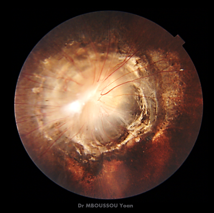
A síndrome da ipoméia (MGS) é uma neuropatia óptica caracterizada por uma escavação congênita em forma de funil do fundo posterior que incorpora a malformação do disco óptico (semelhante à flor da ipomeia). A MGS é geralmente unilateral e pode resultar em uma diminuição na melhor acuidade visual corrigida (BCVA). A MGS ocorre isolada ou associada a outras anomalias oculares ou não oculares.
Introdução
O que você precisa saber de cara
A síndrome da ipoméia (MGS) é uma neuropatia óptica caracterizada por uma escavação congênita em forma de funil do fundo posterior que incorpora a malformação do disco óptico (semelhante à flor da ipomeia). A MGS é geralmente unilateral e pode resultar em uma diminuição na melhor acuidade visual corrigida (BCVA). A MGS ocorre isolada ou associada a outras anomalias oculares ou não oculares.
Escala de raridade
<1/50kMuito rara
1/20kRara
1/10kPouco freq.
1/5kIncomum
1/2k
Encontrou um erro ou informação desatualizada? Sugira uma correção →
Entender a doença
Do básico ao detalhe, leia no seu ritmo
Preparando trilha educativa...
Sinais e sintomas
O que aparece no corpo e com que frequência cada sintoma acontece
Características mais comuns
Os sintomas variam de pessoa para pessoa. Abaixo estão as 7 características clínicas mais associadas, ordenadas por frequência.
Linha do tempo da pesquisa
Encontrou um erro ou informação desatualizada? Sugira uma correção →
Genética e causas
O que está alterado no DNA e como passa nas famílias
Genes associados
1 gene identificado com associação a esta condição.
Transcription factor with important functions in the development of the eye, nose, central nervous system and pancreas. Required for the differentiation of pancreatic islet alpha cells (By similarity). Competes with PAX4 in binding to a common element in the glucagon, insulin and somatostatin promoters. Regulates specification of the ventral neuron subtypes by establishing the correct progenitor domains (By similarity). Acts as a transcriptional repressor of NFATC1-mediated gene expression (By s
Nucleus
Aniridia 1
A congenital, bilateral, panocular disorder characterized by complete absence of the iris or extreme iris hypoplasia. Aniridia is not just an isolated defect in iris development but it is associated with macular and optic nerve hypoplasia, cataract, corneal changes, nystagmus. Visual acuity is generally low but is unrelated to the degree of iris hypoplasia. Glaucoma is a secondary problem causing additional visual loss over time.
Variantes genéticas (ClinVar)
543 variantes patogênicas registradas no ClinVar.
Vias biológicas (Reactome)
5 vias biológicas associadas aos genes desta condição.
Diagnóstico
Os sinais que médicos procuram e os exames que confirmam
Tratamento e manejo
Remédios, cuidados de apoio e o que precisa acompanhar
Onde tratar no SUS
Hospitais de referência no Brasil e o protocolo oficial do SUS (PCDT)
🇧🇷 Atendimento SUS — Síndrome Morning glory
Selecione um estado ou use sua localização para ver resultados.
Dados de DATASUS/CNES, SBGM, ABNeuro e Ministério da Saúde. Sempre confirme a disponibilidade diretamente com o estabelecimento.
Pesquisa ativa
Ensaios clínicos abertos e novidades científicas recentes
Pesquisa e ensaios clínicos
Nenhum ensaio clínico registrado para esta condição.
Publicações mais relevantes
Congenital Optic Nerve Anomalies and Associated Systemic Conditions.
Congenital optic nerve anomalies represent a group of structural malformations that impair vision, increase the risk of ophthalmic complications, and are frequently associated with systemic conditions. We provide a review of major congenital optic disc anomalies, including optic nerve hypoplasia, morning glory disc anomaly, optic disc coloboma, peripapillary staphyloma, persistent fetal vasculature, myelinated nerve fibers, tilted disc syndrome, optic disc pit, papillorenal syndrome, optic disc drusen, and congenital optic disc pigmentation. We will review the definition, epidemiology, pathophysiology, clinical presentation, diagnostic workup, and associated systemic findings. An emphasis is placed on screening and multidisciplinary management aimed at correcting and preserving vision and preventing complications, such as retinal detachment, maculopathy, strabismus, amblyopia, and endocrine disorders. Diagnosis tools, including optical coherence tomography (OCT), B-scan ultrasonography, magnetic resonance imaging (MRI), and computed tomography (CT), are highlighted for their role in screening for these conditions. Many congenital optic nerve anomalies lack a definitive cure, and some conditions are extremely rare and do not have well-defined treatment protocols. However, routine ophthalmic examination, correction of refractive errors, visual surveillance for systemic conditions, and surgical intervention can optimize patient outcomes. Furthermore, multidisciplinary collaboration between ophthalmologists and other physicians, including pediatricians, endocrinologists, neurologists, and geneticists is often needed to facilitate care.
Refractive Error Correction With Glasses in Congenital Ocular Fundus Anomalies: A Retrospective Series of 18 Children With Different Disease Entities Followed Up for More Than 10 Years.
Children with congenital anomalies of the posterior segment of the eye are in the process of visual development, and thus, their refractive errors should be detected by cycloplegic refraction testing to prescribe full-correction glasses, if required, and to help their visual acuity develop with growth. This study aimed to review refractive correction in children with congenital ocular fundus anomalies. A retrospective review was conducted on 18 consecutive children (11 female and seven male children) who were diagnosed with ocular fundus anomalies and followed for 10 years or more by a single ophthalmologist at a referral-based hospital. The age at the initial visit ranged from 10 days after birth to 11 years, with a median of one year and four months, and the age at the last visit ranged from 10 to 32 years, with a median of 15 years. The follow-up periods ranged from 10 to 21 years at a median of 15 years. The diagnoses were familial exudative vitreoretinopathy (FEVR) in eight children, persistent fetal vasculature (PFV) in five, morning glory disc anomaly in two, optic nerve and choroidal coloboma (CHARGE syndrome) in two, and Coats disease in one. Full-correction glasses were prescribed in eight children, while the remaining 10 children did not wear glasses. Among nine children with the uncorrected visual acuity of 1.0 or better in one eye and the visual acuity in the other eye ranging from light perception to 0.01, eight children did not wear glasses, and one child wore glasses with hyperopic correction. The diagnoses in these nine children were PFV in five children, morning glory disc anomaly in two, FEVR in one, and Coats disease in one. In seven children who wore full-correction glasses, the best corrected visual acuity in the better eye ranged from 0.2 to 0.9 at a median of 0.5. In contrast, the visual acuity in the other eye ranged from light perception to 0.1 at a median of 0.03. The diagnoses of these seven children were FEVR in five children and CHARGE syndrome in two. The five children with FEVR showed myopic astigmatism in both eyes, while the two children with CHARGE syndrome showed hyperopic astigmatism in both eyes. Children with unilateral eye anomalies such as PFV and morning glory disc anomaly did not wear glasses since their healthy eyes had good uncorrected visual acuity. In contrast, children with involvement of both eyes in FEVR and CHARGE syndrome wore full-correction glasses. Rough information regarding full-correction glasses in each category would help clinicians cope with rare congenital eye diseases. However, this conclusion is generally applicable to the standard practice of pediatric ophthalmology.
Morning Glory Disc Anomaly With Ipsilateral Carotid Vasculopathy: MRI Characteristics and Clinical Implications.
Morning glory disc anomaly (MGDA) is a rare developmental malformation of the optic nerve that can complicate the diagnosis and treatment of children. We describe a case of a three-year-old girl who presented with a reduction of vision and an exotropia of the right eye. Fundoscopic observation revealed evidence of MGDA, which was confirmed through magnetic resonance imaging (MRI). MRI showed the characteristic triad of orbital appearances: a funnel-shaped morphologic structure of the posterior optic disc and elevation of the adjacent retinal surface, abnormal tissue surrounding the ipsilateral optic nerve distal part with obliteration of the surrounding subarachnoid spaces, and discontinuity of the uveoscleral coat. Post-contrast imaging revealed enhancement in the distal retrobulbar optic nerve area, likely representing displaced choroidal tissue accompanied by glial and fibrous proliferation. Notably, MR angiography showed narrowing of the right internal carotid artery segments (petrous, cavernous, and supraclinoid) with a hypoplastic right A1 segment. This represents an intermediate vascular phenotype--significant vasculopathy without the collateral vessels that define Moyamoya disease. This case highlights the importance of comprehensive neuroimaging in MGDA patients, as early identification of associated vascular anomalies is crucial for appropriate surveillance and management. The consistent MRI findings described can aid in differentiating MGDA from other ocular anomalies, particularly optic nerve coloboma, which has different genetic and prognostic implications.
Hybrid cavitary disc anomaly - A case series and proposal of a novel classification system.
Cavitary disc anomalies, including optic disc pit (ODP), optic nerve coloboma (ODC), and morning glory disc anomaly (MGDA), are congenital conditions with distinct embryological origins and systemic associations. Hybrid features combining these anomalies are rare, complicating diagnosis and management. To describe the clinical spectrum, multimodal imaging characteristics, and management strategies of hybrid cavitary disc anomalies, and to propose a novel classification system based on anatomical and physiological overlap. This retrospective case series included 22 eyes from 17 patients seen at a tertiary care center over 19 months (June 2023-December 2024). All underwent best-corrected visual acuity (BCVA) and fundus examination, multimodal imaging (color fundus photography and [optical coherence tomography] OCT), and neuroimaging when indicated. Findings were confirmed by a senior consultant with over 20 years of experience. Management tailored to visual prognosis and macular status. The median BCVA was Log MAR 0.30 ± 1.13. Various anomalies include ODP with ODC and retinal choroidal coloboma in 10 eyes (45.5%), ODP with MGDA in 3 eyes (13.6%), ODC with RCC and bridging, coloboma in 3 eyes (13.6%), and ODC with MGDA in 2 eyes (9.1%). ODP with ODC, dual ODP with ODC and RCC, and dual ODP with ODC or RCC in 1 eye were each present in 1 eye (4.5). One patient had bilateral hybrid anomalies with maculopathy. Surgical intervention was required in 4 eyes (18.2%). Hybrid cavitary disc anomalies, including MGDA and ODP, present unique diagnostic and management challenges. This study provides insights into these rare conditions, emphasizing the need for further monitoring and research.
Novel compound heterozygous variants in SIX6 cause a PAX2 like Dysplastic Optic Disc with macular abnormalities without coexistent microphthalmia or cataract.
Congenital optic disc dysplasia with coexistent macular abnormalities is seen in Morning-glory disc anomaly, optic disc pit, PAX6-related disc coloboma, and in the PAX2-related Papillo-renal syndrome. We report novel compound heterozygous variants in SIX6 causing optic disc dysplasia and macular abnormality without coexisting cataract or microphthalmia for the first time in Chinese ethnicity and also describe the macular OCT findings. A 4 year-8 months-old female child presented with poor vision, photophobia, and nystagmus noticed 4 months after her birth was diagnosed elsewhere to have isolated cone dystrophy. She was evaluated subsequently by an ocular geneticist. She underwent a complete ophthalmic evaluation including orthoptic evaluation, cycloplegic refraction, and a dilated fundus evaluation. She had fundus photography, macular OCT, and ERG. She had systemic evaluations, relevant systemic investigations, and molecular genetic testing. Whole Exome Sequencing (WES) revealed compound heterozygous variants both in the SIX6 and in the TULP1 gene. No pathogenic variants were identified in the PAX2, PAX6 or in any of the other developmental genes or in the genes currently known to cause cone dystrophy or cone-rod dystrophy. Parents were heterozygous for variants in both SIX6 and TULP1. Homozygous or compound heterozygous pathogenic variants in SIX6 can cause a dysplastic optic disc and coexistent macular abnormalities. This dysplastic disc may be clinically indistinguishable from that seen in the PAX2 related Papillo-renal syndrome. Careful optic disc evaluation of subtle disc dysplasia is critical in differentiating this extremely rare entity from other relatively common causes of isolated cone dystrophies or cone-rod dystrophies.
Publicações recentes
Congenital Optic Nerve Anomalies and Associated Systemic Conditions.
Morning Glory Disc Anomaly-like Optic Disc Changes.
Refractive Error Correction With Glasses in Congenital Ocular Fundus Anomalies: A Retrospective Series of 18 Children With Different Disease Entities Followed Up for More Than 10 Years.
Morning Glory Disc Anomaly With Ipsilateral Carotid Vasculopathy: MRI Characteristics and Clinical Implications.
Hybrid cavitary disc anomaly - A case series and proposal of a novel classification system.
📚 EuropePMC86 artigos no totalmostrando 73
Congenital Optic Nerve Anomalies and Associated Systemic Conditions.
International ophthalmology clinicsMorning Glory Disc Anomaly-like Optic Disc Changes.
OphthalmologyRefractive Error Correction With Glasses in Congenital Ocular Fundus Anomalies: A Retrospective Series of 18 Children With Different Disease Entities Followed Up for More Than 10 Years.
CureusMorning Glory Disc Anomaly With Ipsilateral Carotid Vasculopathy: MRI Characteristics and Clinical Implications.
CureusHybrid cavitary disc anomaly - A case series and proposal of a novel classification system.
Indian journal of ophthalmologyMorning glory disc anomaly and irregular optic nerve thickness: an overlooked association.
Arquivos brasileiros de oftalmologiaPseudo-Morning Glory Disc Anomaly from Trauma.
OphthalmologyNovel compound heterozygous variants in SIX6 cause a PAX2 like Dysplastic Optic Disc with macular abnormalities without coexistent microphthalmia or cataract.
Ophthalmic geneticsThe Evaluation of Ocular Posterior Segment Findings in 5527 Term Infants Using Smartphone-Based Fundus Imaging.
Journal of ophthalmologyMorning Glory Disc Anomaly: Expanding the MR Phenotype.
AJNR. American journal of neuroradiologyRapid Resolution of Serous Retinal Detachment in Morning Glory Disc Anomaly With Oral Acetazolamide Treatment.
Ophthalmic surgery, lasers & imaging retinaMorning Glory Disc Anomaly Associated Transsphenoidal Encephalocele.
Ophthalmology(What's the story) morning glory? MRI findings in morning glory disc anomaly.
NeuroradiologyOptical Coherence Tomography in a Morning Glory Disc Anomaly with a Peripapillary Choroidal Neovascular Membrane.
Neuro-ophthalmology (Aeolus Press)Pediatric orbital lesions: ocular pathologies.
Pediatric radiologyNatural Course for Retinal DetacShment in Morning Glory Disc Anomaly Based on a Grading System.
Asia-Pacific journal of ophthalmology (Philadelphia, Pa.)Pituitary Stalk Duplication: A Radiological Surprise in a Child With Short Stature.
AACE clinical case reportsClinical and Radiologic Findings in Patients With Morning Glory Disc Anomaly and Associated Optic Pathway Enlargement.
Journal of neuro-ophthalmology : the official journal of the North American Neuro-Ophthalmology SocietyMorning glory disc anomaly and its implications in moyamoya arteriopathy: a retrospective case series.
Journal of neurosurgery. PediatricsTotal retinal detachment in a morning glory disc anomaly.
The Pan African medical journalNatural Course for Retinal Detachment in Morning Glory Disc Anomaly Based on a Grading System.
Asia-Pacific journal of ophthalmology (Philadelphia, Pa.)A Case of Morning Glory Disc Associated With Unilateral Retinopathy of Prematurity.
Journal of pediatric ophthalmology and strabismusMorning glory disc anomaly associated with moyamoya disease and pituitary stalk duplication.
American journal of ophthalmology case reportsMorning Glory Disc Anomaly in an Infant.
Journal of pediatric ophthalmology and strabismusCase Report: Multimodal Imaging in a Rare Case of Morning Glory Disc Anomaly Complicated With Choroidal Ossification.
Frontiers in medicineClinical and Echographic Features of Morning Glory Disc Anomaly in Children: A Retrospective Study of 249 Chinese Patients.
Frontiers in medicineA case of anterior persistent hyperplastic primary vitreous associated with morning glory disc anomaly and retinopathy of prematurity like retinopathy in a term-born child.
BMC ophthalmologyOptic Nerve Abnormalities in Morning Glory Disc Anomaly: An MRI Study.
Journal of neuro-ophthalmology : the official journal of the North American Neuro-Ophthalmology SocietySurgical challenges in the management of morning glory disc anomaly-associated retinal detachment.
Indian journal of ophthalmologyOutcomes of vitreoretinal surgery in retinal detachment associated with morning glory disc anomaly.
Indian journal of ophthalmologyCase Report: Fibroglial Retinal Tissue in Contractile Morning Glory Disc Anomaly.
Case reports in ophthalmologyManagement of Retinal Detachment Associated with Morning Glory Disc Syndrome.
Case reports in ophthalmologyCephalic/cardiac neural crest cell and moyamoya disease.
The neuroradiology journalA rare triad of morning glory disc anomaly, moyamoya vasculopathy, and transsphenoidal cephalocele: pathophysiological considerations and surgical management.
Neurological sciences : official journal of the Italian Neurological Society and of the Italian Society of Clinical NeurophysiologyClinical characteristics of morning glory disc anomaly in South India.
Taiwan journal of ophthalmologyMorning Glory Disc Anomaly with Contractile Peripapillary Staphyloma in an 18-Month-Old Girl.
Neuro-ophthalmology (Aeolus Press)Case of peripheral fibrovascular proliferative retinopathy associated with morning glory disc anomaly.
American journal of ophthalmology case reportsAn unusual ophthalmic presentation of Wolf-Hirschhorn syndrome.
Ophthalmic geneticsSalt-and-pepper-like retinopathy in a case of morning glory disc anomaly.
BMJ case reportsMorning glory disc anomaly-associated maculopathy: multimodal imaging.
BMJ case reportsAn unusual association of Morning Glory Syndrome with chronic myeloid leukemia-Philadelphia chromosome.
Journal of family medicine and primary careInternal carotid artery origin of the anterior cerebral artery: A rare anatomic intracranial arterial variation in a child with morning glory disc anomaly and moyamoya vascular pattern; case report and review of literature.
Brain circulationSpotlight on the pediatric eye: a pictorial review of orbital anatomy and congenital orbital pathologies.
The neuroradiology journalUnilateral morning glory disc anomaly in a patient with prenatal Zika virus exposure.
International journal of retina and vitreousMorning glory disc anomaly associated with large facial infantile hemangioma as the presenting signs of PHACE syndrome.
Arquivos brasileiros de oftalmologiaOptic Pathways Enlargement on Magnetic Resonance Imaging in Patients with Morning Glory Disc Anomaly.
OphthalmologyMoyamoya Disease Associated With Morning Glory Disc Anomaly and Other Ophthalmic Findings: A Mini-Review.
Frontiers in neurologyHorizontal Transposition of the Vertical Rectus Muscles to Correct a Head Tilt in 5 Patients With Idiopathic Nystagmus Syndrome.
American journal of ophthalmologyThin-section 3D Steady-State MRI in Optic Nerve Coloboma.
Neuro-ophthalmology (Aeolus Press)Reader response: Teaching NeuroImages: Morning glory disc anomaly.
NeurologyAppearance of pediatric choroidal neovascular membranes on optical coherence tomography angiography.
Graefe's archive for clinical and experimental ophthalmology = Albrecht von Graefes Archiv fur klinische und experimentelle OphthalmologieDelayed presentation of morning glory disc anomaly and transsphenoidal encephalocele: A management dilemma.
Neuro-ophthalmology (Aeolus Press)Postoperative follow-up of a case of atypical morning glory syndrome associated with persistent fetal vasculature.
BMC ophthalmologyOptic Disc Pulsation in a Morning Glory Disc Anomaly.
Ophthalmology. RetinaMarcus Gunn Jaw-Winking Syndrome Associated with Morning Glory Disc Anomaly.
Middle East African journal of ophthalmologyMicrocornea, Posterior Megalolenticonus, Persistent Fetal Vasculature, and Coloboma Syndrome Associated With a New Mutation in ZNF408.
Ophthalmic surgery, lasers & imaging retina[Congenital abnormalities of the optic disc].
Journal francais d'ophtalmologiePersisting Embryonal Infundibular Recess in Morning Glory Syndrome: Clinical Report of a Novel Association.
AJNR. American journal of neuroradiologyMorning glory disc anomaly and facial hemangiomas in a girl with moyamoya syndrome.
Indian journal of ophthalmologyTeaching NeuroImages: Morning glory disc anomaly.
NeurologyA Rare Case of Unilateral Morning Glory Disc Anomaly in a Patient with Turner Syndrome: Report and Review of Posterior Segment Associations.
Case reports in ophthalmological medicineMorning glory syndrome with Moyamoya disease: A rare association with role of imaging.
The Indian journal of radiology & imagingMorning Glory Disc Anomaly in a Child with Esotropia.
The Journal of pediatricsCraniopharyngeal canal, morning glory disc anomaly and hypopituitarism: what do they have in common?
Oxford medical case reportsCase Report: Optical Coherence Tomography Angiography in Morning Glory Disc Anomaly.
Optometry and vision science : official publication of the American Academy of OptometryMorning glory disc anomaly and ipsilateral sporadic optic pathway glioma.
American journal of ophthalmology case reportsAcute retinal detachment induced by the Valsalva manoeuvre in morning glory disc anomaly.
BMJ case reportsCongenital anomalies of the optic disc: insights from optical coherence tomography imaging.
Current opinion in ophthalmologyMorning glory disc anomaly with an ipsilateral enlargement of the optic nerve pathway.
European journal of paediatric neurology : EJPN : official journal of the European Paediatric Neurology SocietyMorning Glory Disc Anomaly Associated with Ipsilateral Optic Nerve and Chiasm Thickening: Three Cases and Review of the Literature.
NeuropediatricsRare bilateral presentation of morning glory disc anomaly.
BMJ case reportsMorning Glory Disc Anomaly, A Report of a Successfully Treated Case of Functional Amblyopia.
Journal of clinical and diagnostic research : JCDRMorning glory disc anomaly in childhood - a population-based study.
Acta ophthalmologicaAssociações
Organizações que acompanham esta doença — pra ter apoio e orientação
Ainda não temos associações cadastradas para Síndrome Morning glory.
É de uma associação que acompanha esta doença? Fale com a gente →
Comunidades
Grupos ativos de quem convive com esta doença aqui no Raras
Ainda não existe comunidade no Raras para Síndrome Morning glory
Pacientes, familiares e cuidadores se organizam em comunidades pra compartilhar experiências, fazer perguntas e se apoiar. Você pode ser o primeiro.
Tire suas dúvidas
Perguntas, dicas e experiências compartilhadas aqui na página
Participe da discussão
Faça login para postar dúvidas, compartilhar experiências e interagir com especialistas.
Fazer loginDoenças relacionadas
Doenças com sintomas parecidos — ajudam quem ainda está buscando diagnóstico
Referências e fontes
Bases de dados externas citadas neste artigo
Publicações científicas
Artigos indexados no PubMed ligados a esta doença no grafo RarasNet — título, periódico e PMID direto da fonte, sem intermediação de IA.
- Congenital Optic Nerve Anomalies and Associated Systemic Conditions.
- Refractive Error Correction With Glasses in Congenital Ocular Fundus Anomalies: A Retrospective Series of 18 Children With Different Disease Entities Followed Up for More Than 10 Years.
- Morning Glory Disc Anomaly With Ipsilateral Carotid Vasculopathy: MRI Characteristics and Clinical Implications.
- Hybrid cavitary disc anomaly - A case series and proposal of a novel classification system.
- Novel compound heterozygous variants in SIX6 cause a PAX2 like Dysplastic Optic Disc with macular abnormalities without coexistent microphthalmia or cataract.
- Morning Glory Disc Anomaly-like Optic Disc Changes.
Bases de dados e fontes oficiais
Identificadores e referências canônicas usadas para montar este verbete.
- ORPHA:35737(Orphanet)
- MONDO:0018169(MONDO)
- GARD:13354(GARD (NIH))
- Variantes catalogadas(ClinVar)
- Busca completa no PubMed(PubMed)
- Artigo Wikipedia(Wikipedia)
Dados compilados pelo RarasNet a partir de fontes abertas (Orphanet, OMIM, MONDO, PubMed/EuropePMC, ClinicalTrials.gov, DATASUS, PCDT/MS). Este conteúdo é informativo e não substitui avaliação médica.
Conteúdo mantido por Agente Raras · Médicos e pesquisadores podem colaborar
Síndrome Morning glory
📋 Origem dos dados
Esta página agrega dados de fontes públicas e oficiais. Dados sobre cobertura no SUS (PCDT, CEAF) são verificados ativamente por agente proativo (ver badge no infobox). Demais dados têm atribuição de fonte + data da última sincronização — clique para abrir o original.
- Doença rara (ontologia)
- fonte: Orphanet
- Identificador unificado
- fonte: MONDO
- Codificação WHO/SUS
- fonte: WHO ICD-10 / DATASUS
- CID-11 (futuro)
- fonte: WHO ICD-11
- NIH/GARD
- fonte: GARD (NIH)