A ataxia espinocerebelar tipo 1 (SCA1) é uma doença autossômica dominante rara que, assim como outras ataxias espinocerebelares, caracteriza-se por sintomas neurológicos que incluem disartria, sacadas hipermétricas e ataxia de marcha e postura. Essa disfunção cerebelar é progressiva e permanente. O início dos sintomas ocorre normalmente entre os 30 e 40 anos de idade, embora o início na juventude possa ocorrer. O óbito ocorre tipicamente dentro de 10 a 30 anos após o início.
Introdução
O que você precisa saber de cara
Visão geral
A ataxia cerebelosa autossômica dominante tipo IV é uma doença neurológica rara que afeta principalmente o cerebelo, a parte do cérebro responsável pela coordenação dos movimentos e pelo equilíbrio. O termo "autossômica dominante" significa que uma única cópia do gene alterado é suficiente para causar a doença, e ela pode ser transmitida de pais para filhos. Esta condição faz parte de um grupo maior de doenças chamadas ataxias espinocerebelares, caracterizadas por dificuldade progressiva de coordenação (ataxia).[1][3]
Sinais e sintomas
Os sintomas da ataxia cerebelosa autossômica dominante tipo IV são variados e podem incluir: ataxia (falta de coordenação dos movimentos), instabilidade postural (dificuldade para manter a postura ereta), incoordenação, disdiadococinesia (dificuldade em realizar movimentos rápidos e alternados, como bater palmas), tremor de ação e tremor cinético (tremor que ocorre durante o movimento voluntário). Também podem ocorrer alterações nos movimentos dos olhos, como nistagmo evocado pelo olhar (movimentos oculares rítmicos e involuntários) e oftalmoparesia (paralisia parcial dos músculos oculares).[1][3]
Outros sinais neurológicos incluem: sinal piramidal anormal e sinal de Babinski (reflexos que indicam lesão no trato piramidal), anormalidade morfológica do trato piramidal, comprometimento cognitivo (dificuldades de raciocínio e memória), apatia, discinesia (movimentos involuntários anormais), coreia (movimentos rápidos e irregulares), distonia oromandibular (contrações musculares involuntárias na boca e mandíbula), propriocepção prejudicada (dificuldade em perceber a posição do corpo no espaço) e polineuropatia (dano aos nervos periféricos).[1][3]
Podem ocorrer crises epilépticas, incluindo crise focal com alteração da consciência e estado de mal epiléptico (crise prolongada). O eletroencefalograma (EEG) pode mostrar descargas epileptiformes generalizadas. A ressonância magnética do cérebro pode revelar hiperintensidade da substância branca cerebral. Além disso, podem surgir anormalidades do sono e incontinência urinária.[1][3]
Causas genéticas
A ataxia cerebelosa autossômica dominante tipo IV está associada a alterações (mutações) em dois genes principais: o gene ATXN10 (que produz a proteína ataxina-10) e o gene ATN1 (que produz a proteína atrofinas-1). Mutações nesses genes são herdadas de forma autossômica dominante, o que significa que uma pessoa afetada tem 50% de chance de transmitir a condição para cada um de seus filhos.[1][4]
Diagnóstico
O diagnóstico é baseado na avaliação clínica dos sintomas neurológicos, história familiar e exames complementares. A ressonância magnética do cérebro pode mostrar alterações na substância branca. O eletroencefalograma (EEG) pode detectar atividade epileptiforme. O diagnóstico definitivo é confirmado por meio de testes genéticos que identificam mutações nos genes ATXN10 ou ATN1. Atualmente, existem 122 testes genéticos disponíveis para esta condição, e 178 variantes genéticas foram registradas no ClinVar, um banco de dados público de variantes genéticas.[1][4][5]
Tratamento e manejo
Atualmente, não há cura para a ataxia cerebelosa autossômica dominante tipo IV. O tratamento é focado no manejo dos sintomas e na melhora da qualidade de vida. Isso pode incluir fisioterapia para melhorar a coordenação e o equilíbrio, terapia ocupacional para auxiliar nas atividades diárias, e fonoaudiologia para dificuldades de fala e deglutição. Para as crises epilépticas, podem ser utilizados medicamentos anticonvulsivantes, sempre sob prescrição e acompanhamento médico. O suporte psicológico e o aconselhamento genético são importantes para o paciente e sua família.[1][5]
Tratamentos citados na literatura
A literatura científica (fonte: PubTator3) menciona associações entre esta doença e diversas substâncias, com base em análises automatizadas de publicações. É importante destacar que estas são associações mineradas da literatura e NÃO representam recomendações de tratamento. As substâncias e o número de publicações associadas são: poliglutamina (63 publicações), Cálcio (35), Lipídios (13), Glicose (12), Levodopa (12), Dopamina (9), Bilirrubina (8), Tiroxina (8), Triiodotironina (8) e Acetazolamida (8). Consulte sempre um médico para decisões sobre tratamento.[5]
Prognóstico e qualidade de vida
O prognóstico da ataxia cerebelosa autossômica dominante tipo IV é variável, dependendo da gravidade dos sintomas e da progressão da doença. Por ser uma condição progressiva, os sintomas tendem a piorar com o tempo, podendo levar a limitações significativas na mobilidade e na independência. O acompanhamento multidisciplinar com neurologista, fisioterapeuta, terapeuta ocupacional e outros especialistas é essencial para otimizar a qualidade de vida e o bem-estar do paciente.[1][3]
Conteúdo informativo gerado e mantido automaticamente a partir de fontes oficiais (Orphanet, HPO, OMIM, SUS). Não substitui avaliação médica.
A ataxia espinocerebelar tipo 1 (SCA1) é uma doença autossômica dominante rara que, assim como outras ataxias espinocerebelares, caracteriza-se por sintomas neurológicos que incluem disartria, sacadas hipermétricas e ataxia de marcha e postura. Essa disfunção cerebelar é progressiva e permanente. O início dos sintomas ocorre normalmente entre os 30 e 40 anos de idade, embora o início na juventude possa ocorrer. O óbito ocorre tipicamente dentro de 10 a 30 anos após o início.
Encontrou um erro ou informação desatualizada? Sugira uma correção →
Entender a doença
Do básico ao detalhe, leia no seu ritmo
Preparando trilha educativa...
Sinais e sintomas
O que aparece no corpo e com que frequência cada sintoma acontece
Visão geral
A ataxia cerebelosa autossômica dominante tipo IV é uma doença neurológica rara que afeta principalmente o cerebelo, a parte do cérebro responsável pela coordenação dos movimentos e pelo equilíbrio. O termo "autossômica dominante" significa que uma única cópia do gene alterado é suficiente para causar a doença, e ela pode ser transmitida de pais para filhos. Esta condição faz parte de um grupo maior de doenças chamadas ataxias espinocerebelares, caracterizadas por dificuldade progressiva de coordenação (ataxia).[1][3]
Sinais e sintomas
Os sintomas da ataxia cerebelosa autossômica dominante tipo IV são variados e podem incluir: ataxia (falta de coordenação dos movimentos), instabilidade postural (dificuldade para manter a postura ereta), incoordenação, disdiadococinesia (dificuldade em realizar movimentos rápidos e alternados, como bater palmas), tremor de ação e tremor cinético (tremor que ocorre durante o movimento voluntário). Também podem ocorrer alterações nos movimentos dos olhos, como nistagmo evocado pelo olhar (movimentos oculares rítmicos e involuntários) e oftalmoparesia (paralisia parcial dos músculos oculares).[1][3]
Outros sinais neurológicos incluem: sinal piramidal anormal e sinal de Babinski (reflexos que indicam lesão no trato piramidal), anormalidade morfológica do trato piramidal, comprometimento cognitivo (dificuldades de raciocínio e memória), apatia, discinesia (movimentos involuntários anormais), coreia (movimentos rápidos e irregulares), distonia oromandibular (contrações musculares involuntárias na boca e mandíbula), propriocepção prejudicada (dificuldade em perceber a posição do corpo no espaço) e polineuropatia (dano aos nervos periféricos).[1][3]
Podem ocorrer crises epilépticas, incluindo crise focal com alteração da consciência e estado de mal epiléptico (crise prolongada). O eletroencefalograma (EEG) pode mostrar descargas epileptiformes generalizadas. A ressonância magnética do cérebro pode revelar hiperintensidade da substância branca cerebral. Além disso, podem surgir anormalidades do sono e incontinência urinária.[1][3]
Causas genéticas
A ataxia cerebelosa autossômica dominante tipo IV está associada a alterações (mutações) em dois genes principais: o gene ATXN10 (que produz a proteína ataxina-10) e o gene ATN1 (que produz a proteína atrofinas-1). Mutações nesses genes são herdadas de forma autossômica dominante, o que significa que uma pessoa afetada tem 50% de chance de transmitir a condição para cada um de seus filhos.[1][4]
Diagnóstico
O diagnóstico é baseado na avaliação clínica dos sintomas neurológicos, história familiar e exames complementares. A ressonância magnética do cérebro pode mostrar alterações na substância branca. O eletroencefalograma (EEG) pode detectar atividade epileptiforme. O diagnóstico definitivo é confirmado por meio de testes genéticos que identificam mutações nos genes ATXN10 ou ATN1. Atualmente, existem 122 testes genéticos disponíveis para esta condição, e 178 variantes genéticas foram registradas no ClinVar, um banco de dados público de variantes genéticas.[1][4][5]
Tratamento e manejo
Atualmente, não há cura para a ataxia cerebelosa autossômica dominante tipo IV. O tratamento é focado no manejo dos sintomas e na melhora da qualidade de vida. Isso pode incluir fisioterapia para melhorar a coordenação e o equilíbrio, terapia ocupacional para auxiliar nas atividades diárias, e fonoaudiologia para dificuldades de fala e deglutição. Para as crises epilépticas, podem ser utilizados medicamentos anticonvulsivantes, sempre sob prescrição e acompanhamento médico. O suporte psicológico e o aconselhamento genético são importantes para o paciente e sua família.[1][5]
Tratamentos citados na literatura
A literatura científica (fonte: PubTator3) menciona associações entre esta doença e diversas substâncias, com base em análises automatizadas de publicações. É importante destacar que estas são associações mineradas da literatura e NÃO representam recomendações de tratamento. As substâncias e o número de publicações associadas são: poliglutamina (63 publicações), Cálcio (35), Lipídios (13), Glicose (12), Levodopa (12), Dopamina (9), Bilirrubina (8), Tiroxina (8), Triiodotironina (8) e Acetazolamida (8). Consulte sempre um médico para decisões sobre tratamento.[5]
Prognóstico e qualidade de vida
O prognóstico da ataxia cerebelosa autossômica dominante tipo IV é variável, dependendo da gravidade dos sintomas e da progressão da doença. Por ser uma condição progressiva, os sintomas tendem a piorar com o tempo, podendo levar a limitações significativas na mobilidade e na independência. O acompanhamento multidisciplinar com neurologista, fisioterapeuta, terapeuta ocupacional e outros especialistas é essencial para otimizar a qualidade de vida e o bem-estar do paciente.[1][3]
Conteúdo informativo gerado e mantido automaticamente a partir de fontes oficiais (Orphanet, HPO, OMIM, SUS). Não substitui avaliação médica.
Partes do corpo afetadas
+ 30 sintomas em outras categorias
Características mais comuns
Os sintomas variam de pessoa para pessoa. Abaixo estão as 66 características clínicas mais associadas, ordenadas por frequência.
Linha do tempo da pesquisa
Encontrou um erro ou informação desatualizada? Sugira uma correção →
Genética e causas
O que está alterado no DNA e como passa nas famílias
Genes associados
2 genes identificados com associação a esta condição.
May play a role in the regulation of cytokinesis (PubMed:21857149, PubMed:25666058). May play a role in signaling by stimulating protein glycosylation. Induces neuritogenesis by activating the Ras-MAP kinase pathway and is necessary for the survival of cerebellar neurons (By similarity). Does not appear to play a major role in ciliogenesis (By similarity)
Cytoplasm, perinuclear regionMidbodyCytoplasm, cytoskeleton, cilium basal bodyCytoplasm, cytoskeleton, microtubule organizing center, centrosome, centriole
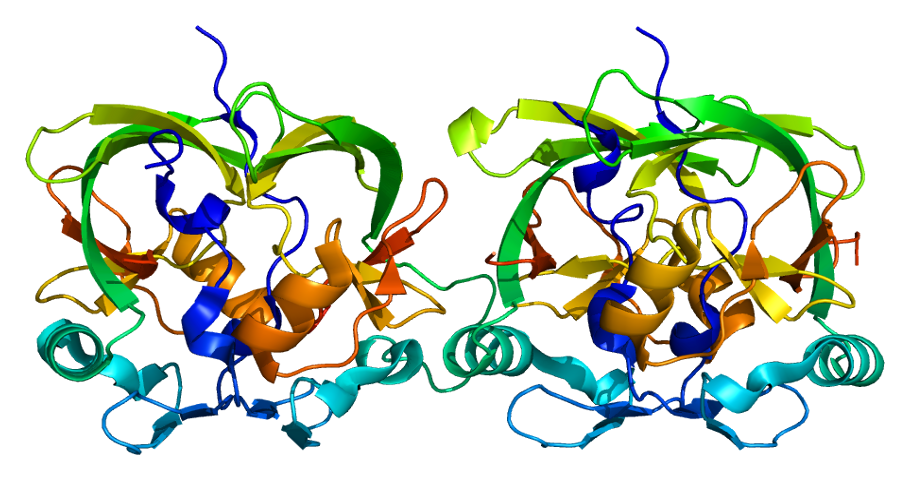
Spinocerebellar ataxia 10
Spinocerebellar ataxia is a clinically and genetically heterogeneous group of cerebellar disorders. Patients show progressive incoordination of gait and often poor coordination of hands, speech and eye movements, due to degeneration of the cerebellum with variable involvement of the brainstem and spinal cord. SCA10 is an autosomal dominant cerebellar ataxia (ADCA).
Transcriptional corepressor. Recruits NR2E1 to repress transcription. Promotes vascular smooth cell (VSMC) migration and orientation (By similarity). Corepressor of MTG8 transcriptional repression. Has some intrinsic repression activity which is independent of the number of poly-Gln (polyQ) repeats
NucleusCytoplasm, perinuclear regionCell junction
Dentatorubral-pallidoluysian atrophy
Autosomal dominant neurodegenerative disorder characterized by a loss of neurons in the dentate nucleus, rubrum, glogus pallidus and Luys'body. Clinical features are myoclonus epilepsy, dementia, and cerebellar ataxia. Onset of the disease occurs usually in the second decade of life and death in the fourth.
Medicamentos aprovados (FDA)
1 medicamento encontrado nos registros da FDA americana.
Variantes genéticas (ClinVar)
178 variantes patogênicas registradas no ClinVar.
Classificação de variantes (ClinVar)
Distribuição de 454 variantes classificadas pelo ClinVar.
Vias biológicas (Reactome)
1 via biológica associada aos genes desta condição.
Diagnóstico
Os sinais que médicos procuram e os exames que confirmam
Tratamento e manejo
Remédios, cuidados de apoio e o que precisa acompanhar
Onde tratar no SUS
Hospitais de referência no Brasil e o protocolo oficial do SUS (PCDT)
🇧🇷 Atendimento SUS — Ataxia cerebelosa autossômica dominante tipo IV
Selecione um estado ou use sua localização para ver resultados.
Dados de DATASUS/CNES, SBGM, ABNeuro e Ministério da Saúde. Sempre confirme a disponibilidade diretamente com o estabelecimento.
Pesquisa ativa
Ensaios clínicos abertos e novidades científicas recentes
Ensaios em destaque
🟢 Recrutando agora
1 pesquisa recrutando participantes. Converse com seu médico sobre a possibilidade de participar.
Outros ensaios clínicos
Publicações mais relevantes
Mostrando amostra de 6 publicações de um total de 85
Cerebellar Ataxia-deafness-narcolepsy (ADCA) syndrome. Description of a variable family phenotype.
Cerebellar ataxia-deafness-narcolepsy syndrome (ADCA-DN) is a very rare polymorphic subtype of autosomal dominant ataxia type 1 (ADCA type 1). It begins in adulthood, and may also be associated with other variable symptoms as optic atrophy, cataracts, psychosis, depression or sensory neuropathy. We present a family diagnosed from the first generation, its phenotypic description and genetic study. We describe three members of a family with ADCA sydrome. Patient II-2 started at 51 years, with ataxia, tremor, epileptic seizures, cerebellar atrophy, narcolepsy without cataplexy, hearing loss, moderate cognitive impairment. Patient III-1 started at 42 years with narcolepsy with cataplexy, hearing loss and tremor, and patient IV-1 started at 4 years, with mild intellectual disability, and narcolepsy without cataplexy. Genetic test showed a mutation in DNMT1 gene: variant c.1709 C > T; p.Ala570Val in the DNMT1 gene. ADCA syndrome has a variable phenotype in a same family. In our experience, this type of ataxia develops narcolepsy as the first sympton in all three cases, with tremor and cognitive impairment, together with tremor and cognitive impairment. Ataxia, despite being a cardinal symptom, does not appear at the onset in younger patients, and hearing loss also seems to develop over the years. Sensory neuropathy is not present in any of the cases studied.
Brain MRI Volumetry Analysis in an Indonesian Family of SCA 3 Patients: A Case-Based Study.
Spinocerebellar ataxia type-3 (SCA3) is an adult-onset autosomal dominant neurodegenerative disease. It is caused by expanding of CAG repeat in ATXN3 gene that later on would affect brain structures. This brain changes could be evaluated using brain MRI volumetric. However, findings across published brain volumetric studies have been inconsistent. Here, we report MRI brain volumetric analysis in a family of SCA 3 patients, which included pre-symptomatic and symptomatic patients. The study included affected and unaffected members from a large six-generation family of SCA 3, genetically confirmed using PolyQ/CAG repeat expansion analysis, Sanger sequencing, and PCR. Clinical evaluation was performed using Scale for the Assessment and Rating of Ataxia (SARA). Subjects' brains were scanned using 3.0-T MRI with a 3D T1 BRAVO sequence. Evaluations were performed by 2 independent neuroradiologists. An automated volumetric analysis was performed using FreeSurfer and CERES (for the cerebellum). We evaluated 7 subjects from this SCA3 family, including 3 subjects with SCA3 and 4 unaffected subjects. The volumetric evaluation revealed smaller brain volumes (p < 0.05) in the corpus callosum, cerebellar volume of lobules I-II, lobule IV, lobule VIIB and lobule IX; and in cerebellar gray matter volume of lobule IV, and VIIIA; in the pathologic/expanded CAG repeat group (SCA3). Brain MRI volumetry of SCA3 subjects showed smaller brain volumes in multiple brain regions including the corpus callosum and gray matter volumes of several cerebellar lobules.
Long-term efficacy of bilateral subthalamic deep brain stimulation in the parkinsonism of SCA 3: A rare case report.
Spinocerebellar ataxia type 3 (SCA3) is an autosomal dominant inherited disorder that manifests as a mixture of cerebellar ataxia, parkinsonism, and polyneuropathy; in type IV SCA3, pure parkinsonism is the only symptom. Currently, no disease-modifying treatment is available, but variable responses to antiparkinsonism agents have been reported. However, the benefits of deep brain stimulation (DBS) for treating parkinsonism in this subtype of SCA3 remain unclear. A 39-year-old male patient with a rare disorder of type IV SCA3 presented with pure parkinsonism including unilateral resting tremor, rigidity, and bradykinesia at the age of 30 years. Young-onset Parkinson disease was diagnosed at the age of 32 years. His family history revealed a mild ataxia in his father since the age of 55 years. Genetic testing confirmed an expanded CAG repeated number, with 66 in this case and 63 in his father for SCA3 mutation. Excellent response to levodopa and dopamine agonists in the first 3 years was noted, but wearing-off phenomena, levodopa-induced dyskinesia, and severe impulse control disorders later developed. To alleviate drug-induced complications, he received bilateral subthalamic nucleus deep brain stimulation (STN-DBS) in the absence of cerebellar signs, depression, and cognitive impairment. As of 2019, no impulsive control disorders, motor fluctuations, or DBS-related complications were observed during a 4-year follow-up, with 66% Unified Parkinson's Disease Rating Scale Part III reduction at medication OFF state noted, whereas levodopa equivalent daily dosage decreased by almost half. STN-DBS may be considered as adjunct treatment for severe dopa-related motor/nonmotor complications in patients with parkinsonian phenotype of SCA 3.
The cerebellar topography of attention sub-components in spinocerebellar ataxia type 2.
Spinocerebellar ataxia type 2 (SCA2) is an autosomal dominant neurodegenerative disease characterized by a progressive cerebellar syndrome and multiple-domain cognitive impairments. The cerebellum is known to contribute to distinct functional networks related to higher-level functions. The aims of the present study were to investigate the different sub-components of attention and to analyse possible correlations between attention deficits and specific cerebellar regions in SCA2 patients. To this purpose, 11 SCA2 patients underwent an exhaustive attention battery that evaluated several attention sub-components. The SCA2 group performed below the normal range in tasks assessing selective attention, divided attention, and sustained attention, obtaining negative Z-scores. These results were confirmed by non-parametric Mann-Whitney U tests that showed significant differences between SCA2 and control subjects in the same sub-components of the attention battery, allowing us to speculate on cerebellar involvement when a high cognitive demand is required (i.e., multisensory integration, sequencing, prediction of events, and inhibition of inappropriate response behaviours). The voxel-based morphometry analysis showed a pattern of significantly reduced grey matter volume in specific cerebellar lobules. In particular, the SCA2 patients showed significant grey matter loss in bilateral regions of the anterior cerebellar hemisphere (IV) and in the posterior lobe (VI-IX) and posterior vermis (VI-IX). Statistical analysis found significant correlations between grey matter reductions in the VIIb/VIIIa cerebellar lobules and impairments in Sustained and Divided Attention tasks and between grey matter reduction in the vermal VI lobule and impairment in the Go/NoGo task. For the first time, the study demonstrated the involvement of specific cerebellar lobules in different sub-components of the attention domain, giving further support to the inclusion of the cerebellum within the attention network.
A mutation in the low voltage-gated calcium channel CACNA1G alters the physiological properties of the channel, causing spinocerebellar ataxia.
Spinocerebellar ataxia (SCA) is a genetically heterogeneous disease. To date, 36 dominantly inherited loci have been reported, and 31 causative genes have been identified. In this study, we analyzed a Japanese family with autosomal dominant SCA using linkage analysis and exome sequencing, and identified CACNA1G, which encodes the calcium channel CaV3.1, as a new causative gene. The same mutation was also found in another family with SCA. Although most patients exhibited the pure form of cerebellar ataxia, two patients showed prominent resting tremor in addition to ataxia. CaV3.1 is classified as a low-threshold voltage-dependent calcium channel (T-type) and is expressed abundantly in the central nervous system, including the cerebellum. The mutation p.Arg1715His, identified in this study, was found to be located at S4 of repeat IV, the voltage sensor of the CaV3.1. Electrophysiological analyses revealed that the membrane potential dependency of the mutant CaV3.1 transfected into HEK293T cells shifted toward a positive potential. We established induced pluripotent stem cells (iPSCs) from fibroblasts of the patient, and to our knowledge, this is the first report of successful differentiation from the patient-derived iPSCs into Purkinje cells. There was no significant difference in the differentiation status between control- and patient-derived iPSCs. To date, several channel genes have been reported as causative genes for SCA. Our findings provide important insights into the pathogenesis of SCA as a channelopathy.
Publicações recentes
Expanding the Genetic Landscape of ATXN2 Variants: Insights From a Biallelic Trinucleotide Repeat Expansion in an Acadian Family.
A case report of spinocerebellar ataxia with TRPC3 gene mutation and review of literature.
Clinical, Genetic, and Imaging Characteristics of SCA27B: Insights from a Large Dutch Cohort.
An R83W mutation in Rab3A causes autosomal-dominant cerebellar ataxia.
Continuous visual stimulus tracking to quantify eye motility in spinocerebellar ataxia type 3.
📚 EuropePMC117 artigos no totalmostrando 6
Cerebellar Ataxia-deafness-narcolepsy (ADCA) syndrome. Description of a variable family phenotype.
Acta neurologica BelgicaBrain MRI Volumetry Analysis in an Indonesian Family of SCA 3 Patients: A Case-Based Study.
Frontiers in neurologyLong-term efficacy of bilateral subthalamic deep brain stimulation in the parkinsonism of SCA 3: A rare case report.
European journal of neurologyThe cerebellar topography of attention sub-components in spinocerebellar ataxia type 2.
Cortex; a journal devoted to the study of the nervous system and behaviorA mutation in the low voltage-gated calcium channel CACNA1G alters the physiological properties of the channel, causing spinocerebellar ataxia.
Molecular brainNovel phenotype associated with a mutation in the KCNA1(Kv1.1) gene.
Frontiers in physiologyAssociações
Organizações que acompanham esta doença — pra ter apoio e orientação
Ainda não temos associações cadastradas para Ataxia cerebelosa autossômica dominante tipo IV.
É de uma associação que acompanha esta doença? Fale com a gente →
Comunidades
Grupos ativos de quem convive com esta doença aqui no Raras
Ainda não existe comunidade no Raras para Ataxia cerebelosa autossômica dominante tipo IV
Pacientes, familiares e cuidadores se organizam em comunidades pra compartilhar experiências, fazer perguntas e se apoiar. Você pode ser o primeiro.
Tire suas dúvidas
Perguntas, dicas e experiências compartilhadas aqui na página
Participe da discussão
Faça login para postar dúvidas, compartilhar experiências e interagir com especialistas.
Fazer loginDoenças relacionadas
Doenças com sintomas parecidos — ajudam quem ainda está buscando diagnóstico
Referências e fontes
Bases de dados externas citadas neste artigo
Publicações científicas
Artigos indexados no PubMed ligados a esta doença no grafo RarasNet — título, periódico e PMID direto da fonte, sem intermediação de IA.
- Cerebellar Ataxia-deafness-narcolepsy (ADCA) syndrome. Description of a variable family phenotype.
- Brain MRI Volumetry Analysis in an Indonesian Family of SCA 3 Patients: A Case-Based Study.
- Long-term efficacy of bilateral subthalamic deep brain stimulation in the parkinsonism of SCA 3: A rare case report.
- The cerebellar topography of attention sub-components in spinocerebellar ataxia type 2.Cortex; a journal devoted to the study of the nervous system and behavior· 2018· PMID 30121445mais citado
- A mutation in the low voltage-gated calcium channel CACNA1G alters the physiological properties of the channel, causing spinocerebellar ataxia.
- Expanding the Genetic Landscape of ATXN2 Variants: Insights From a Biallelic Trinucleotide Repeat Expansion in an Acadian Family.
- A case report of spinocerebellar ataxia with TRPC3 gene mutation and review of literature.
- Clinical, Genetic, and Imaging Characteristics of SCA27B: Insights from a Large Dutch Cohort.
- An R83W mutation in Rab3A causes autosomal-dominant cerebellar ataxia.
- Continuous visual stimulus tracking to quantify eye motility in spinocerebellar ataxia type 3.
Bases de dados e fontes oficiais
Identificadores e referências canônicas usadas para montar este verbete.
- ORPHA:94149(Orphanet)
- MONDO:0019794(MONDO)
- GARD:19254(GARD (NIH))
- Variantes catalogadas(ClinVar)
- Busca completa no PubMed(PubMed)
- Q55346089(Wikidata)
Dados compilados pelo RarasNet a partir de fontes abertas (Orphanet, OMIM, MONDO, PubMed/EuropePMC, ClinicalTrials.gov, DATASUS, PCDT/MS). Este conteúdo é informativo e não substitui avaliação médica.
Conteúdo mantido por Agente Raras · Médicos e pesquisadores podem colaborar
Ataxia cerebelosa autossômica dominante tipo IV
📋 Origem dos dados
Esta página agrega dados de fontes públicas e oficiais. Dados sobre cobertura no SUS (PCDT, CEAF) são verificados ativamente por agente proativo (ver badge no infobox). Demais dados têm atribuição de fonte + data da última sincronização — clique para abrir o original.
- Doença rara (ontologia)
- fonte: Orphanet
- Identificador unificado
- fonte: MONDO
- NIH/GARD
- fonte: GARD (NIH)
- Dado público estruturado
- fonte: Wikidata
- Medicamentos aprovados FDA
- fonte: FDA OpenFDA