Introdução
O que você precisa saber de cara
Visão geral
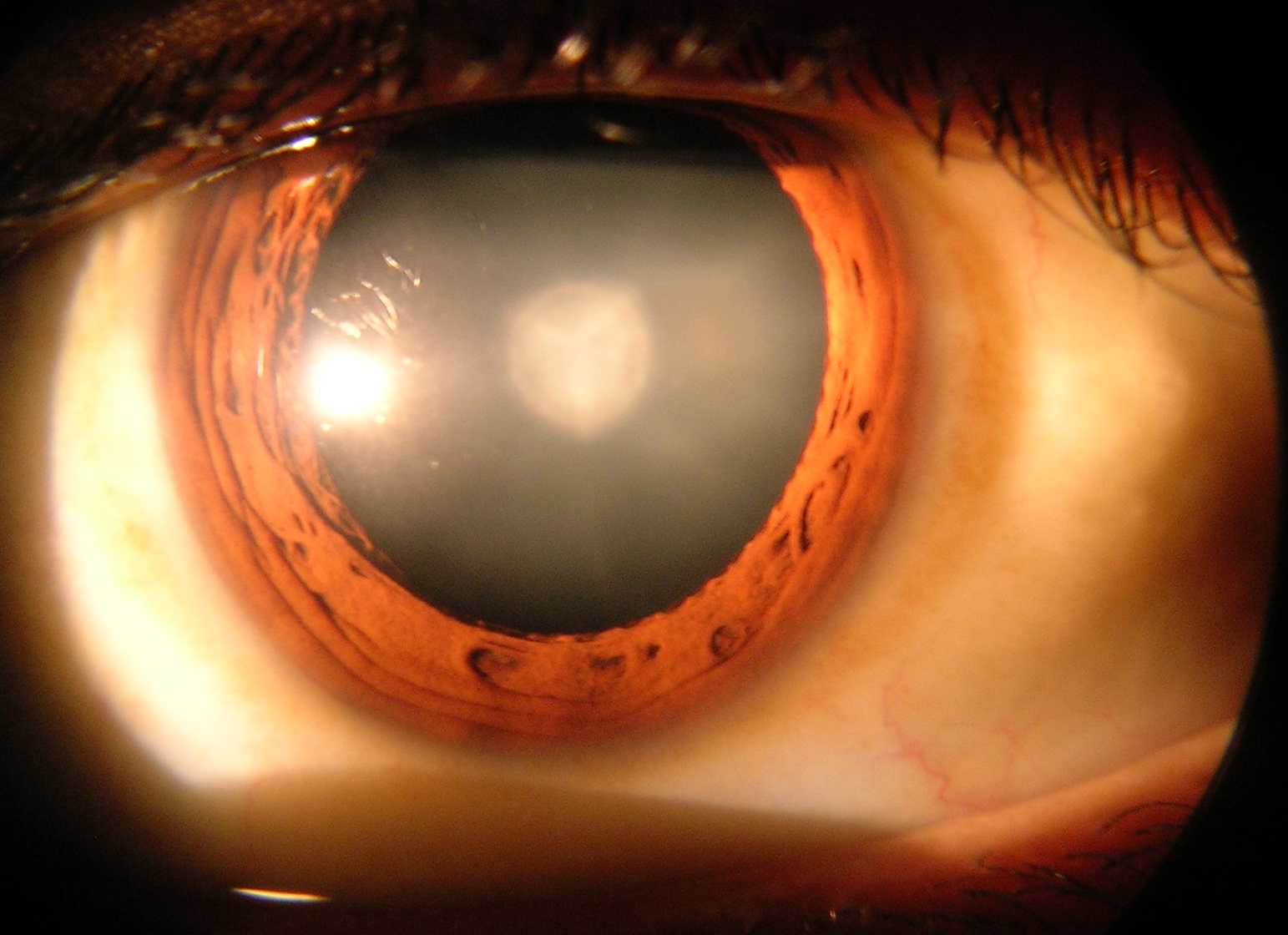
A catarata nuclear de início precoce é uma condição rara que afeta a transparência do cristalino do olho, especificamente na região central (núcleo), e se manifesta já nos primeiros dias ou meses de vida (período neonatal ou na infância). A opacidade do cristalino prejudica a passagem da luz até a retina, podendo comprometer o desenvolvimento visual se não for tratada adequadamente.[1][3]
Sinais e sintomas
O principal sinal é a presença de catarata nuclear, ou seja, uma opacidade localizada no centro do cristalino. Em bebês, isso pode ser percebido como um reflexo esbranquiçado na pupila (leucocoria) ou dificuldade para fixar o olhar em objetos. A catarata pode ser unilateral ou bilateral e, se não for corrigida precocemente, pode levar à ambliopia (olho preguiçoso).[1][3]
Causas genéticas
A catarata nuclear de início precoce é causada por alterações (variantes patogênicas) em genes que codificam proteínas essenciais para a estrutura e a transparência do cristalino. Os genes já associados a esta condição incluem: NHS, CRYBA2, UNC45B, CRYBB1, FYCO1, GJA8, CRYBB3, CRYGD, GJA3, CRYAB, WFS1 e CRYAA. Muitos desses genes produzem cristalinas (proteínas que mantêm o cristalino transparente) ou proteínas de comunicação entre as células do cristalino.[1][4]
Diagnóstico
O diagnóstico é feito por um oftalmologista por meio do exame de fundo de olho com lâmpada de fenda, que permite visualizar a opacidade nuclear. Para confirmar a causa genética, podem ser solicitados exames como cariótipo (bandas G, Q ou R), pesquisa de microdeleções/microduplicações por FISH, sequenciamento completo do exoma (WES) e dosagem de alfa-fetoproteína. Atualmente, existem 336 testes genéticos disponíveis para esta condição e 154 variantes patogênicas registradas no ClinVar.[1][4]
Tratamento e manejo
O tratamento principal é cirúrgico: a remoção do cristalino opaco (facoemulsificação ou facectomia) e, em crianças, a correção da visão com lentes de contato ou óculos após a cirurgia. O acompanhamento precoce é essencial para prevenir a ambliopia. No Brasil, o Sistema Único de Saúde (SUS) oferece cobertura mínima para esta condição, incluindo procedimentos como atendimento em reabilitação para doenças raras e os exames genéticos mencionados no diagnóstico.[1]
Prognóstico e qualidade de vida
Quando diagnosticada e tratada precocemente, a catarata nuclear de início precoce tem bom prognóstico visual. Atrasos no tratamento podem resultar em perda permanente da visão (ambliopia). O suporte de uma equipe multidisciplinar (oftalmologista, geneticista, terapeuta ocupacional) é importante para o desenvolvimento global da criança.[1]
Conteúdo informativo gerado e mantido automaticamente a partir de fontes oficiais (Orphanet, HPO, OMIM, SUS). Não substitui avaliação médica.
Catarata nuclear de início precoce é uma opacificação do cristalino que afeta a visão central desde a infância ou adolescência. Pode ser causada por mutações em genes como EPHA2, MIP, CRYGC, CRYBB2 e BFSP1.
Encontrou um erro ou informação desatualizada? Sugira uma correção →
Entender a doença
Do básico ao detalhe, leia no seu ritmo
Preparando trilha educativa...
Sinais e sintomas
O que aparece no corpo e com que frequência cada sintoma acontece
Visão geral
A catarata nuclear de início precoce é uma condição rara que afeta a transparência do cristalino do olho, especificamente na região central (núcleo), e se manifesta já nos primeiros dias ou meses de vida (período neonatal ou na infância). A opacidade do cristalino prejudica a passagem da luz até a retina, podendo comprometer o desenvolvimento visual se não for tratada adequadamente.[1][3]
Sinais e sintomas
O principal sinal é a presença de catarata nuclear, ou seja, uma opacidade localizada no centro do cristalino. Em bebês, isso pode ser percebido como um reflexo esbranquiçado na pupila (leucocoria) ou dificuldade para fixar o olhar em objetos. A catarata pode ser unilateral ou bilateral e, se não for corrigida precocemente, pode levar à ambliopia (olho preguiçoso).[1][3]
Causas genéticas
A catarata nuclear de início precoce é causada por alterações (variantes patogênicas) em genes que codificam proteínas essenciais para a estrutura e a transparência do cristalino. Os genes já associados a esta condição incluem: NHS, CRYBA2, UNC45B, CRYBB1, FYCO1, GJA8, CRYBB3, CRYGD, GJA3, CRYAB, WFS1 e CRYAA. Muitos desses genes produzem cristalinas (proteínas que mantêm o cristalino transparente) ou proteínas de comunicação entre as células do cristalino.[1][4]
Diagnóstico
O diagnóstico é feito por um oftalmologista por meio do exame de fundo de olho com lâmpada de fenda, que permite visualizar a opacidade nuclear. Para confirmar a causa genética, podem ser solicitados exames como cariótipo (bandas G, Q ou R), pesquisa de microdeleções/microduplicações por FISH, sequenciamento completo do exoma (WES) e dosagem de alfa-fetoproteína. Atualmente, existem 336 testes genéticos disponíveis para esta condição e 154 variantes patogênicas registradas no ClinVar.[1][4]
Tratamento e manejo
O tratamento principal é cirúrgico: a remoção do cristalino opaco (facoemulsificação ou facectomia) e, em crianças, a correção da visão com lentes de contato ou óculos após a cirurgia. O acompanhamento precoce é essencial para prevenir a ambliopia. No Brasil, o Sistema Único de Saúde (SUS) oferece cobertura mínima para esta condição, incluindo procedimentos como atendimento em reabilitação para doenças raras e os exames genéticos mencionados no diagnóstico.[1]
Prognóstico e qualidade de vida
Quando diagnosticada e tratada precocemente, a catarata nuclear de início precoce tem bom prognóstico visual. Atrasos no tratamento podem resultar em perda permanente da visão (ambliopia). O suporte de uma equipe multidisciplinar (oftalmologista, geneticista, terapeuta ocupacional) é importante para o desenvolvimento global da criança.[1]
Conteúdo informativo gerado e mantido automaticamente a partir de fontes oficiais (Orphanet, HPO, OMIM, SUS). Não substitui avaliação médica.
Características mais comuns
Os sintomas variam de pessoa para pessoa. Abaixo estão as 2 características clínicas mais associadas, ordenadas por frequência.
Linha do tempo da pesquisa
Encontrou um erro ou informação desatualizada? Sugira uma correção →
Genética e causas
O que está alterado no DNA e como passa nas famílias
Genes associados
18 genes identificados com associação a esta condição.
May function in cell morphology by maintaining the integrity of the circumferential actin ring and controlling lamellipod formation. Involved in the regulation eye, tooth, brain and craniofacial development
Apical cell membraneCell projection, lamellipodiumCell junction, tight junctionCell junction, focal adhesionCytoplasm
Nance-Horan syndrome
Rare X-linked disorder characterized by congenital cataracts, dental anomalies, dysmorphic features, and, in some cases, intellectual disability. Distinctive dental anomalies are seen in affected males, including supernumerary incisors and crown shaped permanent teeth. Characteristic facial features are anteverted pinnae, long face, and prominent nasal bridge and nose. Carrier females display milder variable symptoms of disease with lens opacities often involving the posterior Y sutures, and on occasion dental anomalies and the characteristic facial features described.
Crystallins are the dominant structural components of the vertebrate eye lens
Cataract 42
An opacification of the crystalline lens of the eye that frequently results in visual impairment or blindness. Opacities vary in morphology, are often confined to a portion of the lens, and may be static or progressive. In general, the more posteriorly located and dense an opacity, the greater the impact on visual function.
Acts as a co-chaperone for HSP90 and is required for proper folding of the myosin motor domain. Plays a role in sarcomere formation during muscle cell development. Is necessary for normal early lens development
Cytoplasm, myofibril, sarcomere, Z lineCytoplasm, myofibril, sarcomere, A bandCytoplasm, perinuclear regionCytoplasm, cytosol
Cataract 43
An opacification of the crystalline lens of the eye that frequently results in visual impairment or blindness. Opacities vary in morphology, are often confined to a portion of the lens, and may be static or progressive. In general, the more posteriorly located and dense an opacity, the greater the impact on visual function.
Crystallins are the dominant structural components of the vertebrate eye lens
Cataract 17, multiple types
An opacification of the crystalline lens of the eye that frequently results in visual impairment or blindness. Opacities vary in morphology, are often confined to a portion of the lens, and may be static or progressive. In general, the more posteriorly located and dense an opacity, the greater the impact on visual function. CTRCT17 includes nuclear and pulverulent cataracts, among others. Nuclear cataracts affect the central nucleus of the eye, are often not highly visually significant. The density of the opacities varies greatly from fine dots to a dense, white and chalk-like, central cataract. The condition is usually bilateral. Nuclear cataracts are often combined with opacified cortical fibers encircling the nuclear opacity, which are referred to as cortical riders. Pulverulent cataracts are characterized by a dust-like, 'pulverised' appearance of the opacities which can be found in any part of the lens.
May mediate microtubule plus end-directed vesicle transport
Cytoplasmic vesicle, autophagosomeEndosomeLysosome
Cataract 18
An opacification of the crystalline lens of the eye becoming evident at birth or in infancy. It frequently results in visual impairment or blindness. Opacities vary in morphology, are often confined to a portion of the lens, and may be static or progressive. In general, the more posteriorly located and dense an opacity, the greater the impact on visual function.
Structural component of eye lens gap junctions (PubMed:18006672, PubMed:19756179). Gap junctions are dodecameric channels that connect the cytoplasm of adjoining cells. They are formed by the docking of two hexameric hemichannels, one from each cell membrane (By similarity). Small molecules and ions diffuse from one cell to a neighboring cell via the central pore (PubMed:18006672, PubMed:19756179)
Cell membraneCell junction, gap junction
Cataract 1, multiple types
An opacification of the crystalline lens of the eye that frequently results in visual impairment or blindness. Opacities vary in morphology, are often confined to a portion of the lens, and may be static or progressive. CTRCT1 includes congenital, zonular pulverulent, nuclear progressive, nuclear pulverulent, nuclear total, total, and posterior subcapsular types of cataract. Zonular or lamellar cataracts are opacities, broad or narrow, usually consisting of powdery white dots affecting only certain layers or zones between the cortex and nucleus of an otherwise clear lens. The opacity may be so dense as to render the entire central region of the lens completely opaque, or so translucent that vision is hardly if at all impeded. Zonular cataracts generally do not involve the embryonic nucleus, though sometimes they involve the fetal nucleus. Usually sharply separated from a clear cortex outside them, they may have projections from their outer edges known as riders or spokes. In some cases cataract is associated with microcornea without any other systemic anomaly or dysmorphism. Microcornea is defined by a corneal diameter inferior to 10 mm in both meridians in an otherwise normal eye.
Crystallins are the dominant structural components of the vertebrate eye lens
Cataract 22, multiple types
An opacification of the crystalline lens of the eye that frequently results in visual impairment or blindness. Opacities vary in morphology, are often confined to a portion of the lens, and may be static or progressive. In general, the more posteriorly located and dense an opacity, the greater the impact on visual function. CTRCT22 includes nuclear cataract among others. Nuclear cataracts affect the central nucleus of the eye, and are often not highly visually significant. The density of the opacities varies greatly from fine dots to a dense, white and chalk-like, central cataract. The condition is usually bilateral. Nuclear cataracts are often combined with opacified cortical fibers encircling the nuclear opacity, which are referred to as cortical riders.
Crystallins are the dominant structural components of the vertebrate eye lens
Cataract 4, multiple types
An opacification of the crystalline lens of the eye that frequently results in visual impairment or blindness. Opacities vary in morphology, are often confined to a portion of the lens, and may be static or progressive. CTRCT4 includes crystalline aculeiform, congenital cerulean and non-nuclear polymorphic cataracts, among others. Crystalline aculeiform cataract is characterized by fiberglass-like or needle-like crystals projecting in different directions, through or close to the axial region of the lens. Non-nuclear polymorphic cataract is a partial opacity with variable location between the fetal nucleus of the lens and the equator. The fetal nucleus is normal. The opacities are irregular and look similar to a bunch of grapes and may be present simultaneously in different lens layers. Congenital cerulean cataract is characterized by peripheral bluish and white opacifications organized in concentric layers with occasional central lesions arranged radially. The opacities are observed in the superficial layers of the fetal nucleus as well as the adult nucleus of the lens. Involvement is usually bilateral. Visual acuity is only mildly reduced in childhood. In adulthood, the opacifications may progress, making lens extraction necessary. Histologically the lesions are described as fusiform cavities between lens fibers which contain a deeply staining granular material. Although the lesions may take on various colors, a dull blue is the most common appearance and is responsible for the designation cerulean cataract.
Structural component of lens fiber gap junctions (PubMed:30044662). Gap junctions are dodecameric channels that connect the cytoplasm of adjoining cells (By similarity). They are formed by the docking of two hexameric hemichannels, one from each cell membrane. Small molecules and ions diffuse from one cell to a neighboring cell via the central pore (PubMed:30044662)
Cell membraneCell junction, gap junction
Cataract 14, multiple types
An opacification of the crystalline lens of the eye that frequently results in visual impairment or blindness. Opacities vary in morphology, are often confined to a portion of the lens, and may be static or progressive. CTRCT14 includes zonular pulverulent cataract, among others. Zonular or lamellar cataracts are opacities, broad or narrow, usually consisting of powdery white dots affecting only certain layers or zones between the cortex and nucleus of an otherwise clear lens. The opacity may be so dense as to render the entire central region of the lens completely opaque, or so translucent that vision is hardly if at all impeded. Usually sharply separated from a clear cortex outside them, they may have projections from their outer edges known as riders or spokes.
May contribute to the transparency and refractive index of the lens. Has chaperone-like activity, preventing aggregation of various proteins under a wide range of stress conditions. In lens epithelial cells, stabilizes the ATP6V1A protein, preventing its degradation by the proteasome (By similarity)
CytoplasmNucleusSecretedLysosome
Myopathy, myofibrillar, 2A, adult-onset
A form of myofibrillar myopathy, a group of chronic neuromuscular disorders characterized at ultrastructural level by disintegration of the sarcomeric Z disk and myofibrils, and replacement of the normal myofibrillar markings by small dense granules, or larger hyaline masses, or amorphous material. MFM2A is an autosomal dominant form characterized by weakness of the proximal and distal limb muscles, weakness of the neck, velopharynx and trunk muscles, respiratory insufficiency, hypertrophic cardiomyopathy, and cataract.
Participates in the regulation of cellular Ca(2+) homeostasis, at least partly, by modulating the filling state of the endoplasmic reticulum Ca(2+) store (PubMed:16989814). Negatively regulates the ER stress response and positively regulates the stability of V-ATPase subunits ATP6V1A and ATP1B1 by preventing their degradation through an unknown proteasome-independent mechanism (PubMed:23035048)
Endoplasmic reticulum membraneCytoplasmic vesicle, secretory vesicle
Wolfram syndrome 1
A rare disorder characterized by juvenile-onset insulin-dependent diabetes mellitus with optic atrophy. Other manifestations include diabetes insipidus, sensorineural deafness, dementia, psychiatric illnesses.
Contributes to the transparency and refractive index of the lens (PubMed:18302245). In its oxidized form (absence of intramolecular disulfide bond), acts as a chaperone, preventing aggregation of various proteins under a wide range of stress conditions (PubMed:18199971, PubMed:19595763, PubMed:22120592, PubMed:31792453). Required for the correct formation of lens intermediate filaments as part of a complex composed of BFSP1, BFSP2 and CRYAA (PubMed:28935373)
CytoplasmNucleus
Crystallins are the dominant structural components of the vertebrate eye lens
Cataract 10, multiple types
An opacification of the crystalline lens of the eye that frequently results in visual impairment or blindness. Opacities vary in morphology, are often confined to a portion of the lens, and may be static or progressive. CTRCT10 includes congenital zonular with sutural opacities, among others. This is a form of zonular cataract with an erect Y-shaped anterior and an inverted Y-shaped posterior sutural opacities. Zonular or lamellar cataracts are opacities, broad or narrow, usually consisting of powdery white dots affecting only certain layers or zones between the cortex and nucleus of an otherwise clear lens. The opacity may be so dense as to render the entire central region of the lens completely opaque, or so translucent that vision is hardly if at all impeded. Zonular cataracts generally do not involve the embryonic nucleus, though sometimes they involve the fetal nucleus. Usually sharply separated from a clear cortex outside them, they may have projections from their outer edges known as riders or spokes.
Required for the correct formation of lens intermediate filaments as part of a complex composed of BFSP1, BFSP2 and CRYAA (PubMed:28935373). Involved in altering the calcium regulation of MIP water permeability (PubMed:30790544)
Cell membraneCytoplasmCytoplasm, cytoskeletonCytoplasm, cell cortex
Cataract 33, multiple types
An opacification of the crystalline lens of the eye that frequently results in visual impairment or blindness. Opacities vary in morphology, are often confined to a portion of the lens, and may be static or progressive. In general, the more posteriorly located and dense an opacity, the greater the impact on visual function. CTRCT33 has juvenile-onset and the opacities are restricted to the cortex of the lens, not involving the nucleus.
Crystallins are the dominant structural components of the vertebrate eye lens
Cataract 3, multiple types
An opacification of the crystalline lens of the eye that frequently results in visual impairment or blindness. Opacities vary in morphology, are often confined to a portion of the lens, and may be static or progressive. CTRCT3 includes congenital cerulean and sutural cataract with punctate and cerulean opacities, among others. Cerulean cataract is characterized by peripheral bluish and white opacifications organized in concentric layers with occasional central lesions arranged radially. The opacities are observed in the superficial layers of the fetal nucleus as well as the adult nucleus of the lens. Involvement is usually bilateral. Visual acuity is only mildly reduced in childhood. In adulthood, the opacifications may progress, making lens extraction necessary. Histologically the lesions are described as fusiform cavities between lens fibers which contain a deeply staining granular material. Although the lesions may take on various colors, a dull blue is the most common appearance and is responsible for the designation cerulean cataract. Sutural cataract with punctate and cerulean opacities is characterized by white opacification around the anterior and posterior Y sutures, and grayish and bluish, spindle shaped, oval punctate and cerulean opacities of various sizes arranged in lamellar form. The spots are more concentrated towards the peripheral layers and do not delineate the embryonal or fetal nucleus. Phenotypic variation with respect to the size and density of the sutural opacities as well as the number and position of punctate and cerulean spots is observed among affected subjects.
Crystallins are the dominant structural components of the vertebrate eye lens
Cataract 2, multiple types
An opacification of the crystalline lens of the eye that frequently results in visual impairment or blindness. Opacities vary in morphology, are often confined to a portion of the lens, and may be static or progressive. CTRCT2 includes Coppock-like cataract, among others. Coppock-like cataract is a congenital pulverulent disk-like opacity involving the embryonic nucleus with many tiny white dots in the lamellar portion of the lens. It is usually bilateral and dominantly inherited. In some cases, CTRCT2 is associated with microcornea without any other systemic anomaly or dysmorphism. Microcornea is defined by a corneal diameter inferior to 10 mm in both meridians in an otherwise normal eye.
Aquaporins form homotetrameric transmembrane channels, with each monomer independently mediating water transport across the plasma membrane along its osmotic gradient (PubMed:11001937, PubMed:24120416). Specifically expressed in lens fiber cells, this aquaporin is crucial for maintaining lens water homeostasis and transparency. Beyond water permeability, it also acts as a cell-to-cell adhesion molecule, forming thin junctions between lens fiber cells that are essential for maintaining the ordere
Cell membraneCell junction
Cataract 15, multiple types
An opacification of the crystalline lens of the eye that frequently results in visual impairment or blindness. Opacities vary in morphology, are often confined to a portion of the lens, and may be static or progressive. CTRCT15 includes polymorphic, progressive punctate lamellar, cortical, anterior and posterior polar, nonprogressive lamellar with sutural opacities, embryonic nuclear, and pulverulent cortical, among others.
Receptor tyrosine kinase which binds promiscuously membrane-bound ephrin-A family ligands residing on adjacent cells, leading to contact-dependent bidirectional signaling into neighboring cells. The signaling pathway downstream of the receptor is referred to as forward signaling while the signaling pathway downstream of the ephrin ligand is referred to as reverse signaling. Activated by the ligand ephrin-A1/EFNA1 regulates migration, integrin-mediated adhesion, proliferation and differentiation
Cell membraneCell projection, ruffle membraneCell projection, lamellipodium membraneCell junction, focal adhesion
Cataract 6, multiple types
An opacification of the crystalline lens of the eye that frequently results in visual impairment or blindness. Opacities vary in morphology, are often confined to a portion of the lens, and may be static or progressive. CTRCT6 includes posterior polar and age-related cortical cataracts, among others. Posterior polar cataract is a subcapsular opacity, usually disk-shaped, located at the back of the lens. Age-related cortical cataract is a developmental punctate opacity restricted to the cortex. The cataract is white or cerulean, increases in number with age, but rarely affects vision.
Variantes genéticas (ClinVar)
154 variantes patogênicas registradas no ClinVar.
Vias biológicas (Reactome)
18 vias biológicas associadas aos genes desta condição.
Diagnóstico
Os sinais que médicos procuram e os exames que confirmam
Tratamento e manejo
Remédios, cuidados de apoio e o que precisa acompanhar
Onde tratar no SUS
Hospitais de referência no Brasil e o protocolo oficial do SUS (PCDT)
🇧🇷 Atendimento SUS — Catarata nuclear de início precoce
Selecione um estado ou use sua localização para ver resultados.
Dados de DATASUS/CNES, SBGM, ABNeuro e Ministério da Saúde. Sempre confirme a disponibilidade diretamente com o estabelecimento.
Pesquisa ativa
Ensaios clínicos abertos e novidades científicas recentes
Pesquisa e ensaios clínicos
Nenhum ensaio clínico registrado para esta condição.
Publicações mais relevantes
Senescence-Driven IL-17A Inflammatory Circuit Promotes Epithelial-Mesenchymal Transition (EMT) and Progression in Age-Related Posterior Subcapsular Cataracts.
Cataract is a leading cause of visual impairment worldwide, and its prevalence is increasing with population aging. The majority are age-related cataracts (ARC), clinically classified into nuclear, cortical, and posterior subcapsular cataracts (PSC) by opacity location. Mechanisms of cataractogenesis remain incompletely understood. While cortical and nuclear cataracts are largely attributed to crystallin aggregation, such protein-centric mechanisms fail to explain the early onset and axial location of PSC. Morphologically, PSC resembles posterior capsule opacification (PCO), a secondary cataract driven by epithelial-mesenchymal transition (EMT) of lens epithelial cells (LECs), suggesting LEC-EMT may also contribute to PSC. Using clinically stratified human lens samples, we confirmed EMT marker expression across PSC subtypes. Transcriptomic profiling revealed that LECs, the lens's sole metabolically active cells, in age-related PSC (ARC-PSC), compared with age-related nuclear cataract (ANC) and clear lenses, exhibit elevated EMT signatures tightly linked to senescence-associated inflammatory signaling. Aqueous humor (AqH) profiling demonstrated a pro-inflammatory milieu across PSC subtypes, with IL-17A uniquely elevated in ARC-PSC, consistent with transcriptomic findings. Integrated analyses support a model in which senescence functions as an upstream driver, whereby senescent LECs release SASP factors, including IL-17A, that activate NF-κB signaling to amplify inflammation, reinforce senescence, and drive EMT. In vitro, senolysis and IL-17A blockade disrupted this loop, attenuating senescence- and EMT-associated phenotypes. Collectively, our study demonstrates that senescent LECs sustain an IL-17A-NF-κB circuit that drives EMT and accelerates ARC-PSC progression, positioning ARC-PSC as a unique human context to study senescence-induced epithelial remodeling in aging tissues.
TGF-β1-induced m6A modifications accelerate onset of nuclear cataract in high myopia by modulating the PCP pathway.
High myopia is an important cause of visual impairment worldwide, characterized by early-onset nuclear cataracts, whose underlying mechanisms remain largely unexplained. Here, we identify conspicuously polarized and compacted lens fiber alignment, along with a simultaneous rise in N6-methyladenosine (m6A) modifications in patients with highly myopic cataracts (HMC), which is confirmed to be induced by elevated transforming growth factor-β1 (TGF-β1) in lens. Mechanistically, methyltransferase METTL3 and m6A reader insulin-like growth factor 2 mRNA binding protein 3 synergistically enhance planar cell polarity (PCP) signaling by affecting mRNA stability of dishevelled 2. This, in turn, alters proliferation, migration, and polarity formation of human lens epithelial cells. Moreover, Mettl3 conditional knockdown in mice leads to disrupted lens fiber arrangement and alleviates TGF-β1-induced increase in lens nuclear density. Collectively, these findings highlight the significance of m6A-modified PCP pathway in regulating postnatal lens fiber organization, which may hold great promise as a therapeutic target for HMC.
Experimental models of myopia development: A review of literature.
Myopia is one of the most prevalent refractive errors and one of the leading causes of visual impairment and blindness worldwide. It results from a mismatch between the axial length and optical power of the eye, resulting in a focal plane that lies in front of the retina. In children and young adults, myopia is most commonly caused by excessive elongation of the eyeball during development - a hallmark of school-age and some early-onset genetic forms of myopia. However, myopic refractive error can also result from other mechanisms, such as increased lens power in age-related nuclear cataracts or corneal steepening in keratoconus, which are not associated with axial elongation. The prevalence of myopia in young Asian adults has increased from 20-30% to 80-85% over the last 50 years. In contrast, recent meta-analytic data for European young adults, emphasizing studies with cycloplegic refraction essential for accuracy, indicate myopia prevalence rates of approximately 19-24%. The prevalence of high myopia (greater than or equal to-6.0 diopters) has increased disproportionately to myopia in the last 50 years, from 1-5% to 10-20% and became a global problem. The reason for this state of affairs is believed to be lifestyle and prolonged near vision activities. Although refractive error can be corrected, sight-threatening pathologies such as retinal detachment, macular degeneration, glaucoma, and cataracts are more challenging to control. Owing to years of research, the biological mechanisms of eye growth and refractive development are increasingly elucidated. The signaling cascade mechanisms that link the retinal image processing and alterations in choroidal thickness and scleral development have also been studied. While the retina can detect defocus and changes in defocus, decades of research have led to a growing understanding of the fundamental pathways in visually guided eye growth, yet the precise initial mechanisms by which the retina senses and transduces these optical signals continue to be an active and important area of investigation. Animal studies have demonstrated that the retina can locally regulate visually guided eye growth through intrinsic mechanisms, even in the absence of direct input from the brain. The precise molecular mechanisms underlying common forms of myopia, particularly those involving axial elongation, are yet to be fully elucidated. This reflects the complexity and multifactorial influences inherent even in these prevalent forms, alongside the challenges posed by experimental models in completely recapitulating all aspects of the human condition.
Should XYY syndrome be considered in the differential diagnosis of syndromic myopia? Apropos of a case.
XYY syndrome is relatively common and misdiagnosed. In addition high myopia appears to be more frequent in this syndrome than in the general population. Therefore, the objective of the present report is to emphasize the association of XYY syndrome and high myopia. Case report of a single patient. An 8-year-old male patient with a history of XYY syndrome was examined. Physical examination revealed tall stature, low-set auricular pavilion implantation, mid-face hypoplasia, horizontal palpebral fissures. Ophthalmological evaluation showed early-onset high myopia in both eyes and a complete nuclear cataract and chronic retinal detachment in the right eye. Patients with XYY syndrome appear to exhibit a high prevalence of myopia. Although the pathophysiology of myopia in patients with XYY syndrome is not yet fully understood, it should be considered as a differential diagnosis for syndromic myopia.
Pathobiology of the crystalline lens in Stickler syndrome.
The Stickler syndromes are a group of connective tissue disorders characterised by congenital myopia, giant retinal tear and retinal detachment, cleft palate, hearing loss and premature arthropathy. Patients with Stickler syndrome are also susceptible to abnormalities of the crystalline lens. Since neither type II or type XI collagen (those typically affected in the vast majority of Stickler patients) are highly expressed in the lens, this observational cohort study explores potential alternative mechanisms to explain why patients frequently exhibit such unusual but characteristic types of cataract. Author observations drawn from a cohort of over 1800 patients with genetically confirmed Stickler syndrome. 3 distinct lens pathologies were identified. Firstly, a congenital quadrantic lamellar opacity. This can be present in both type 1 (COL2A1) and type 2 (COL11A1) Stickler syndrome. Secondly, early onset Pantone 557 C blue-green nuclear cataract. Thirdly, congenital lens coloboma associated with localised zonule deficiency. The characteristic quadrantic lamellar lens opacity can be helpful in alerting to the possible diagnosis, particularly in sub-groups with an ocular-only phenotype. Temporal and spatial signalling pathways shared embryologically by both the developing vitreous body and crystalline lens suggest an ancillary role of the fibrillar collagens in cell signalling beyond their basic structural function. A common pathway of TGFβ/BMP super-family dysregulation may be shared with allied disorders associated with both retinal detachment and cataract as well as the pathobiology linking retinal detachment and cataract in the population at large. Congenital lens coloboma associated with localised zonule deficiency can increase the difficulty and risks of cataract surgery. Strategies to mitigate such risks are presented.
Publicações recentes
TGF-β1-induced m6A modifications accelerate onset of nuclear cataract in high myopia by modulating the PCP pathway.
Pathobiology of the crystalline lens in Stickler syndrome.
Association between single nucleotide polymorphisms in exon 3 of the alpha-A-crystallin gene and susceptibility to age-related cataract.
Racemization in cataractous lens from diabetic and aging individuals: analysis of Asp 58 residue in αA-crystallin.
Cataract formation in transgenic HO-1 G143H mutant mice: Involvement of oxidative stress and endoplasmic reticulum stress.
📚 EuropePMCmostrando 26
Senescence-Driven IL-17A Inflammatory Circuit Promotes Epithelial-Mesenchymal Transition (EMT) and Progression in Age-Related Posterior Subcapsular Cataracts.
Aging cellExperimental models of myopia development: A review of literature.
Journal of physiology and pharmacology : an official journal of the Polish Physiological SocietyShould XYY syndrome be considered in the differential diagnosis of syndromic myopia? Apropos of a case.
Archivos de la Sociedad Espanola de OftalmologiaTGF-β1-induced m6A modifications accelerate onset of nuclear cataract in high myopia by modulating the PCP pathway.
Nature communicationsPathobiology of the crystalline lens in Stickler syndrome.
Progress in retinal and eye researchImpairment of flicker-induced increase in retinal blood flow in diabetic pigs.
Japanese journal of ophthalmologyHereditary Hyperferritinemia-Cataract Syndrome in a Family With HFE-H63D Mutation.
CureusAssociation between single nucleotide polymorphisms in exon 3 of the alpha-A-crystallin gene and susceptibility to age-related cataract.
Ophthalmic geneticsWerner syndrome in a Lebanese family.
American journal of medical genetics. Part ARacemization in cataractous lens from diabetic and aging individuals: analysis of Asp 58 residue in αA-crystallin.
AgingCataract formation in transgenic HO-1 G143H mutant mice: Involvement of oxidative stress and endoplasmic reticulum stress.
Biochemical and biophysical research communicationsCataract in You-Hoover-Fong syndrome: TELO2 deficiency.
Ophthalmic geneticsHereditary cataract in the Bengal cat in Poland.
BMC veterinary researchIn-vivo anterior segment OCT imaging provides unique insight into cerulean blue-dot opacities and cataracts in Down syndrome.
Scientific reportsProteasome subunit PSMC3 variants cause neurosensory syndrome combining deafness and cataract due to proteotoxic stress.
EMBO molecular medicineThe Tudor-domain protein TDRD7, mutated in congenital cataract, controls the heat shock protein HSPB1 (HSP27) and lens fiber cell morphology.
Human molecular geneticsConformational dynamics study on human γS-crystallin as an efficient route to childhood blindness.
Biochemical and biophysical research communicationsCombined loss of LAP1B and LAP1C results in an early onset multisystemic nuclear envelopathy.
Nature communicationsDNA hypermethylation-mediated downregulation of antioxidant genes contributes to the early onset of cataracts in highly myopic eyes.
Redox biologyAntagonising Wnt/β-catenin signalling ameliorates lens-capsulotomy-induced retinal degeneration in a mouse model of diabetes.
DiabetologiaRacemization at the Asp 58 residue in αA-crystallin from the lens of high myopic cataract patients.
Journal of cellular and molecular medicineConcomitant Presence of Carcinoma of Breast in a Patient with Early-Onset Cataract: First Case Report of a Potential Association.
Journal of clinical and diagnostic research : JCDRA novel missense mutation of Mip causes semi-dominant cataracts in the Nat mouse.
Experimental animalsDJ-1 modulates mitochondrial response to oxidative stress: clues from a novel diagnosis of PARK7.
Clinical geneticsLEBER CONGENITAL AMAUROSIS WITH LARGE RETINAL PIGMENT CLUMPS CAUSED BY COMPOUND HETEROZYGOUS MUTATIONS IN KCNJ13.
Retinal cases & brief reportsHypoglycemic action of vitamin K1 protects against early-onset diabetic nephropathy in streptozotocin-induced rats.
Nutrition (Burbank, Los Angeles County, Calif.)Associações
Organizações que acompanham esta doença — pra ter apoio e orientação
Ainda não temos associações cadastradas para Catarata nuclear de início precoce.
É de uma associação que acompanha esta doença? Fale com a gente →
Comunidades
Grupos ativos de quem convive com esta doença aqui no Raras
Ainda não existe comunidade no Raras para Catarata nuclear de início precoce
Pacientes, familiares e cuidadores se organizam em comunidades pra compartilhar experiências, fazer perguntas e se apoiar. Você pode ser o primeiro.
Tire suas dúvidas
Perguntas, dicas e experiências compartilhadas aqui na página
Participe da discussão
Faça login para postar dúvidas, compartilhar experiências e interagir com especialistas.
Fazer loginDoenças relacionadas
Doenças com sintomas parecidos — ajudam quem ainda está buscando diagnóstico
Referências e fontes
Bases de dados externas citadas neste artigo
Publicações científicas
Artigos indexados no PubMed ligados a esta doença no grafo RarasNet — título, periódico e PMID direto da fonte, sem intermediação de IA.
- Senescence-Driven IL-17A Inflammatory Circuit Promotes Epithelial-Mesenchymal Transition (EMT) and Progression in Age-Related Posterior Subcapsular Cataracts.
- TGF-β1-induced m6A modifications accelerate onset of nuclear cataract in high myopia by modulating the PCP pathway.
- Experimental models of myopia development: A review of literature.Journal of physiology and pharmacology : an official journal of the Polish Physiological Society· 2025· PMID 41335529mais citado
- Should XYY syndrome be considered in the differential diagnosis of syndromic myopia? Apropos of a case.
- Pathobiology of the crystalline lens in Stickler syndrome.
- Association between single nucleotide polymorphisms in exon 3 of the alpha-A-crystallin gene and susceptibility to age-related cataract.
- Racemization in cataractous lens from diabetic and aging individuals: analysis of Asp 58 residue in αA-crystallin.
- Cataract formation in transgenic HO-1 G143H mutant mice: Involvement of oxidative stress and endoplasmic reticulum stress.
Bases de dados e fontes oficiais
Identificadores e referências canônicas usadas para montar este verbete.
- ORPHA:98991(Orphanet)
- MONDO:0020376(MONDO)
- GARD:16887(GARD (NIH))
- Variantes catalogadas(ClinVar)
- Busca completa no PubMed(PubMed)
- Q56014424(Wikidata)
Dados compilados pelo RarasNet a partir de fontes abertas (Orphanet, OMIM, MONDO, PubMed/EuropePMC, ClinicalTrials.gov, DATASUS, PCDT/MS). Este conteúdo é informativo e não substitui avaliação médica.
Conteúdo mantido por Agente Raras · Médicos e pesquisadores podem colaborar
Catarata nuclear de início precoce
📋 Origem dos dados
Esta página agrega dados de fontes públicas e oficiais. Dados sobre cobertura no SUS (PCDT, CEAF) são verificados ativamente por agente proativo (ver badge no infobox). Demais dados têm atribuição de fonte + data da última sincronização — clique para abrir o original.
- Doença rara (ontologia)
- fonte: Orphanet
- Identificador unificado
- fonte: MONDO
- Codificação WHO/SUS
- fonte: WHO ICD-10 / DATASUS
- CID-11 (futuro)
- fonte: WHO ICD-11
- NIH/GARD
- fonte: GARD (NIH)
- Indexação biomédica
- fonte: MeSH (NLM)
- Dado público estruturado
- fonte: Wikidata