O forame parietal aumentado (FPE) é um defeito de desenvolvimento, caracterizado por defeitos variáveis de ossificação intramembranosa dos ossos parietais, que pode ser assintomático, sintomático (dores de cabeça, náuseas, vômitos, deficiência intelectual) ou associado a outras patologias.
Introdução
O que você precisa saber de cara
O forame parietal aumentado (FPE) é um defeito de desenvolvimento, caracterizado por defeitos variáveis de ossificação intramembranosa dos ossos parietais, que pode ser assintomático, sintomático (dores de cabeça, náuseas, vômitos, deficiência intelectual) ou associado a outras patologias.
Escala de raridade
<1/50kMuito rara
1/20kRara
1/10kPouco freq.
1/5kIncomum
1/2k
Encontrou um erro ou informação desatualizada? Sugira uma correção →
Entender a doença
Do básico ao detalhe, leia no seu ritmo
Preparando trilha educativa...
Sinais e sintomas
O que aparece no corpo e com que frequência cada sintoma acontece
Partes do corpo afetadas
+ 10 sintomas em outras categorias
Características mais comuns
Os sintomas variam de pessoa para pessoa. Abaixo estão as 28 características clínicas mais associadas, ordenadas por frequência.
Linha do tempo da pesquisa
Encontrou um erro ou informação desatualizada? Sugira uma correção →
Genética e causas
O que está alterado no DNA e como passa nas famílias
Genes associados
2 genes identificados com associação a esta condição. Padrão de herança: Autosomal dominant.
Curadoria gene-doença
fontes oficiaisTranscription factor involved in skull and limb development. Plays an essential role in craniofacial development, skin and hair follicle development
Nucleus
Parietal foramina 2
Autosomal dominant disease characterized by oval defects of the parietal bones caused by deficient ossification around the parietal notch, which is normally obliterated during the fifth fetal month. PFM2 is also a clinical feature of Potocki-Shaffer syndrome.
Acts as a transcriptional regulator in bone development. Represses the ALPL promoter activity and antagonizes the stimulatory effect of DLX5 on ALPL expression during osteoblast differentiation. Probable morphogenetic role. May play a role in limb-pattern formation. In osteoblasts, suppresses transcription driven by the osteocalcin FGF response element (OCFRE). Binds to the homeodomain-response element of the ALPL promoter
Nucleus
Parietal foramina 1
Autosomal dominant disease characterized by oval defects of the parietal bones caused by deficient ossification around the parietal notch, which is normally obliterated during the fifth fetal month.
Variantes genéticas (ClinVar)
97 variantes patogênicas registradas no ClinVar.
Vias biológicas (Reactome)
1 via biológica associada aos genes desta condição.
Diagnóstico
Os sinais que médicos procuram e os exames que confirmam
Tratamento e manejo
Remédios, cuidados de apoio e o que precisa acompanhar
Onde tratar no SUS
Hospitais de referência no Brasil e o protocolo oficial do SUS (PCDT)
🇧🇷 Atendimento SUS — Forame parietal
Selecione um estado ou use sua localização para ver resultados.
Dados de DATASUS/CNES, SBGM, ABNeuro e Ministério da Saúde. Sempre confirme a disponibilidade diretamente com o estabelecimento.
Pesquisa ativa
Ensaios clínicos abertos e novidades científicas recentes
Pesquisa e ensaios clínicos
Nenhum ensaio clínico registrado para esta condição.
Publicações mais relevantes
New insights into enlarged parietal foramina: an anatomical, radiological, and histological study.
Enlarged parietal foramina (EPF) are rare, symmetric calvarial defects. Although typically benign, they are clinically significant due to possible associations with venous malformations and inherited genetic variants. However, the anatomical, radiological, and histological details of these entities are scant in the medical literature. Two hundred and fifty adult human skulls from multiple anatomical collections were examined for EPF. Gross morphometric data were supplemented by high-resolution microcomputed tomography (microCT) and histological analysis (H&E, PAS, and Masson's trichrome). Morphology, cortical continuity, and edge characteristics were evaluated to distinguish developmental defects from acquired bone lesions. Bilateral EPF were identified in two (0.8%) specimens. MicroCT revealed smooth, corticated margins without lytic or reactive changes. Histological sections demonstrated abrupt cortical thinning and fibrovascular connective tissue at the defect edge, without evidence of osteoclastic activity, inflammation, or abnormal deposition. Surrounding bone exhibited normal cortical and trabecular architecture. Our findings support the theory that EPF represents a localized congenital ossification defect of the parietal bone rather than a pathologic erosion. Their developmental origin likely reflects aberrant parietal notch closure and persistent falcine venous structures. Understanding their embryologic and genetic basis enhances clinical recognition, guides genetic counseling, and informs surgical management strategies.
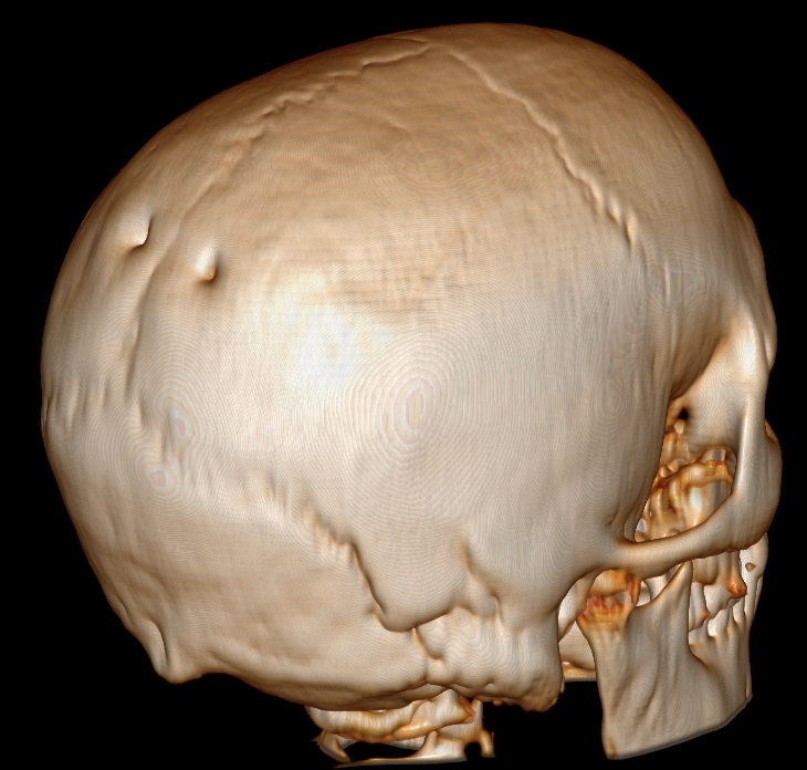
AI-Based CT Image Recognition With Med-Gemini-3D in the Diagnosis of a Rare Craniofacial Condition: A Catlin Mark Skull.
Artificial intelligence (AI) is increasingly applied in diagnostic imaging to enhance pattern recognition and support clinical decision-making. In 2024, Google introduced Med-Gemini-3D, a multimodal platform capable of interpreting 3-dimensional computed tomography scans and generating radiologist-level reports. Although not yet approved for independent clinical use, such systems may assist in identifying rare conditions that are unfamiliar to clinicians. The authors describe a 22-month-old girl who presented with persistent bilateral parietal skull defects and global developmental delay. Computed tomography demonstrated symmetric ossification defects adjacent to the sagittal suture that were not initially recognized by the treating physician. The patient's mother used a smartphone application powered by Med-Gemini-3D to analyze a 3D-CT reconstruction image, which suggested a diagnosis of "Catlin mark skull," a historical term for Enlarged Parietal Foramina (EPF). This prompted genetic evaluation and identification of a CDC42BPB variant associated with Chilton-Okur-Chung neurodevelopmental syndrome-a finding not previously reported in association with EPF. Establishing the diagnosis facilitated earlier therapeutic interventions and informed long-term management. This case underscores the potential role of AI-assisted tools in recognizing rare craniofacial anomalies. While such technologies cannot replace clinical expertise and remain limited by variable accuracy, they may help expand differential diagnoses, expedite referrals, and improve outcomes through earlier intervention. Continued research is needed to validate their reliability and to define their optimal integration into clinical practice. Enlarged parietal foramina are characteristic symmetric, paired radiolucencies of the parietal bones, located close to the intersection of the sagittal and lambdoid sutures, caused by deficient ossification around the parietal notch. Enlarged parietal foramina are usually asymptomatic. Meningeal, cortical, and vascular malformations of the posterior fossa occasionally accompany the bone defects and may predispose to epilepsy. In a minority of individuals, headaches, vomiting, or intense local pain are sometimes associated with the defects, especially on application of mild pressure to the unprotected cerebral cortex. The diagnosis of enlarged parietal foramina is established in a proband with characteristic clinical and imaging findings and a heterozygous pathogenic variant in ALX4 or MSX2 identified by molecular genetic testing. Treatment of manifestations: Treatment is generally conservative. Persistent cranium bifidum may warrant operative closure. The risk for penetrating injury to the brain is small but may cause anxiety; education of parents, teachers, and the affected child to avoid risky behaviors that could result in injury suffices in most circumstances. Treatment of seizures per neurologist; symptomatic treatment of headaches. Surveillance: Assess osseous defect every six to 12 months until natural history is established; assess for seizures, headache, or other concerning clinical manifestations if symptomatic. Agents/circumstances to avoid: Contact sports in those with a persistent midline bony defect. Enlarged parietal foramina are inherited in an autosomal dominant manner. Most individuals diagnosed with enlarged parietal foramina have an affected parent. The proportion of individuals with enlarged parietal foramina caused by a de novo pathogenic variant appears to be small. Each child of an individual with enlarged parietal foramina has a 50% chance of inheriting the pathogenic variant. Detailed fetal ultrasound examination at 18 to 20 weeks' gestation can usually detect the defects in a fetus at risk; fetal MRI is also an option. If the pathogenic variant has been identified in an affected family member, prenatal and preimplantation genetic testing are possible.
12mm wide parietal foramina: Morphological aspects and forensic application-A case report.
The enlarged parietal foramina (EPF) consist of a rare anatomical variation resulting from an autosomal dominant mutation, typically measuring over 5mm. Although often asymptomatic, this condition can be relevant in forensic anthropology for both human identification and differential diagnosis. This case report describes the presence of bilateral EPF in the skull of an adult male discovered in an advanced state of decomposition in Central-Western Brazil. Each foramen measured approximately 12mm and was detected during routine anthropological analysis. Potentially mimicking old gunshot wounds (especially when unilateral), the EPF was identified by its blunt margins, bilateral distribution in the parietal bone, and lack of macroscopically visible firearm residue and radiating fractures. This case underscores the importance of recognizing rare anatomical variants to avoid misinterpretation during medicolegal evaluations. When available, antemortem records may support its use as an identifying feature. The EPF remains an underreported condition, particularly in forensic settings, warranting further attention.
A case of enlarged parietal foramina or foramina parietalia permagna in an individual from the Chinchorro Culture of northern Chile (4000 BP).
The goal of this study was to analyze and differentially diagnose the presence of two large holes noted in the parietal bones of an individual and the presence of traumatic lesions. A partially mummified young adult female associated with the Chinchorro culture, 4000 BP, from the coast of the Atacama Desert (northern Chile). The bone lesions were evaluated macroscopically and radiologically. In addition, Sr isotopic analyses were performed on 62 individuals from eight sites associated with the Chinchorro culture. The parietal orifices are compatible with a rare anomaly of genetic origin known as foramina parietalia permagna (FPP). In addition, the cranial fracture pattern appear compatible with perimortem trauma, and Sr isotopes indicate a marine signal for Chinchorro populations. This case serves as evidence that FPP was present in the early Andean populations and that endogamy and mutagenic factors might have contributed to its presence. This paper expands our knowledge of the genetic anomalies that affected past populations and may contribute to our understanding of the etiologies of the condition. The absence of comparative FPP data inhibits comparative studies (with the exception of cases from California, USA). To explore in depth the genetic component of this condition in the Chinchorro populations.
Repair of Congenital Enlarged Parietal Foramina With Porous Polyethylene Implants.
Enlarged biparietal foramina is an autosomal dominant disorder that is caused by a failure of completion of ossification within the parietal bones. Enlarged parietal foramina measuring more than a few millimeters are uncommon. Even though spontaneous regression has been described, closure is rarely complete, and depending on the size of the resulting defect, an unprotected brain is a concern. There are few reports on the surgical management of persistent enlarged biparietal foramina. This is the first report describing our experience with a custom porous polyethylene implant.
Publicações recentes
New insights into enlarged parietal foramina: an anatomical, radiological, and histological study.
AI-Based CT Image Recognition With Med-Gemini-3D in the Diagnosis of a Rare Craniofacial Condition: A Catlin Mark Skull.
Enlarged Parietal Foramina.
12mm wide parietal foramina: Morphological aspects and forensic application-A case report.
A case of enlarged parietal foramina or foramina parietalia permagna in an individual from the Chinchorro Culture of northern Chile (4000 BP).
📚 EuropePMC35 artigos no totalmostrando 21
New insights into enlarged parietal foramina: an anatomical, radiological, and histological study.
Child's nervous system : ChNS : official journal of the International Society for Pediatric NeurosurgeryAI-Based CT Image Recognition With Med-Gemini-3D in the Diagnosis of a Rare Craniofacial Condition: A Catlin Mark Skull.
The Journal of craniofacial surgery12mm wide parietal foramina: Morphological aspects and forensic application-A case report.
Morphologie : bulletin de l'Association des anatomistesA case of enlarged parietal foramina or foramina parietalia permagna in an individual from the Chinchorro Culture of northern Chile (4000 BP).
International journal of paleopathologyRepair of Congenital Enlarged Parietal Foramina With Porous Polyethylene Implants.
The Journal of craniofacial surgeryA retrospective study of incidental findings occurring in a consecutive case series of lateral cephalograms of 12- to 20-year-old patients referred for routine orthodontic treatment.
Imaging science in dentistryMultiple Osteochondromas Comorbid With Enlarged Parietal Foramina, Elongated Styloid Processes, and Tibiofibular Synostosis.
American journal of clinical pathologyVertical transmission of a large calvarial ossification defect due to heterozygous variants of ALX4 and TWIST1.
American journal of medical genetics. Part AA Rare Congenital Cause of Epilepsy.
CureusCaput membranaceum: A novel clinical presentation of ZIC1 related skull malformation and craniosynostosis.
American journal of medical genetics. Part ATeaching NeuroImages: Enlarged parietal foramina inadvertently labeled as burr holes.
NeurologyDe novo truncating variants in PHF21A cause intellectual disability and craniofacial anomalies.
European journal of human genetics : EJHGAsymmetrically enlarged parietal foramina in a rare case of Goldenhar syndrome with a possible etiopathogenesis.
OncotargetCranium Bifidum Occultum Associated with Hypertelorism Treated with Posterior Vault Reconstruction and Orbital Box Osteotomies: Case Report and Technical Note.
World neurosurgery'Holes in the head': a case of enlarged parietal foramina.
Archives of disease in childhoodForamina parietalia permagna: familial and radiological evaluation of two cases and review of literature.
Child's nervous system : ChNS : official journal of the International Society for Pediatric Neurosurgery[Mother and son with enlarged parietal foramina, persistent fetal vein, and ALX4 mutation].
No to hattatsu = Brain and developmentEnlarged parietal foramina presenting as scalp swelling in an infant.
The Medical journal of MalaysiaEnlarged parietal foramina: a rare forensic autopsy finding.
International journal of legal medicineMultiple occipital, parietal, temporal, and frontal foramina: a variant of enlarged parietal foramina in an infant.
Balkan medical journalThe first Korean patient with Potocki-Shaffer syndrome: a rare cause of multiple exostoses.
Journal of Korean medical scienceAssociações
Organizações que acompanham esta doença — pra ter apoio e orientação
Ainda não temos associações cadastradas para Forame parietal.
É de uma associação que acompanha esta doença? Fale com a gente →
Comunidades
Grupos ativos de quem convive com esta doença aqui no Raras
Ainda não existe comunidade no Raras para Forame parietal
Pacientes, familiares e cuidadores se organizam em comunidades pra compartilhar experiências, fazer perguntas e se apoiar. Você pode ser o primeiro.
Tire suas dúvidas
Perguntas, dicas e experiências compartilhadas aqui na página
Participe da discussão
Faça login para postar dúvidas, compartilhar experiências e interagir com especialistas.
Fazer loginDoenças relacionadas
Doenças com sintomas parecidos — ajudam quem ainda está buscando diagnóstico
Referências e fontes
Bases de dados externas citadas neste artigo
Publicações científicas
Artigos indexados no PubMed ligados a esta doença no grafo RarasNet — título, periódico e PMID direto da fonte, sem intermediação de IA.
- New insights into enlarged parietal foramina: an anatomical, radiological, and histological study.Child's nervous system : ChNS : official journal of the International Society for Pediatric Neurosurgery· 2026· PMID 41832921mais citado
- AI-Based CT Image Recognition With Med-Gemini-3D in the Diagnosis of a Rare Craniofacial Condition: A Catlin Mark Skull.
- 12mm wide parietal foramina: Morphological aspects and forensic application-A case report.
- A case of enlarged parietal foramina or foramina parietalia permagna in an individual from the Chinchorro Culture of northern Chile (4000 BP).
- Repair of Congenital Enlarged Parietal Foramina With Porous Polyethylene Implants.
- Enlarged Parietal Foramina.
Bases de dados e fontes oficiais
Identificadores e referências canônicas usadas para montar este verbete.
- ORPHA:60015(Orphanet)
- MONDO:0018953(MONDO)
- GARD:16662(GARD (NIH))
- Variantes catalogadas(ClinVar)
- Busca completa no PubMed(PubMed)
- Q18987133(Wikidata)
Dados compilados pelo RarasNet a partir de fontes abertas (Orphanet, OMIM, MONDO, PubMed/EuropePMC, ClinicalTrials.gov, DATASUS, PCDT/MS). Este conteúdo é informativo e não substitui avaliação médica.
Conteúdo mantido por Agente Raras · Médicos e pesquisadores podem colaborar
Forame parietal
📋 Origem dos dados
Esta página agrega dados de fontes públicas e oficiais. Dados sobre cobertura no SUS (PCDT, CEAF) são verificados ativamente por agente proativo (ver badge no infobox). Demais dados têm atribuição de fonte + data da última sincronização — clique para abrir o original.
- Doença rara (ontologia)
- fonte: Orphanet
- Identificador unificado
- fonte: MONDO
- Codificação WHO/SUS
- fonte: WHO ICD-10 / DATASUS
- CID-11 (futuro)
- fonte: WHO ICD-11
- NIH/GARD
- fonte: GARD (NIH)
- Indexação biomédica
- fonte: MeSH (NLM)
- Dado público estruturado
- fonte: Wikidata