Malformação do SNC é um grupo de distúrbios congênitos afetando o desenvolvimento cerebral e espinhal. Manifesta-se com fraqueza muscular, problemas de memória, dor nas costas e alterações faciais, frequentemente associados a cistos aracnoides e nefropatia.
Introdução
O que você precisa saber de cara
Visão geral
Sinais e sintomas
Os sinais e manifestações clínicas documentados incluem alterações neurológicas e estruturais, tais como lesão cística intracraniana, cisto espinharacnoide, cisto inter-hemisférico, morfologia anormal da meninge espinhal e hemorragia subaracnoidea.[1][2]
Podem ocorrer fraqueza muscular do membro inferior, dor no membro inferior, dor nas costas, ciática e dificuldade para ficar em pé. No aspecto oftalmológico, descrevem-se visão turva, midríase e paralisia do olhar para cima.[1][2]
Manifestações comportamentais, cognitivas e sistêmicas também são observadas, incluindo comprometimento da memória, desinibição, choro inapropriado, mania, insônia, autonegligência, deterioração social e ocupacional, além de face longa, apetite pobre, dor abdominal, nefropatia e fisiologia anormal do intestino grosso.[1][2]
Causas genéticas
Diagnóstico
Tratamento e manejo
Tratamentos citados na literatura
A literatura científica apresenta associações com determinados fármacos e substâncias obtidas por mineração automatizada de dados (PubTator3). É importante ressaltar que essas menções representam apenas termos identificados em publicações científicas e NÃO constituem recomendação de uso clínico: Sirolimus (85 publicações), Enbucrilate (64 publicações), Onyx 18 (34 publicações), Indocyanine Green (32 publicações), ethylene vinyl alcohol copolymer (24 publicações), Propranolol (15 publicações), Steroids (15 publicações), Polyvinyl Alcohol (14 publicações), Titanium (11 publicações) e Alpelisib (9 publicações). Há também 1 ensaio clínico ativo registrado.[4]
Conteúdo informativo gerado e mantido automaticamente a partir de fontes oficiais (Orphanet, HPO, OMIM, SUS). Não substitui avaliação médica.
A **malformação do sistema nervoso central**, também chamada de **más-formações cefálicas congênitas**, é uma categoria que reúne várias alterações estruturais no cérebro e na medula espinhal que acontecem antes do nascimento. As pessoas afetadas podem manifestar dificuldades motoras como **tetraparesia** (fraqueza nos braços e pernas), problemas de coordenação, alterações na visão e disfunções no controle da bexiga e do intestino. O quadro clínico é investigado por meio de exames de imagem, como ultrassonografia, e por testes genéticos especializados. No Brasil, a condição não conta com um PCDT oficial de doença rara no SUS, de modo que o cuidado e as terapias de suporte são organizados individualmente pelas equipes de referência da rede pública.
Malformação do SNC é um grupo de distúrbios congênitos afetando o desenvolvimento cerebral e espinhal. Manifesta-se com fraqueza muscular, problemas de memória, dor nas costas e alterações faciais, frequentemente associados a cistos aracnoides e nefropatia.
O que está sendo pesquisado
Encontrou um erro ou informação desatualizada? Sugira uma correção →
Entender a doença
Do básico ao detalhe, leia no seu ritmo
Preparando trilha educativa...
Sinais e sintomas
O que aparece no corpo e com que frequência cada sintoma acontece
Partes do corpo afetadas
+ 454 sintomas em outras categorias
Características mais comuns
Os sintomas variam de pessoa para pessoa. Abaixo estão as 1133 características clínicas mais associadas, ordenadas por frequência.
Linha do tempo da pesquisa
Encontrou um erro ou informação desatualizada? Sugira uma correção →
Genética e causas
O que está alterado no DNA e como passa nas famílias
Genes associados
72 genes identificados com associação a esta condição.
Curadoria gene-doença
fontes oficiaisIntellectual developmental disorder, X-linked, syndromic, Billuart type
A disorder characterized by significantly below average general intellectual functioning associated with impairments in adaptive behavior and manifested during the developmental period. MRXSBL patients manifest intellectual disability associated with cerebellar hypoplasia and distinctive facial dysmorphism.
Agenesis of the corpus callosum, with facial anomalies and cerebellar ataxia
An autosomal recessive intellectual disability syndrome characterized by congenital microcephaly, low anterior hairline, bitemporal narrowing, low-set protruding ears, strabismus and tented thick eyebrows with sparse hair in their medial segment.
Craniosynostosis 6
A form of craniosynostosis, a primary abnormality of skull growth involving premature fusion of one or more cranial sutures. The growth velocity of the skull often cannot match that of the developing brain resulting in an abnormal head shape and, in some cases, increased intracranial pressure, which must be treated promptly to avoid permanent neurodevelopmental disability.
Joubert syndrome 36
A form of Joubert syndrome, a disorder presenting with cerebellar ataxia, oculomotor apraxia, hypotonia, neonatal breathing abnormalities and psychomotor delay. Neuroradiologically, it is characterized by cerebellar vermian hypoplasia/aplasia, thickened and reoriented superior cerebellar peduncles, and an abnormally large interpeduncular fossa, giving the appearance of a molar tooth on transaxial slices (molar tooth sign). Additional variable features include retinal dystrophy, renal disease, liver fibrosis, and polydactyly. JBTS36 inheritance is autosomal recessive.
Orofaciodigital syndrome 1
A form of orofaciodigital syndrome, a group of heterogeneous disorders characterized by abnormalities in the oral cavity, face, and digits and associated phenotypic abnormalities that lead to the delineation of various subtypes. OFD1 is X-linked dominant syndrome, lethal in males. Craniofacial findings consist of facial asymmetry, hypertelorism, median cleft, or pseudocleft of the upper lip, hypoplasia of the alae nasi, oral clefts and abnormal frenulea, tongue anomalies (clefting, cysts, hamartoma), and anomalous dentition involving missing or extra teeth. Asymmetric brachydactyly and/or syndactyly of the fingers and toes occur frequently. Approximately 50% of OFD1 females have some degree of intellectual disability. Some patients have structural central nervous system anomalies such as agenesis of the corpus callosum, cerebellar agenesis, or a Dandy-Walker malformation. Patients with OFD1 can develop fibrocystic disease of the liver and pancreas, in addition to polycystic kidneys.
Orofaciodigital syndrome 4
A form of orofaciodigital syndrome, a group of heterogeneous disorders characterized by malformations of the oral cavity, face and digits, and associated phenotypic abnormalities that lead to the delineation of various subtypes. OFD4 patients have tongue nodules, multiple frenulae, broad flat nose, hypertelorism, and short rib polydactyly with tibial dysplasia (Majewski syndrome). The presence of severe tibial aplasia differentiates OFD4 from OFD1. Additional features of cystic dysplastic kidneys and brain malformation, including occipital encephalocele, are observed in severely affected patients.
Orofaciodigital syndrome 15
A form of orofaciodigital syndrome, a group of heterogeneous disorders characterized by malformations of the oral cavity, face and digits, and associated phenotypic abnormalities that lead to the delineation of various subtypes. OFD15 features include facial dysmorphism, lobulated tongue, clefting of the alveolar ridges, left hand postaxial polydactyly, broad right hallux and left hallux duplication, and intermittent respiratory difficulty. Brain anomalies include vermis hypoplasia with molar tooth sign, agenesis of corpus callosum, and ventricular dilation. OFD15 inheritance is autosomal recessive.
Neurodegeneration, childhood-onset, with cerebellar atrophy
An autosomal recessive disorder characterized by early onset of progressive neurodegeneration affecting the central and peripheral nervous systems. Clinical features include global developmental delay, impaired intellectual development, poor or absent speech, and motor abnormalities. Brain imaging shows cerebellar atrophy. Death in childhood may occur.
Thyroid cancer, non-medullary, 2
A form of non-medullary thyroid cancer (NMTC), a cancer characterized by tumors originating from the thyroid follicular cells. NMTCs represent approximately 95% of all cases of thyroid cancer and are classified into papillary, follicular, Hurthle cell, and anaplastic neoplasms.
Congenital disorder of glycosylation 2N
A form of congenital disorder of glycosylation, a genetically heterogeneous group of autosomal recessive, multisystem disorders caused by a defect in glycoprotein biosynthesis and characterized by under-glycosylated serum glycoproteins. Congenital disorders of glycosylation result in a wide variety of clinical features, such as defects in the nervous system development, psychomotor retardation, dysmorphic features, hypotonia, coagulation disorders, and immunodeficiency. The broad spectrum of features reflects the critical role of N-glycoproteins during embryonic development, differentiation, and maintenance of cell functions.
Joubert syndrome 2
A disorder presenting with cerebellar ataxia, oculomotor apraxia, hypotonia, neonatal breathing abnormalities and psychomotor delay. Neuroradiologically, it is characterized by cerebellar vermian hypoplasia/aplasia, thickened and reoriented superior cerebellar peduncles, and an abnormally large interpeduncular fossa, giving the appearance of a molar tooth on transaxial slices (molar tooth sign). Additional variable features include retinal dystrophy and renal disease.
Lissencephaly 3
A classic type lissencephaly associated with psychomotor retardation and seizures. Features include agyria or pachygyria or laminar heterotopia, severe intellectual disability, motor delay, variable presence of seizures, and abnormalities of corpus callosum, hippocampus, cerebellar vermis and brainstem.
Cortical dysplasia, complex, with other brain malformations 7
A malformation of the cortex in which the brain surface is irregular and characterized by an excessive number of small gyri with abnormal lamination. Polymicrogyria is a heterogeneous disorder, considered to be the result of postmigratory abnormal cortical organization.
Cerebellar atrophy, visual impairment, and psychomotor retardation
An autosomal recessive, neurodegenerative disorder characterized by developmental delay, intellectual disability, hypotonia, scoliosis, cerebellar atrophy, and variable dysmorphic features.
Congenital disorder of glycosylation 2M
An X-linked dominant, severe neurologic disorder characterized by developmental delay, hypotonia, ocular anomalies, and brain malformations. Othere more variable clinical features included seizures, hypsarrhythmia, poor feeding, microcephaly, recurrent infections, dysmorphic features, shortened limbs, and coagulation defects. Congenital disorders of glycosylation are caused by a defect in glycoprotein biosynthesis and characterized by under-glycosylated serum glycoproteins and a wide variety of clinical features. The broad spectrum of features reflects the critical role of N-glycoproteins during embryonic development, differentiation, and maintenance of cell functions.
Meckel syndrome 6
A disorder characterized by a combination of renal cysts and variably associated features including developmental anomalies of the central nervous system (typically encephalocele), hepatic ductal dysplasia and cysts, and polydactyly.
Pontocerebellar hypoplasia 9
A form of pontocerebellar hypoplasia, a disorder characterized by structural defects of the pons and cerebellum, evident upon brain imaging. PCH9 features include severely delayed psychomotor development, progressive microcephaly, spasticity, seizures, and brain abnormalities, including brain atrophy, thin corpus callosum, and delayed myelination.
Autism, X-linked 5
A complex multifactorial, pervasive developmental disorder characterized by impairments in reciprocal social interaction and communication, restricted and stereotyped patterns of interests and activities, and the presence of developmental abnormalities by 3 years of age. Most individuals with autism also manifest moderate intellectual disability.
Neural tube defects
Congenital malformations of the central nervous system and adjacent structures related to defective neural tube closure during the first trimester of pregnancy. Failure of neural tube closure can occur at any level of the embryonic axis. Common NTD forms include anencephaly, myelomeningocele and spina bifida, which result from the failure of fusion in the cranial and spinal region of the neural tube. NTDs have a multifactorial etiology encompassing both genetic and environmental components.
Poretti-Boltshauser syndrome
An autosomal recessive disorder characterized by cerebellar dysplasia, cerebellar vermis atrophy, cerebellar cysts in most patients, high myopia, variable retinal dystrophy, and eye movement abnormalities including strabismus, ocular apraxia, nystagmus. Affected individuals have ataxia, delayed motor development, language impairment, and intellectual disability with variable severity.
Pontocerebellar hypoplasia 1A
A form of pontocerebellar hypoplasia, a disorder characterized by structural defects of the pons and cerebellum, evident upon brain imaging. PCH1A is an autosomal recessive form characterized by an abnormally small cerebellum and brainstem, central and peripheral motor dysfunction from birth, gliosis and spinal cord anterior horn cells degeneration resembling infantile spinal muscular atrophy. Additional features include muscle hypotonia, congenital contractures and respiratory insufficiency that is evident at birth.
Congenital disorder of glycosylation 2D
A multisystem disorder caused by a defect in glycoprotein biosynthesis and characterized by under-glycosylated serum glycoproteins. Congenital disorders of glycosylation result in a wide variety of clinical features, such as defects in the nervous system development, psychomotor retardation, dysmorphic features, hypotonia, coagulation disorders, and immunodeficiency. The broad spectrum of features reflects the critical role of N-glycoproteins during embryonic development, differentiation, and maintenance of cell functions.
Neural tube defects
Congenital malformations of the central nervous system and adjacent structures related to defective neural tube closure during the first trimester of pregnancy. Failure of neural tube closure can occur at any level of the embryonic axis. Common NTD forms include anencephaly, myelomeningocele and spina bifida, which result from the failure of fusion in the cranial and spinal region of the neural tube. NTDs have a multifactorial etiology encompassing both genetic and environmental components.
Pontocerebellar hypoplasia 7
A form of pontocerebellar hypoplasia, a group of related disorders characterized by underdevelopment of the pons and the cerebellum. Pontocerebellar hypoplasia also causes impaired growth of other parts of the brain, leading to an unusually small head size. PCH7 patients manifest delayed psychomotor development, hypotonia, breathing abnormalities, and gonadal abnormalities.
Pancreatic and cerebellar agenesis
A disease characterized by neonatal diabetes mellitus, cerebellar agenesis or hypoplasia, severe intrauterine growth retardation, the presence of very little subcutaneous fat, and dysmorphic facial features.
Neurodegeneration, childhood-onset, with brain atrophy
An autosomal dominant neurodegenerative disease with onset in childhood, characterized by progressive cortical atrophy, developmental delay, developmental regression, loss of motor skills and ambulation, absence of language, and intellectual disability.
Spinocerebellar ataxia, autosomal recessive, 20
A form of spinocerebellar ataxia, a clinically and genetically heterogeneous group of cerebellar disorders due to degeneration of the cerebellum with variable involvement of the brainstem and spinal cord. SCAR20 is characterized by cerebellar atrophy, ataxia, coarsened facial features, severely delayed psychomotor development with poor or absent speech, and intellectual disability.
Neuropathy, hereditary motor and sensory, 6B, with optic atrophy
An autosomal recessive neurologic disorder characterized by early-onset optic atrophy, progressive visual loss, and peripheral sensorimotor neuropathy manifesting as axonal Charcot-Marie-Tooth disease, with variable age at onset and severity. Charcot-Marie-Tooth disease is a disorder of the peripheral nervous system, characterized by progressive weakness and atrophy, initially of the peroneal muscles and later of the distal muscles of the arms. It is classified in two main groups on the basis of electrophysiologic properties and histopathology: primary peripheral demyelinating neuropathies and primary peripheral axonal neuropathies. Peripheral axonal neuropathies are characterized by signs of axonal regeneration in the absence of obvious myelin alterations, and normal or slightly reduced nerve conduction velocities.
Pontocerebellar hypoplasia 2F
A neurodevelopmental disorder characterized by progressive microcephaly, cognitive and motor delay, poor or absent speech, seizures, and spasticity. PCH2F inheritance is autosomal recessive.
Pontocerebellar hypoplasia 1D
An autosomal recessive neurologic disorder with onset at birth or in infancy, and characterized by progressive axonal motor neuronopathy, severe generalized hypotonia, respiratory insufficiency, and cerebellar atrophy. Death in childhood may occur.
Pontocerebellar hypoplasia 1C
A severe autosomal recessive neurodegenerative disease characterized by cerebellar and corpus callosum hypoplasia, abnormal myelination of the central nervous system, and spinal motor neuron disease. Affected individuals manifest failure to thrive, severe muscle weakness, spasticity and psychomotor retardation. Vision and hearing are impaired.
Neurodegeneration with brain iron accumulation 6
A neurodegenerative disorder associated with iron accumulation in the brain, primarily in the basal ganglia. It is characterized by progressive motor and cognitive dysfunction beginning in childhood or young adulthood. Patients show extrapyramidal motor signs, such as spasticity, dystonia, and parkinsonism.
Parietal foramina 2
Autosomal dominant disease characterized by oval defects of the parietal bones caused by deficient ossification around the parietal notch, which is normally obliterated during the fifth fetal month. PFM2 is also a clinical feature of Potocki-Shaffer syndrome.
Joubert syndrome 17
A disorder presenting with cerebellar ataxia, oculomotor apraxia, hypotonia, neonatal breathing abnormalities and psychomotor delay. Neuroradiologically, it is characterized by cerebellar vermian hypoplasia/aplasia, thickened and reoriented superior cerebellar peduncles, and an abnormally large interpeduncular fossa, giving the appearance of a molar tooth on transaxial slices (molar tooth sign). Additional variable features include retinal dystrophy and renal disease.
Joubert syndrome 3
A disorder presenting with cerebellar ataxia, oculomotor apraxia, hypotonia, neonatal breathing abnormalities and psychomotor delay. Neuroradiologically, it is characterized by cerebellar vermian hypoplasia/aplasia, thickened and reoriented superior cerebellar peduncles, and an abnormally large interpeduncular fossa, giving the appearance of a molar tooth on transaxial slices (molar tooth sign). Additional variable features include retinal dystrophy and renal disease. Joubert syndrome type 3 shows minimal extra central nervous system involvement and appears not to be associated with renal dysfunction.
Megalencephaly-polymicrogyria-polydactyly-hydrocephalus syndrome 1
A syndrome characterized by megalencephaly, hydrocephalus, and polymicrogyria; polydactyly may also be seen. There is considerable phenotypic similarity between this disorder and the megalencephaly-capillary malformation syndrome.
Joubert syndrome 16
An autosomal recessive disorder characterized by oculomotor apraxia, variable coloboma, and rare kidney involvement. Neuroradiologically, it is characterized by an abnormally large interpeduncular fossa, giving the appearance of a molar tooth on transaxial slices (molar tooth sign). Additional variable features include retinal dystrophy and polydactyly.
Pontocerebellar hypoplasia 3
A form of pontocerebellar hypoplasia, a disorder characterized by structural defects of the pons and cerebellum. Brain MRI shows an abnormally small cerebellum and brainstem, decreased cerebral white matter, and a thin corpus callosum. PCH3 features include seizures, short stature, optic atrophy, progressive microcephaly, severe developmental delay.
Pontocerebellar hypoplasia 2B
A disorder characterized by an abnormally small cerebellum and brainstem, and progressive microcephaly from birth combined with extrapyramidal dyskinesia. Severe chorea occurs and epilepsy is frequent. There are no signs of spinal cord anterior horn cells degeneration.
Cerebral arteriopathy, autosomal dominant, with subcortical infarcts and leukoencephalopathy, 1
A cerebrovascular disease characterized by multiple subcortical infarcts, pseudobulbar palsy, dementia, and the presence of granular deposits in small cerebral arteries producing ischemic stroke.
Joubert syndrome 5
A disorder presenting with cerebellar ataxia, oculomotor apraxia, hypotonia, neonatal breathing abnormalities and psychomotor delay. Neuroradiologically, it is characterized by cerebellar vermian hypoplasia/aplasia, thickened and reoriented superior cerebellar peduncles, and an abnormally large interpeduncular fossa, giving the appearance of a molar tooth on transaxial slices (molar tooth sign). Additional variable features include retinal dystrophy and renal disease. Joubert syndrome type 5 shares the neurologic and neuroradiologic features of Joubert syndrome together with severe retinal dystrophy and/or progressive renal failure characterized by nephronophthisis.
Meckel syndrome 1
A disorder characterized by a combination of renal cysts and variably associated features including developmental anomalies of the central nervous system (typically encephalocele), hepatic ductal dysplasia and cysts, and polydactyly.
Joubert syndrome 20
A disorder presenting with cerebellar ataxia, oculomotor apraxia, hypotonia, neonatal breathing abnormalities and psychomotor delay. Neuroradiologically, it is characterized by cerebellar vermian hypoplasia/aplasia, thickened and reoriented superior cerebellar peduncles, and an abnormally large interpeduncular fossa, giving the appearance of a molar tooth on transaxial slices (molar tooth sign). Additional variable features include retinal dystrophy and renal disease.
Tuberous sclerosis 1
An autosomal dominant multi-system disorder that affects especially the brain, kidneys, heart, and skin. It is characterized by hamartomas (benign overgrowths predominantly of a cell or tissue type that occurs normally in the organ) and hamartias (developmental abnormalities of tissue combination). Clinical manifestations include epilepsy, learning difficulties, behavioral problems, and skin lesions. Seizures can be intractable and premature death can occur from a variety of disease-associated causes.
Ritscher-Schinzel syndrome 2
A form of Ritscher-Schinzel syndrome, a developmental malformation syndrome characterized by cerebellar brain malformations, congenital heart defects, and craniofacial abnormalities. RTSC2 is an X-linked recessive form characterized by intellectual disability associated with posterior fossa defects, cardiac malformations, and minor abnormalities of the face and distal extremities.
Pontocerebellar hypoplasia 13
A form of pontocerebellar hypoplasia, a disorder characterized by structural defects of the pons and cerebellum, evident upon brain imaging. PCH13 is an autosomal recessive form characterized by delayed psychomotor development, absent speech, severe intellectual disability and postnatal microcephaly, with brain malformations consisting of cerebellar atrophy and hypoplastic corpus callosum. Additional features, including seizures and visual impairment, are variable.
Pontocerebellar hypoplasia 2D
A disorder characterized by postnatal onset of progressive atrophy of the cerebrum and cerebellum, microcephaly, profound intellectual disability, spasticity, and variable seizures.
Parietal foramina 1
Autosomal dominant disease characterized by oval defects of the parietal bones caused by deficient ossification around the parietal notch, which is normally obliterated during the fifth fetal month.
Nephronophthisis 3
An autosomal recessive disorder resulting in end-stage renal disease. It is characterized by polyuria, polydipsia, anemia. Onset of terminal renal failure occurr significantly later (median age, 19 years) than in juvenile nephronophthisis. Renal pathology is characterized by alterations of tubular basement membranes, tubular atrophy and dilation, sclerosing tubulointerstitial nephropathy, and renal cyst development predominantly at the corticomedullary junction.
Ritscher-Schinzel syndrome 4
An autosomal dominant form of Ritscher-Schinzel syndrome, a developmental malformation syndrome characterized by cerebellar brain anomalies associated with global developmental delay and impaired intellectual development, congenital heart defects, and craniofacial abnormalities.
Pontocerebellar hypoplasia 1B
A severe autosomal recessive neurologic disorder characterized by a combination of cerebellar and spinal motor neuron degeneration beginning at birth. There is diffuse muscle weakness, progressive microcephaly, global developmental delay, and brainstem involvement.
Cerebellar dysfunction, impaired intellectual development, and hypogonadotropic hypogonadism
An autosomal recessive disorder characterized by delayed motor development, ataxia with cerebellar hypoplasia, severe progressive scoliosis, moderate to severe intellectual disability, and delayed puberty with congenital hypogonadotropic hypogonadism.
Pettigrew syndrome
An X-linked syndrome characterized by intellectual disability and additional highly variable features, including choreoathetosis, hydrocephalus, Dandy-Walker malformation, seizures, and iron or calcium deposition in the brain. Intellectual disability is characterized by significantly below average general intellectual functioning associated with impairments in adaptive behavior and manifested during the developmental period.
Smith-Kingsmore syndrome
An autosomal dominant syndrome characterized by intellectual disability, macrocephaly, seizures, umbilical hernia, and facial dysmorphic features.
You-Hoover-Fong syndrome
A syndrome characterized by severe global developmental delay, intellectual disability, dysmorphic facial features, microcephaly, abnormal movements, congenital heart disease comprising developmental abnormalities of the great vessels, and abnormal auditory and visual function. The transmission pattern is consistent with autosomal recessive inheritance.
Pontocerebellar hypoplasia 2E
An autosomal recessive neurodegenerative disorder characterized by progressive cerebello-cerebral atrophy, profound intellectual disability, progressive microcephaly, spasticity, and early-onset epilepsy.
Megalencephaly-polymicrogyria-polydactyly-hydrocephalus syndrome 3
A syndrome characterized by megalencephaly, ventriculomegaly that may lead to hydrocephalus, and polymicrogyria; polydactyly may also be seen. There is considerable phenotypic similarity between this disorder and the megalencephaly-capillary malformation syndrome.
Leukodystrophy, hypomyelinating, 8, with or without oligodontia and/or hypogonadotropic hypogonadism
An autosomal recessive neurodegenerative disorder characterized by early childhood onset of cerebellar ataxia and mild intellectual disabilities associated with diffuse hypomyelination apparent on brain MRI. Variable features include oligodontia and/or hypogonadotropic hypogonadism.
Joubert syndrome 14
An autosomal recessive disorder characterized by severe intellectual disability, hypotonia, breathing abnormalities in infancy, and dysmorphic facial features. Neuroradiologically, it is characterized by cerebellar vermian hypoplasia/aplasia, thickened and reoriented superior cerebellar peduncles, and an abnormally large interpeduncular fossa, giving the appearance of a molar tooth on transaxial slices (molar tooth sign). Additional variable features include renal disease, abnormal eye movements, and postaxial polydactyly.
Ritscher-Schinzel syndrome 3
A form of Ritscher-Schinzel syndrome, a developmental malformation syndrome characterized by cerebellar brain malformations, congenital heart defects, and craniofacial abnormalities. RTSC3 is an autosomal recessive form. Affected individuals show cranio-cerebello-cardiac anomalies, coloboma, microphthalmia, chondrodysplasia punctata, complicated skeletal malformations, periventricular nodular heterotopia and proteinuria.
Spastic paraplegia 8, autosomal dominant
A form of spastic paraplegia, a neurodegenerative disorder characterized by a slow, gradual, progressive weakness and spasticity of the lower limbs. Rate of progression and the severity of symptoms are quite variable. Initial symptoms may include difficulty with balance, weakness and stiffness in the legs, muscle spasms, and dragging the toes when walking. In some forms of the disorder, bladder symptoms (such as incontinence) may appear, or the weakness and stiffness may spread to other parts of the body.
Pontocerebellar hypoplasia 8
An autosomal recessive neurodevelopmental disorder characterized by severe psychomotor retardation, abnormal movements, hypotonia, spasticity, and variable visual defects. Brain MRI shows pontocerebellar hypoplasia, decreased cerebral white matter, and a thin corpus callosum.
Pontocerebellar hypoplasia 6
A disorder characterized by an abnormally small cerebellum and brainstem, infantile encephalopathy, generalized hypotonia, lethargy and poor feeding. Recurrent apnea, intractable seizures occur early in the course of this condition.
Pontocerebellar hypoplasia 10
A form of pontocerebellar hypoplasia, a disorder characterized by structural defects of the pons and cerebellum, evident upon brain imaging. PCH10 features include cortical dysgenesis marked by a simplified gyral pattern, cortical atrophy, mild or focal cerebellar vermian volume loss, delayed myelination, progressive microcephaly, global growth and developmental delays, severe intellectual disabilities, and seizures refractory to treatment.
Pontocerebellar hypoplasia 4
A disorder characterized by an abnormally small cerebellum and brainstem, severe neonatal encephalopathy, microcephaly, myoclonus and muscular hypertonia. There is a severe inferior olivary and pontine neuronal loss and a diffuse white matter gliosis.
Joubert syndrome 15
An autosomal recessive disorder presenting with cerebellar ataxia, oculomotor apraxia, hypotonia, neonatal breathing abnormalities and psychomotor delay. Neuroradiologically, it is characterized by cerebellar vermian hypoplasia/aplasia, thickened and reoriented superior cerebellar peduncles, and an abnormally large interpeduncular fossa, giving the appearance of a molar tooth on transaxial slices (molar tooth sign). Additional variable features include retinal dystrophy, renal disease, liver fibrosis and polydactyly.
Medicamentos e terapias
Mecanismo: Estrogen receptor alpha agonist
Mecanismo: Progesterone receptor agonist
Mecanismo: PI3-kinase p110-alpha subunit inhibitor
Variantes genéticas (ClinVar)
337 variantes patogênicas registradas no ClinVar.
Vias biológicas (Reactome)
226 vias biológicas associadas aos genes desta condição.
Diagnóstico
Os sinais que médicos procuram e os exames que confirmam
Tratamento e manejo
Remédios, cuidados de apoio e o que precisa acompanhar
Onde tratar no SUS
Hospitais de referência no Brasil e o protocolo oficial do SUS (PCDT)
🇧🇷 Atendimento SUS — Malformação do sistema nervoso central
Centros de Referência SUS
24 centros habilitados pelo SUS para Malformação do sistema nervoso central
Centros para Malformação do sistema nervoso central
Detalhes dos centros
Hospital Universitário Prof. Edgard Santos (HUPES)
R. Dr. Augusto Viana, s/n - Canela, Salvador - BA, 40110-060 · CNES 0003808
Serviço de Referência
Hospital Infantil Albert Sabin
R. Tertuliano Sales, 544 - Vila União, Fortaleza - CE, 60410-794 · CNES 2407876
Serviço de Referência
Hospital de Apoio de Brasília (HAB)
AENW 3 Lote A Setor Noroeste - Plano Piloto, Brasília - DF, 70684-831 · CNES 0010456
Serviço de Referência
Hospital Estadual Infantil e Maternidade Alzir Bernardino Alves (HIABA)
Av. Min. Salgado Filho, 918 - Soteco, Vila Velha - ES, 29106-010 · CNES 6631207
Serviço de Referência
Hospital das Clínicas da UFG
Rua 235 QD. 68 Lote Área, Nº 285, s/nº - Setor Leste Universitário, Goiânia - GO, 74605-050 · CNES 2338424
Serviço de Referência
Hospital Universitário da UFJF
R. Catulo Breviglieri, Bairro - s/n - Santa Catarina, Juiz de Fora - MG, 36036-110 · CNES 2297442
Atenção Especializada
Hospital das Clínicas da UFMG
Av. Prof. Alfredo Balena, 110 - Santa Efigênia, Belo Horizonte - MG, 30130-100 · CNES 2280167
Serviço de Referência
Hospital Universitário Julio Müller (HUJM)
R. Luis Philippe Pereira Leite, s/n - Alvorada, Cuiabá - MT, 78048-902 · CNES 2726092
Atenção Especializada
Hospital Universitário João de Barros Barreto
R. dos Mundurucus, 4487 - Guamá, Belém - PA, 66073-000 · CNES 2337878
Serviço de Referência
Hospital Universitário Lauro Wanderley (HULW)
R. Tabeliao Estanislau Eloy, 585 - Castelo Branco, João Pessoa - PB, 58050-585 · CNES 0002470
Atenção Especializada
Instituto de Medicina Integral Prof. Fernando Figueira (IMIP)
R. dos Coelhos, 300 - Boa Vista, Recife - PE, 50070-902 · CNES 0000647
Serviço de Referência
Hospital Pequeno Príncipe
R. Des. Motta, 1070 - Água Verde, Curitiba - PR, 80250-060 · CNES 3143805
Serviço de Referência
Hospital Universitário Regional de Maringá (HUM)
Av. Mandacaru, 1590 - Parque das Laranjeiras, Maringá - PR, 87083-240 · CNES 2216108
Atenção Especializada
Hospital de Clínicas da UFPR
R. Gen. Carneiro, 181 - Alto da Glória, Curitiba - PR, 80060-900 · CNES 2364980
Serviço de Referência
Hospital Universitário Pedro Ernesto (HUPE-UERJ)
Blvd. 28 de Setembro, 77 - Vila Isabel, Rio de Janeiro - RJ, 20551-030 · CNES 2280221
Serviço de Referência
Instituto Nacional de Saúde da Mulher, da Criança e do Adolescente Fernandes Figueira (IFF/Fiocruz)
Av. Rui Barbosa, 716 - Flamengo, Rio de Janeiro - RJ, 22250-020 · CNES 2269988
Serviço de Referência
Hospital São Lucas da PUCRS
Av. Ipiranga, 6690 - Jardim Botânico, Porto Alegre - RS, 90610-000 · CNES 2232928
Serviço de Referência
Hospital de Clínicas de Porto Alegre (HCPA)
Rua Ramiro Barcelos, 2350 Bloco A - Av. Protásio Alves, 211 - Bloco B e C - Santa Cecília, Porto Alegre - RS, 90035-903 · CNES 2237601
Serviço de Referência
Hospital Universitário da UFSC (HU-UFSC)
R. Profa. Maria Flora Pausewang - Trindade, Florianópolis - SC, 88036-800 · CNES 2560356
Serviço de Referência
Hospital das Clínicas da FMUSP
R. Dr. Ovídio Pires de Campos, 225 - Cerqueira César, São Paulo - SP, 05403-010 · CNES 2077485
Serviço de Referência
Hospital de Base de São José do Rio Preto
Av. Brg. Faria Lima, 5544 - Vila Sao Jose, São José do Rio Preto - SP, 15090-000 · CNES 2079798
Atenção Especializada
Hospital de Clínicas da UNICAMP
R. Vital Brasil, 251 - Cidade Universitária, Campinas - SP, 13083-888 · CNES 2748223
Serviço de Referência
Hospital de Clínicas de Ribeirão Preto (HCRP-USP)
R. Ten. Catão Roxo, 3900 - Vila Monte Alegre, Ribeirão Preto - SP, 14015-010 · CNES 2082187
Serviço de Referência
UNIFESP / Hospital São Paulo
R. Napoleão de Barros, 715 - Vila Clementino, São Paulo - SP, 04024-002 · CNES 2688689
Serviço de Referência
Dados de DATASUS/CNES, SBGM, ABNeuro e Ministério da Saúde. Sempre confirme a disponibilidade diretamente com o estabelecimento.
Pesquisa ativa
Ensaios clínicos abertos e novidades científicas recentes
Ensaios em destaque
Com ensaio aberto em 2 países. O ponto verde marca onde há vaga agora.
🟢 Recrutando agora
1 pesquisa recrutando participantes. Converse com seu médico sobre a possibilidade de participar.
Outros ensaios clínicos
3 ensaios clínicos encontrados, 1 ativos.
Publicações mais relevantes
13q Deletion Syndrome Presenting with Lymphopenia Detected Through Newborn Screening for Primary Immunodeficiencies.
The expanded newborn screening (NBS) program in the Russian Federation, launched in 2023, includes the detection of severe forms of T- and B-cell immunodeficiencies via TREC/KREC quantification. We report a rare case of a male infant having multiple congenital anomalies and lymphopenia identified through this program. Genetic testing revealed a 25.8 Mb terminal deletion spanning 13q31.2-qter, consistent with 13q deletion syndrome. Initial NBS revealed reduced TREC levels, prompting further evaluation. The patient exhibited a complex phenotype, including central nervous system malformation (alobar holoprosencephaly), severe congenital heart disease, renal hypoplasia, limb and genitourinary anomalies, and facial dysmorphism. Postnatal complications included pneumonia, pleuritis, and chylothorax. Flow cytometry demonstrated mild T- and B-cell lymphopenia. The genomic defect was characterized using long-read third-generation sequencing, enabling precise breakpoint identification and accurate mapping of deleted genes. The deletion was confirmed via subtelomeric FISH analysis. The patient died at 7 months of age due to the progression of underlying congenital anomalies and associated complications. Our findings broaden the clinical characterization of distal 13q deletion syndrome and demonstrate the value of long-read sequencing in structural chromosomal analysis. They further highlight the difficulties of caring for neonates having complex malformations and immune dysfunction. Given the potential for both primary and secondary immune disturbances, comprehensive immunological evaluation should be considered in patients having 13q deletion syndrome to improve diagnostic accuracy and inform appropriate clinical management.
Dandy-Walker Malformation and Optic Nerve Hypoplasia: A Developmental Case Highlighting the Psychiatric Impact of Central Nervous System Malformation.
Dandy-Walker malformation (DWM) and optic nerve hypoplasia (ONH) are rare congenital anomalies associated with neuropsychiatric morbidity, but their co-occurrence in adults has not been described. We present an adult patient with both DWM and unilateral ONH who developed auditory hypersensitivity, visual agnosia, phonological dyslexia, dysgraphia, and longstanding academic challenges, later evolving into recurrent depression, anxiety, and insomnia. Neuroimaging revealed a Dandy-Walker variant with cerebellar atrophy and ONH, and her presentation was consistent with cerebellar cognitive affective syndrome. Notably, despite the established risk of autism spectrum-like traits in both DWM and ONH, she did not demonstrate autistic features. This case highlights how overlapping congenital anomalies may synergistically increase vulnerability to psychiatric illness and underscores the importance of early recognition and tailored interventions. To our knowledge, this is among the first adult cases describing co-occurring DWM and ONH with a longitudinal psychiatric course.
Poland Anomaly and Atretic Cephalocele in the Same Child: Coincidence or Association?
Poland Anomaly is a rare congenital disorder typically characterized by hypoplasia or agenesis of pectoral muscle with or without ipsilateral limb hypoplasia. The association of central nervous system malformation with Poland Anomaly has been rarely reported and includes craniofacial dysplasia, microcephaly, and Dandy-Walker malformation. However, no anomalies in neural tube closure have been described in association with Poland Anomaly. Atretic cephalocele is another rare diagnosis, which is an involuted congenital herniation of intracranial contents through defects in the skull. Herein, we present the case of a child affected with Poland Anomaly in whom an atretic cephalocele has been diagnosed at the age of 2 months.
Risk factors associated with congenital central nervous system abnormalities in the National Hospital of Zinder, Niger.
Congenital malformations of the central nervous system (CNS) are morphological abnormalities of the brain and spinal cord that occur during fetal development. They constitute the second most common congenital disability, after congenital cardiac defects. Many risk factors have been identified; however, these studies included various types of congenital abnormality. Furthermore, there is a lack of information on risk factors for congenital CNS malformation, and notably in the Zinder region of Niger. This study aimed to identify the risk factors associated with congenital CNS malformations in the Zinder region. In a case-control design, patients with congenital CNS malformation were enrolled between June 2022 and April 2023 in the Department of Neurosurgery of the National Hospital of Zinder. Family history of malformation (aOR:3.31, 95% CI:1.25-8.78) and consanguine marriage (aOR:2.28, 95% CI:1.23-4.20) were significantly associated with congenital CNS malformation. In contrast, folic acid supplementation (aOR:0.34, 95% CI:0.13, 0.89), multiparity (aOR:0.34, 95% CI:0.13, 0.89), and grand multiparity (aOR, 0.47; 95% CI:0.23, 0.97) had a protective effect. Risk factors such as family malformation history and consanguine marriage increased the risk of developing congenital malformations of the central nervous system. In contrast, folic acid supplementation in the index period and multiparity had a significant protective effect.
Levetiracetam prescription profile in children younger than 4 years treated at a tertiary care hospital in Chile.
Introduction. Levetiracetam (LEV) is an antiepileptic drug approved by the Chilean Institute of Public Health as concomitant therapy for epileptic seizures in children older than 4 years of age. However, it is widely prescribed from the neonatal period, which makes it necessary to evaluate its off-label use. Objective. To determine the prescription-indication profile of LEV in the treatment of epileptic seizures in children younger than 4 years in a tertiary care hospital in southern Chile. Population and method. Observational, descriptive, and retrospective study. The medical records of patients who started treatment with LEV between 2014 and 2019 were reviewed, and data on sociodemographic, pharmacological, and clinical variables were collected. The analysis was based on the description of the profile of patients, prescriptions, follow-up, and safety. Results. A total of 68 patients were included: 40 (58.8%) were males, 49 (72.1%) were born at a gestational age ≥ 37 weeks. The main etiology of epilepsy was structural (35.3%); LEV was mostly used in children diagnosed with central nervous system malformation (17.6%), and monotherapy was the prevailing dosage (55.9%). LEV was used for focal seizures in 50% of cases. Five children (7.3%) had psychiatric disorders, classified as probable adverse drug reactions. Conclusion. LEV was used in children with various diagnoses, with a low rate of adverse events. The profile of drug use varied in the different age groups. Future studies are needed to identify effectiveness, especially in newborn infants and patients with refractory epilepsy. Introducción. El levetiracetam (LEV) es un antiepiléptico aprobado por el Instituto de Salud Pública de Chile como terapia concomitante en crisis epilépticas en niños mayores de cuatro años. Sin embargo, es ampliamente indicado desde el periodo neonatal, lo que hace necesario evaluar su utilización fuera de ficha técnica. Objetivo. Determinar el perfil de prescripción-indicación de LEV en el tratamiento de las crisis epilépticas en menores de cuatro años en un hospital de alta complejidad del sur de Chile. Población y método. Estudio observacional, descriptivo y retrospectivo. Se revisaron las historias clínicas de quienes iniciaron tratamiento con LEV entre 2014 y 2019, y se recopilaron datos sobre variables sociodemográficas, farmacológicas y clínicas. El análisis se basó en la descripción del perfil de los pacientes, prescripción, seguimiento y seguridad. Resultados. Se incluyeron 68 pacientes: 40 (58,8 %) de sexo masculino, 49 (72,1 %) con edad gestacional ≥ 37 semanas. La etiología principal de la epilepsia fue de tipo estructural (35,3 %); el LEV se utilizó principalmente en niños diagnosticados con malformación del sistema nervioso central (17,6 %) y predominó la monoterapia (55,9 %). En el 50 % se usó LEV para crisis focales. Cinco niños (7,3 %) presentaron trastornos de tipo psiquiátrico clasificados como probables reacciones adversas al medicamento. Conclusión. El LEV se utilizó en niños con diferentes diagnósticos con baja frecuencia de eventos adversos. El perfil de utilización varió en los diferentes grupos etarios. Es necesario identificar en futuros estudios la efectividad especialmente en el recién nacido y en epilepsias refractarias.
Publicações recentes
Dandy-Walker Malformation and Optic Nerve Hypoplasia: A Developmental Case Highlighting the Psychiatric Impact of Central Nervous System Malformation.
13q Deletion Syndrome Presenting with Lymphopenia Detected Through Newborn Screening for Primary Immunodeficiencies.
Poland Anomaly and Atretic Cephalocele in the Same Child: Coincidence or Association?
Risk factors associated with congenital central nervous system abnormalities in the National Hospital of Zinder, Niger.
Levetiracetam prescription profile in children younger than 4 years treated at a tertiary care hospital in Chile.
📚 EuropePMC15 artigos no totalmostrando 40
Dandy-Walker Malformation and Optic Nerve Hypoplasia: A Developmental Case Highlighting the Psychiatric Impact of Central Nervous System Malformation.
Cureus13q Deletion Syndrome Presenting with Lymphopenia Detected Through Newborn Screening for Primary Immunodeficiencies.
International journal of molecular sciencesPoland Anomaly and Atretic Cephalocele in the Same Child: Coincidence or Association?
American journal of medical genetics. Part ARisk factors associated with congenital central nervous system abnormalities in the National Hospital of Zinder, Niger.
Neuro-ChirurgieLevetiracetam prescription profile in children younger than 4 years treated at a tertiary care hospital in Chile.
Archivos argentinos de pediatriaDandy-Walker Malformation with Neonatal Meningitis: A Case Report.
JNMA; journal of the Nepal Medical AssociationWhole-genome sequencing in prenatally detected congenital malformations: prospective cohort study in clinical setting.
Ultrasound in obstetrics & gynecology : the official journal of the International Society of Ultrasound in Obstetrics and GynecologyEpilepsy Frequency and Risk Factors Three Years After Neonatal Seizures.
Pediatric neurologyThe Relationship Between Fetal Central Nervous System Malformations and Modified Myocardial Performance Index.
CureusThe impact of epilepsy and antiseizure medications on pregnancy and neonatal outcomes: A nationwide cohort study.
Brain and behaviorVascular anomaly, lipoma, and polymicrogyria associated with schizencephaly: developmental and diagnostic insights. Illustrative case.
Journal of neurosurgery. Case lessonsAutosomal Recessive Primary Microcephaly (MCPH) and Novel Pathogenic Variants in ASPM and WDR62 Genes.
Molecular syndromologyUltrasound Appearance of Fetal Posterior Fossa and a Case Report of Prenatal Diagnosis of Dandy-Walker Malformation.
MaedicaOpioid exposure during pregnancy and the risk of congenital malformation: a meta-analysis of cohort studies.
BMC pregnancy and childbirthCase Report: Preimplantation Genetic Testing for Meckel Syndrome Induced by Novel Compound Heterozygous Mutations of MKS1.
Frontiers in geneticsAlobar Holoprosencephaly in an Aborted American Quarter Horse Fetus.
Journal of equine veterinary scienceUse of real-time artificial intelligence in detection of abnormal image patterns in standard sonographic reference planes in screening for fetal intracranial malformations.
Ultrasound in obstetrics & gynecology : the official journal of the International Society of Ultrasound in Obstetrics and GynecologyTraumatic epidural hematoma in a patient with severe schizencephaly.
Clinical case reportsAccuracy of ultrasonography in diagnosis of fetal central nervous system malformation.
World journal of clinical casesCarbamazepine-Responsive Chorea in a Toddler with Semilobar Holoprosencephaly: Case Report.
Journal of pediatric neurosciencesPrenatal diagnosis of the Dandy-Walker malformation associated with partial trisomy 12p and distal 15q deletion.
Journal of geneticsPrenatal Diagnosis of Acromelic Frontonasal Dysostosis.
Molecular syndromologyMassive posterior cranial vault erosion and its reconstruction: A peculiar presentation of "mega cisterna magna".
Journal of oral biology and craniofacial researchApplication of Ultrasonic Doppler Technology Based on Wavelet Threshold Denoising Algorithm in Fetal Heart Rate and Central Nervous System Malformation Detection.
World neurosurgeryEpilepsy in paediatric patients with schizencephaly.
Annals of agricultural and environmental medicine : AAEMPoland Syndrome with Atypical Malformations Associated to a de novo 1.5 Mb Xp22.31 Duplication.
NeuropediatricsMalignant Hyperthermia and Cerebral Venous Sinus Thrombosis After Ventriculoperitoneal Shunt in Infant with Schizencephaly and COL4A1 Mutation.
World neurosurgeryAmphiphilic block copolymer delivery of a DNA vaccine against Zika virus.
VaccineTwo-year postnatal outcome of 263 cases of fetal ventriculomegaly.
The journal of maternal-fetal & neonatal medicine : the official journal of the European Association of Perinatal Medicine, the Federation of Asia and Oceania Perinatal Societies, the International Society of Perinatal ObstetriciansCranial neural tube defect after trimethoprim exposure.
BMC research notesNeurologic Involvement in Primary Immunodeficiency Disorders.
Journal of child neurologyCentral nervous system malformation associated with methamphetamine abuse during pregnancy.
Clinical toxicology (Philadelphia, Pa.)Retinoic Acid Embryopathy.
International journal of applied & basic medical researchPerinatal and follow-up outcome study of fetal anomalies with multidisciplinary consultation.
Therapeutics and clinical risk managementSpina Bifida: Pathogenesis, Mechanisms, and Genes in Mice and Humans.
ScientificaArthrogryposis-renal tubular dysfunction-cholestasis syndrome: a cause of neonatal cholestasis. Case report.
Archivos argentinos de pediatriaMultifetal gestations with assisted reproductive technique before the single-embryo transfer legislation: obstetric, neonatal outcomes and congenital anomalies.
The journal of maternal-fetal & neonatal medicine : the official journal of the European Association of Perinatal Medicine, the Federation of Asia and Oceania Perinatal Societies, the International Society of Perinatal ObstetriciansUrodynamic Findings and Renal Function in Children with Neurogenic Bladder after Myelomeningocele.
Urologia internationalisMeckel-Gruber Syndrome with unilateral renal agenesis.
Journal of the College of Physicians and Surgeons--Pakistan : JCPSPEvaluation of the association of zoonotic Ljungan virus with perinatal deaths and fetal malformation.
Birth defects research. Part C, Embryo today : reviewsAssociações
Organizações que acompanham esta doença — pra ter apoio e orientação
Ainda não temos associações cadastradas para Malformação do sistema nervoso central.
É de uma associação que acompanha esta doença? Fale com a gente →
Doença com base genética
Um médico geneticista pode ajudar no diagnóstico de Malformação do sistema nervoso central e no aconselhamento genético da família.
Doenças relacionadas
Doenças com sintomas parecidos — ajudam quem ainda está buscando diagnóstico
Perguntas frequentes
O que as famílias mais perguntam sobre esta doença — cada resposta com a fonte de onde saiu
Trata-se de um grupo amplo de anomalias congênitas estruturais que atingem o encéfalo e a medula espinhal. Na classificação internacional de doenças, o conjunto é catalogado sob o código CID-10 Q00-Q07 e ORPHA:98044.
Referências
Fontes citadas no texto, publicações do grafo e bases de dados usadas neste verbete
5 fontes citadas no texto · 5 publicações do grafo RarasNet (PubMed) · 6 bases de dados. Títulos, periódicos e PMIDs vêm direto da fonte, sem intermediação de IA.
Dados compilados pelo RarasNet a partir de fontes abertas (Orphanet, OMIM, MONDO, PubMed/EuropePMC, ClinicalTrials.gov, DATASUS, PCDT/MS). Este conteúdo é informativo e não substitui avaliação médica.
Citar este verbete
Raras. (2026). Malformação do sistema nervoso central. Em Raras — Enciclopédia de Doenças Raras do Brasil. https://raras.org/doenca/malformacao-do-sistema-nervoso-central
Formato APA. Conteúdo sob CC BY 4.0 — reuso livre com atribuição.
Conteúdo mantido por Agente Raras · Médicos e pesquisadores podem colaborar
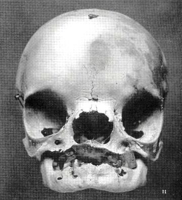
Malformação do sistema nervoso central
📋 Origem dos dados
Esta página agrega dados de fontes públicas e oficiais. Dados sobre cobertura no SUS (PCDT, CEAF) são verificados ativamente por agente proativo (ver badge no infobox). Demais dados têm atribuição de fonte + data da última sincronização — clique para abrir o original.
- Doença rara (ontologia)
- fonte: Orphanet
- Identificador unificado
- fonte: MONDO
- Codificação WHO/SUS
- fonte: WHO ICD-10 / DATASUS
- NIH/GARD
- fonte: GARD (NIH)
- Indexação biomédica
- fonte: MeSH (NLM)
- Dado público estruturado
- fonte: Wikidata
- Moléculas estudadas na doença
- fonte: OpenTargets
- Ensaios clínicos
- fonte: ClinicalTrials.gov

