A síndrome de sinostoses múltiplas (SMS) é um distúrbio raro do desenvolvimento ósseo caracterizado por simfalangismo proximal dos dedos das mãos e/ou pés, frequentemente associado à fusão das articulações do carpo e do tarso, umeroradial e da coluna cervical.
Introdução
O que você precisa saber de cara
A síndrome de sinostoses múltiplas (SMS) é um distúrbio raro do desenvolvimento ósseo caracterizado por simfalangismo proximal dos dedos das mãos e/ou pés, frequentemente associado à fusão das articulações do carpo e do tarso, umeroradial e da coluna cervical.
Escala de raridade
<1/50kMuito rara
1/20kRara
1/10kPouco freq.
1/5kIncomum
1/2k
Encontrou um erro ou informação desatualizada? Sugira uma correção →
Entender a doença
Do básico ao detalhe, leia no seu ritmo
Preparando trilha educativa...
Sinais e sintomas
O que aparece no corpo e com que frequência cada sintoma acontece
Partes do corpo afetadas
+ 33 sintomas em outras categorias
Características mais comuns
Os sintomas variam de pessoa para pessoa. Abaixo estão as 79 características clínicas mais associadas, ordenadas por frequência.
Linha do tempo da pesquisa
Encontrou um erro ou informação desatualizada? Sugira uma correção →
Genética e causas
O que está alterado no DNA e como passa nas famílias
Genes associados
4 genes identificados com associação a esta condição. Padrão de herança: Autosomal dominant.
Curadoria gene-doença
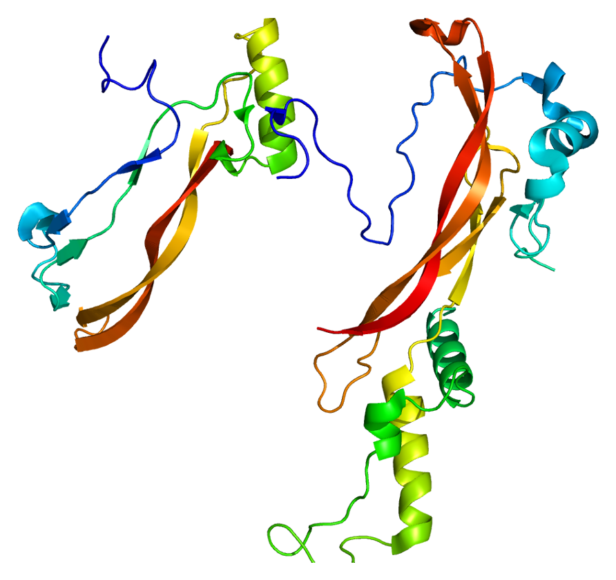
fontes oficiaisGrowth factor involved in bone and cartilage formation. During cartilage development regulates differentiation of chondrogenic tissue through two pathways. Firstly, positively regulates differentiation of chondrogenic tissue through its binding of high affinity with BMPR1B and of less affinity with BMPR1A, leading to induction of SMAD1-SMAD5-SMAD8 complex phosphorylation and then SMAD protein signaling transduction (PubMed:15530414, PubMed:21976273, PubMed:24098149, PubMed:25092592). Secondly, n
SecretedCell membrane
Acromesomelic dysplasia 2A
A form of acromesomelic dysplasia, a skeletal disorder characterized by short stature, very short limbs and hand/foot malformations. The severity of limb abnormalities increases from proximal to distal with profoundly affected hands and feet showing brachydactyly and/or rudimentary fingers (knob-like fingers). AMD2A is an autosomal recessive form characterized by normal axial skeletons and missing or fused skeletal elements within the hands and feet.
Plays an important role in the regulation of embryonic development, cell proliferation, cell differentiation and cell migration. May have a role in glial cell growth and differentiation during development, gliosis during repair and regeneration of brain tissue after damage, differentiation and survival of neuronal cells, and growth stimulation of glial tumors
Secreted
Multiple synostoses syndrome 3
A bone disease characterized by multiple progressive joint fusions that commonly involve proximal interphalangeal, tarsal-carpal, humeroradial and cervical spine joints. Additional features can include progressive conductive deafness and facial dysmorphism.
Growth factor that controls proliferation and cellular differentiation in the retina and bone formation. Plays a key role in regulating apoptosis during retinal development. Establishes dorsal-ventral positional information in the retina and controls the formation of the retinotectal map (PubMed:23307924). Required for normal formation of bones and joints in the limbs, skull, digits and axial skeleton. Plays a key role in establishing boundaries between skeletal elements during development. Regu
Secreted
Klippel-Feil syndrome 1, autosomal dominant
A skeletal disorder characterized by congenital fusion of cervical vertebrae. It is due to a failure in the normal segmentation of vertebrae during the early weeks of fetal development. The clinical triad consists of short neck, low posterior hairline, and limited neck movement. Deafness is a feature in some cases and may be of sensorineural, conductive, or mixed type.
Inhibitor of bone morphogenetic proteins (BMP) signaling which is required for growth and patterning of the neural tube and somite. Essential for cartilage morphogenesis and joint formation. Inhibits chondrocyte differentiation through its interaction with GDF5 and, probably, GDF6 (PubMed:21976273, PubMed:26643732)
Secreted
Symphalangism, proximal 1A
A disease characterized by the hereditary absence of the proximal interphalangeal joints. Distal interphalangeal joints are less frequently involved and metacarpophalangeal joints are rarely affected whereas carpal bone malformation and fusion are common. In the lower extremities, tarsal bone coalition is common. Conductive hearing loss is seen and is due to fusion of the stapes to the petrous part of the temporal bone.
Variantes genéticas (ClinVar)
197 variantes patogênicas registradas no ClinVar.
Classificação de variantes (ClinVar)
Distribuição de 146 variantes classificadas pelo ClinVar.
Vias biológicas (Reactome)
33 vias biológicas associadas aos genes desta condição.
Diagnóstico
Os sinais que médicos procuram e os exames que confirmam
Tratamento e manejo
Remédios, cuidados de apoio e o que precisa acompanhar
Onde tratar no SUS
Hospitais de referência no Brasil e o protocolo oficial do SUS (PCDT)
🇧🇷 Atendimento SUS — Síndrome de sinostoses múltiplas
Selecione um estado ou use sua localização para ver resultados.
Dados de DATASUS/CNES, SBGM, ABNeuro e Ministério da Saúde. Sempre confirme a disponibilidade diretamente com o estabelecimento.
Pesquisa ativa
Ensaios clínicos abertos e novidades científicas recentes
Ensaios em destaque
🟢 Recrutando agora
1 pesquisa recrutando participantes. Converse com seu médico sobre a possibilidade de participar.
Outros ensaios clínicos
0 ensaios clínicos encontrados.
Publicações mais relevantes
Management of a four-generation family affected by GDF6 multiple synostoses syndrome type 4.
Humeroradial Synostosis: An Updated Classification and Differential Diagnosis Based on Genetic Aetiology.
Humeroradial synostosis (HRS) is a rare congenital limb malformation, characterised by fusion of the humeral and radial bones, leading to functional disability of the elbow joint. HRS may be reported in familial or sporadic cases and observed either isolated or as part of a syndromic condition. According to an extensive review of the literature, a dozen known conditions may comprise an HRS. The present review aims to propose an updated classification based on molecular pathways (chondrogenesis and osteogenesis; limb development and patterning; genome regulation), combined with a concise overview of the conditions associated with HRS. This knowledge could guide molecular analyses, patient management and genetic counselling. As some cases remain unexplained, further genetic and epidemiological studies are required to evaluate the contribution of genetic and environmental factors in HRS physiopathology.
Genetics, epidemiology and management of clubfoot and related disorders.
Clubfoot, medically termed congenital talipes equinovarus (CTEV), is a prevalent musculoskeletal birth defect, affecting approximately 0.3% of all live births. This serious congenital anomaly results from structural abnormalities in the foot and lower leg, leading to abnormal positioning of the ankle and foot joints. This review provides a comprehensive overview of the causative factors associated with CTEV and evaluates current therapeutic approaches. Although variations in genes encoding contractile proteins of skeletal myofibers have been proposed as contributors to the etiology of CTEV, no definitive candidate genes have been conclusively linked to increased risk. However, genes such as TBX4, PITX1, and members of the HOXA, HOXC, and HOXD clusters, as well as NAT2, have been implicated in the condition's development, playing critical roles in limb development, muscle formation, and tissue differentiation. Also, Axin1 plays a key role in joint formation and skeletal development by inhibiting β-catenin-BMP signaling. It could significantly serve as a therapeutic target for fibular hemimelia and multiple synostoses syndrome. The exact mechanisms and the extent of their physical and genetic interactions remain subjects of ongoing research. Understanding the genetic determinants and cellular pathways involved in CTEV is crucial for unravelling the pathophysiology of this complex deformity.
Transforming growth factor beta signaling and craniofacial development: modeling human diseases in zebrafish.
Humans and other jawed vertebrates rely heavily on their craniofacial skeleton for eating, breathing, and communicating. As such, it is vital that the elements of the craniofacial skeleton develop properly during embryogenesis to ensure a high quality of life and evolutionary fitness. Indeed, craniofacial abnormalities, including cleft palate and craniosynostosis, represent some of the most common congenital abnormalities in newborns. Like many other organ systems, the development of the craniofacial skeleton is complex, relying on specification and migration of the neural crest, patterning of the pharyngeal arches, and morphogenesis of each skeletal element into its final form. These processes must be carefully coordinated and integrated. One way this is achieved is through the spatial and temporal deployment of cell signaling pathways. Recent studies conducted using the zebrafish model underscore the importance of the Transforming Growth Factor Beta (TGF-β) and Bone Morphogenetic Protein (BMP) pathways in craniofacial development. Although both pathways contain similar components, each pathway results in unique outcomes on a cellular level. In this review, we will cover studies conducted using zebrafish that show the necessity of these pathways in each stage of craniofacial development, starting with the induction of the neural crest, and ending with the morphogenesis of craniofacial elements. We will also cover human skeletal and craniofacial diseases and malformations caused by mutations in the components of these pathways (e.g., cleft palate, craniosynostosis, etc.) and the potential utility of zebrafish in studying the etiology of these diseases. We will also briefly cover the utility of the zebrafish model in joint development and biology and discuss the role of TGF-β/BMP signaling in these processes and the diseases that result from aberrancies in these pathways, including osteoarthritis and multiple synostoses syndrome. Overall, this review will demonstrate the critical roles of TGF-β/BMP signaling in craniofacial development and show the utility of the zebrafish model in development and disease.
A missense GDF5 variant causes brachydactyly type A1 and multiple-synostoses syndrome 2.
This study aimed to identify the molecular defects and clinical manifestations in a Chinese family with brachydactyly (BD) type A1 (BDA1) and multiple-synostoses syndrome 2 (SYNS2). A Chinese family with BDA1 and SYNS2 was enrolled in this study. Whole-exome sequencing was used to analyze the gene variants in the proband. The sequences of the candidate pathogenic variant in GDF5 was validated via Sanger sequencing. I-TASSER and PyMOL were used to analyze the functional domains of the corresponding mutant proteins. The family was found to have an autosomal-dominantly inherited combination of BDA1 and SYNS2 caused by the S475N variant in the GDF5 gene. The variant was located within the functional region, and the mutated residue was found to be highly conserved among species. Via bioinformatic analyses, we predicted this variant to be deleterious, which perturb the protein function. The substitution of the negatively charged amino acid S475 with the neutral N475 was predicted to disrupt the formation of salt bridges with Y487 and impair the structure, stability, and function of the protein, consequently, the abnormalities in cartilage and bone development ensue. A single genetic variant (S475N) which disrupt the formation of salt bridges with Y487, in the interface of the antagonist- and receptor-binding sites of GDF5 concurrently causes two pathological mechanisms. This is the first report of this variant, identified in a Chinese family with BDA1 and SYNS2.
Publicações recentes
Management of a four-generation family affected by GDF6 multiple synostoses syndrome type 4.
Genetics, epidemiology and management of clubfoot and related disorders.
Humeroradial Synostosis: An Updated Classification and Differential Diagnosis Based on Genetic Aetiology.
Transforming growth factor beta signaling and craniofacial development: modeling human diseases in zebrafish.
A missense GDF5 variant causes brachydactyly type A1 and multiple-synostoses syndrome 2.
📚 EuropePMC21 artigos no totalmostrando 23
Management of a four-generation family affected by GDF6 multiple synostoses syndrome type 4.
Clinical dysmorphologyGenetics, epidemiology and management of clubfoot and related disorders.
Genes & diseasesHumeroradial Synostosis: An Updated Classification and Differential Diagnosis Based on Genetic Aetiology.
Clinical geneticsTransforming growth factor beta signaling and craniofacial development: modeling human diseases in zebrafish.
Frontiers in cell and developmental biologyA missense GDF5 variant causes brachydactyly type A1 and multiple-synostoses syndrome 2.
JOR spineThe fundamentals of fibroblast growth factor 9.
Differentiation; research in biological diversity[Analysis of phenotype and pathogenic variants in a Chinese pedigree affected with Multiple synostoses syndrome type 1].
Zhonghua yi xue yi chuan xue za zhi = Zhonghua yixue yichuanxue zazhi = Chinese journal of medical geneticsFGF9-Associated Multiple Synostoses Syndrome Type 3 in a Multigenerational Family.
GenesDefective Joint Development and Maintenance in GDF6-Related Multiple Synostoses Syndrome.
Journal of bone and mineral research : the official journal of the American Society for Bone and Mineral ResearchClinical observation and genetic analysis of a SYNS1 family caused by novel NOG gene mutation.
Molecular genetics & genomic medicineNovel FGF9 variant contributes to multiple synostoses syndrome 3.
American journal of medical genetics. Part AGDF6 Knockdown in a Family with Multiple Synostosis Syndrome and Speech Impairment.
GenesBMP antagonists in tissue development and disease.
Matrix biology plusMultiple synostoses syndrome: Radiological findings and orthopedic management in a single institution cohort.
Journal of pediatric rehabilitation medicineNonlethal presentations of CYP26B1-related skeletal anomalies and multiple synostoses syndrome.
American journal of medical genetics. Part AA novel heterozygous variant in FGF9 associated with previously unreported features of multiple synostosis syndrome 3.
Clinical geneticsMultiple synostoses syndrome: Clinical report and retrospective analysis.
American journal of medical genetics. Part AModified French Osteotomy for Humeroradial Synostosis in a Child with Multiple Synostoses Syndrome: A Case Report.
Malaysian orthopaedic journalA Novel GDF6 Mutation in a Family with Multiple Synostoses Syndrome without Hearing Loss.
Molecular syndromologyFurther delineation of the GDF6 related multiple synostoses syndrome.
American journal of medical genetics. Part AA point mutation in Fgf9 impedes joint interzone formation leading to multiple synostoses syndrome.
Human molecular geneticsA New Subtype of Multiple Synostoses Syndrome Is Caused by a Mutation in GDF6 That Decreases Its Sensitivity to Noggin and Enhances Its Potency as a BMP Signal.
Journal of bone and mineral research : the official journal of the American Society for Bone and Mineral ResearchNovel NOG mutation in Japanese patients with stapes ankylosis with broad thumbs and toes.
European journal of medical geneticsAssociações
Organizações que acompanham esta doença — pra ter apoio e orientação
Ainda não temos associações cadastradas para Síndrome de sinostoses múltiplas.
É de uma associação que acompanha esta doença? Fale com a gente →
Comunidades
Grupos ativos de quem convive com esta doença aqui no Raras
Ainda não existe comunidade no Raras para Síndrome de sinostoses múltiplas
Pacientes, familiares e cuidadores se organizam em comunidades pra compartilhar experiências, fazer perguntas e se apoiar. Você pode ser o primeiro.
Tire suas dúvidas
Perguntas, dicas e experiências compartilhadas aqui na página
Participe da discussão
Faça login para postar dúvidas, compartilhar experiências e interagir com especialistas.
Fazer loginDoenças relacionadas
Doenças com sintomas parecidos — ajudam quem ainda está buscando diagnóstico
Referências e fontes
Bases de dados externas citadas neste artigo
Publicações científicas
Artigos indexados no PubMed ligados a esta doença no grafo RarasNet — título, periódico e PMID direto da fonte, sem intermediação de IA.
- Management of a four-generation family affected by GDF6 multiple synostoses syndrome type 4.
- Humeroradial Synostosis: An Updated Classification and Differential Diagnosis Based on Genetic Aetiology.
- Genetics, epidemiology and management of clubfoot and related disorders.
- Transforming growth factor beta signaling and craniofacial development: modeling human diseases in zebrafish.
- A missense GDF5 variant causes brachydactyly type A1 and multiple-synostoses syndrome 2.
Bases de dados e fontes oficiais
Identificadores e referências canônicas usadas para montar este verbete.
- ORPHA:3237(Orphanet)
- MONDO:0017923(MONDO)
- GARD:3836(GARD (NIH))
- Variantes catalogadas(ClinVar)
- Busca completa no PubMed(PubMed)
- Artigo Wikipedia(Wikipedia)
Dados compilados pelo RarasNet a partir de fontes abertas (Orphanet, OMIM, MONDO, PubMed/EuropePMC, ClinicalTrials.gov, DATASUS, PCDT/MS). Este conteúdo é informativo e não substitui avaliação médica.
Conteúdo mantido por Agente Raras · Médicos e pesquisadores podem colaborar
Síndrome de sinostoses múltiplas
📋 Origem dos dados
Esta página agrega dados de fontes públicas e oficiais. Dados sobre cobertura no SUS (PCDT, CEAF) são verificados ativamente por agente proativo (ver badge no infobox). Demais dados têm atribuição de fonte + data da última sincronização — clique para abrir o original.
- Doença rara (ontologia)
- fonte: Orphanet
- Identificador unificado
- fonte: MONDO
- Codificação WHO/SUS
- fonte: WHO ICD-10 / DATASUS
- CID-11 (futuro)
- fonte: WHO ICD-11
- NIH/GARD
- fonte: GARD (NIH)