A displasia do tipo Schneckenbecken é uma condição hereditária autossômica recessiva rara, fatal no período pré-natal, que afeta os ossos e o crescimento pré-natal.
Introdução
O que você precisa saber de cara
Visão geral
A displasia espondilodisplásica é uma doença genética rara que afeta o desenvolvimento dos ossos, especialmente da coluna vertebral e dos membros. Ela faz parte de um grupo de condições conhecidas como displasias esqueléticas, que comprometem a formação e o crescimento do esqueleto. A condição pode ser grave e, em muitos casos, é identificada ainda antes do nascimento ou nos primeiros dias de vida.[1][3]
Sinais e sintomas
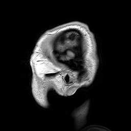
Os sinais e sintomas da displasia espondilodisplásica incluem baixa estatura com membros curtos desde o nascimento, alterações na coluna (como vértebras achatadas — platispondilia fina como uma bolacha — e estreitamento do canal espinhal na região do pescoço), pelve pequena e com formato anormal (ilíacos em forma de caracol), e ossos do crânio com testa proeminente. Podem ocorrer também alterações nas mãos, como dedos tortos (clinodactilia) e curvatura anormal (camptodactilia), além de deformidade em valgo da mão. Em alguns casos, há atraso global grave do desenvolvimento, ausência de interação social, e problemas neurológicos como hipoplasia cerebelar e meningocele lombossacral. Achados pré-natais incluem ausência de ossificação intrauterina das costelas e osso nasal fetal ausente. A condição pode evoluir para parada cardiorrespiratória e é considerada uma displasia esquelética letal.[1][3]
Causas genéticas
A displasia espondilodisplásica está associada a alterações (mutações) em diversos genes que desempenham papéis importantes no desenvolvimento e na manutenção dos ossos e cartilagens. Os genes já identificados incluem: GDF5, GPX4, EXTL3, TRPV4, COL2A1, BMPER, TRIP11, INPPL1, PAPSS2, MYH3, PAM16 e SLC35D1. Cada um desses genes fornece instruções para a produção de proteínas essenciais para o crescimento esquelético. Mutações nesses genes podem interromper esses processos, levando às características da doença.[1][4]
Diagnóstico
O diagnóstico da displasia espondilodisplásica é baseado na avaliação clínica dos sinais e sintomas, exames de imagem (como radiografias que mostram as alterações ósseas características) e, principalmente, por meio de testes genéticos. Atualmente, existem 336 testes genéticos disponíveis para essa condição, e 215 variantes genéticas associadas foram registradas no banco de dados ClinVar. A confirmação genética é importante para diferenciar essa displasia de outras condições esqueléticas semelhantes.[1][4]
Tratamento e manejo
Não há cura para a displasia espondilodisplásica, e o tratamento é focado no manejo dos sintomas e na melhora da qualidade de vida. O acompanhamento deve ser multidisciplinar, envolvendo ortopedistas, geneticistas, neurologistas e fisioterapeutas. Medidas de suporte podem incluir cirurgias ortopédicas para correção de deformidades, fisioterapia para estimular o desenvolvimento motor e suporte respiratório quando necessário. Cada caso deve ser avaliado individualmente por uma equipe médica especializada. No Brasil, a doença não possui cobertura específica pelo Sistema Único de Saúde (SUS).[1]
Prognóstico e qualidade de vida
O prognóstico da displasia espondilodisplásica é geralmente reservado, sendo classificada como uma displasia esquelética letal em muitos casos. A gravidade varia conforme as alterações genéticas e a presença de complicações como parada cardiorrespiratória ou meningocele. O suporte médico intensivo desde o nascimento pode melhorar a sobrevida e a qualidade de vida, mas o acompanhamento deve ser contínuo e adaptado às necessidades de cada paciente.[1][3]
Conteúdo informativo gerado e mantido automaticamente a partir de fontes oficiais (Orphanet, HPO, OMIM, SUS). Não substitui avaliação médica.
A displasia do tipo Schneckenbecken é uma condição hereditária autossômica recessiva rara, fatal no período pré-natal, que afeta os ossos e o crescimento pré-natal.
Encontrou um erro ou informação desatualizada? Sugira uma correção →
Entender a doença
Do básico ao detalhe, leia no seu ritmo
Preparando trilha educativa...
Sinais e sintomas
O que aparece no corpo e com que frequência cada sintoma acontece
Visão geral
A displasia espondilodisplásica é uma doença genética rara que afeta o desenvolvimento dos ossos, especialmente da coluna vertebral e dos membros. Ela faz parte de um grupo de condições conhecidas como displasias esqueléticas, que comprometem a formação e o crescimento do esqueleto. A condição pode ser grave e, em muitos casos, é identificada ainda antes do nascimento ou nos primeiros dias de vida.[1][3]
Sinais e sintomas
Os sinais e sintomas da displasia espondilodisplásica incluem baixa estatura com membros curtos desde o nascimento, alterações na coluna (como vértebras achatadas — platispondilia fina como uma bolacha — e estreitamento do canal espinhal na região do pescoço), pelve pequena e com formato anormal (ilíacos em forma de caracol), e ossos do crânio com testa proeminente. Podem ocorrer também alterações nas mãos, como dedos tortos (clinodactilia) e curvatura anormal (camptodactilia), além de deformidade em valgo da mão. Em alguns casos, há atraso global grave do desenvolvimento, ausência de interação social, e problemas neurológicos como hipoplasia cerebelar e meningocele lombossacral. Achados pré-natais incluem ausência de ossificação intrauterina das costelas e osso nasal fetal ausente. A condição pode evoluir para parada cardiorrespiratória e é considerada uma displasia esquelética letal.[1][3]
Causas genéticas
A displasia espondilodisplásica está associada a alterações (mutações) em diversos genes que desempenham papéis importantes no desenvolvimento e na manutenção dos ossos e cartilagens. Os genes já identificados incluem: GDF5, GPX4, EXTL3, TRPV4, COL2A1, BMPER, TRIP11, INPPL1, PAPSS2, MYH3, PAM16 e SLC35D1. Cada um desses genes fornece instruções para a produção de proteínas essenciais para o crescimento esquelético. Mutações nesses genes podem interromper esses processos, levando às características da doença.[1][4]
Diagnóstico
O diagnóstico da displasia espondilodisplásica é baseado na avaliação clínica dos sinais e sintomas, exames de imagem (como radiografias que mostram as alterações ósseas características) e, principalmente, por meio de testes genéticos. Atualmente, existem 336 testes genéticos disponíveis para essa condição, e 215 variantes genéticas associadas foram registradas no banco de dados ClinVar. A confirmação genética é importante para diferenciar essa displasia de outras condições esqueléticas semelhantes.[1][4]
Tratamento e manejo
Não há cura para a displasia espondilodisplásica, e o tratamento é focado no manejo dos sintomas e na melhora da qualidade de vida. O acompanhamento deve ser multidisciplinar, envolvendo ortopedistas, geneticistas, neurologistas e fisioterapeutas. Medidas de suporte podem incluir cirurgias ortopédicas para correção de deformidades, fisioterapia para estimular o desenvolvimento motor e suporte respiratório quando necessário. Cada caso deve ser avaliado individualmente por uma equipe médica especializada. No Brasil, a doença não possui cobertura específica pelo Sistema Único de Saúde (SUS).[1]
Prognóstico e qualidade de vida
O prognóstico da displasia espondilodisplásica é geralmente reservado, sendo classificada como uma displasia esquelética letal em muitos casos. A gravidade varia conforme as alterações genéticas e a presença de complicações como parada cardiorrespiratória ou meningocele. O suporte médico intensivo desde o nascimento pode melhorar a sobrevida e a qualidade de vida, mas o acompanhamento deve ser contínuo e adaptado às necessidades de cada paciente.[1][3]
Conteúdo informativo gerado e mantido automaticamente a partir de fontes oficiais (Orphanet, HPO, OMIM, SUS). Não substitui avaliação médica.
Partes do corpo afetadas
+ 163 sintomas em outras categorias
Características mais comuns
Os sintomas variam de pessoa para pessoa. Abaixo estão as 411 características clínicas mais associadas, ordenadas por frequência.
Linha do tempo da pesquisa
Encontrou um erro ou informação desatualizada? Sugira uma correção →
Genética e causas
O que está alterado no DNA e como passa nas famílias
Genes associados
15 genes identificados com associação a esta condição.
Growth factor involved in bone and cartilage formation. During cartilage development regulates differentiation of chondrogenic tissue through two pathways. Firstly, positively regulates differentiation of chondrogenic tissue through its binding of high affinity with BMPR1B and of less affinity with BMPR1A, leading to induction of SMAD1-SMAD5-SMAD8 complex phosphorylation and then SMAD protein signaling transduction (PubMed:15530414, PubMed:21976273, PubMed:24098149, PubMed:25092592). Secondly, n
SecretedCell membrane
Acromesomelic dysplasia 2A
A form of acromesomelic dysplasia, a skeletal disorder characterized by short stature, very short limbs and hand/foot malformations. The severity of limb abnormalities increases from proximal to distal with profoundly affected hands and feet showing brachydactyly and/or rudimentary fingers (knob-like fingers). AMD2A is an autosomal recessive form characterized by normal axial skeletons and missing or fused skeletal elements within the hands and feet.
Essential antioxidant peroxidase that directly reduces phospholipid hydroperoxide even if they are incorporated in membranes and lipoproteins (PubMed:40281343). Can also reduce cholesterol hydroperoxide and thymine hydroperoxide (By similarity). Plays a key role in protecting cells from oxidative damage by preventing membrane lipid peroxidation (PubMed:40281343). Required to prevent cells from ferroptosis, a non-apoptotic cell death resulting from an iron-dependent accumulation of lipid reactive
MitochondrionCytoplasm
Spondylometaphyseal dysplasia, Sedaghatian type
A form of spondylometaphyseal dysplasia, a group of short stature disorders distinguished by abnormalities in the vertebrae and the metaphyses of the tubular bones. SMDS is a neonatal lethal form characterized by severe metaphyseal chondrodysplasia with mild limb shortening, platyspondyly, cardiac conduction defects, and central nervous system abnormalities.
Glycosyltransferase which regulates the biosynthesis of heparan sulfate (HS) (PubMed:28132690, PubMed:28148688). Initiates HS synthesis by transferring the first N-acetyl-alpha-D-glucosamine (alpha-GlcNAc) residue (GlcNAcT-I activity) to the tetrasaccharide linker (GlcA-Gal-Gal-Xyl-)Ser core linker (PubMed:11390981, PubMed:35676258). May also transfer alpha-GlcNAc residues during HS elongation (GlcNAcT-II activity) (PubMed:11390981, PubMed:35676258). Lacks glucuronyl transferase II (GlcAT-II) ac
Endoplasmic reticulum membraneGolgi apparatusCell membraneNucleus
Immunoskeletal dysplasia with neurodevelopmental abnormalities
An autosomal recessive disorder characterized by variable skeletal abnormalities and neurodevelopmental defects. Neurologic manifestations include intellectual disability and motor delay. Some patients manifest hypotonia and seizures. Skeletal features include disproportionate short stature, cervical malformations, epiphyseal and metaphyseal dysplasia, and rarely premature craniosynostosis with progressive microcephaly. Severe combined immunodeficiency with a complete absence of T cells is observed in some patients.
Non-selective calcium permeant cation channel involved in osmotic sensitivity and mechanosensitivity (PubMed:16293632, PubMed:18695040, PubMed:18826956, PubMed:22526352, PubMed:23136043, PubMed:29899501). Activation by exposure to hypotonicity within the physiological range exhibits an outward rectification (PubMed:18695040, PubMed:18826956, PubMed:29899501). Also activated by heat, low pH, citrate and phorbol esters (PubMed:16293632, PubMed:18695040, PubMed:18826956, PubMed:20037586, PubMed:219
Cell membraneApical cell membraneCell junction, adherens junctionCell projection, ciliumEndoplasmic reticulum
Brachyolmia 3
A form of brachyolmia, a clinically and genetically heterogeneous skeletal dysplasia primarily affecting the spine and characterized by a short trunk, short stature, and platyspondyly. BCYM3 is an autosomal dominant form with severe scoliosis with or without kyphosis, and flattened irregular cervical vertebrae.
Type II collagen is specific for cartilaginous tissues. It is essential for the normal embryonic development of the skeleton, for linear growth and for the ability of cartilage to resist compressive forces
Secreted, extracellular space, extracellular matrix
Spondyloepiphyseal dysplasia congenital type
Disorder characterized by disproportionate short stature and pleiotropic involvement of the skeletal and ocular systems.
Inhibitor of bone morphogenetic protein (BMP) function, it may regulate BMP responsiveness of osteoblasts and chondrocytes
Secreted
Diaphanospondylodysostosis
A rare, recessively inherited, perinatal lethal skeletal disorder. The primary skeletal characteristics of the phenotype include a small chest, abnormal vertebral segmentation, and posterior rib gaps containing incompletely differentiated mesenchymal tissue. Consistent craniofacial features include ocular hypertelorism, epicanthal folds, a depressed nasal bridge with a short nose, and low-set ears. The most commonly described extraskeletal finding is nephroblastomatosis with cystic kidneys, but other visceral findings have been described in some cases.
Is a membrane tether required for vesicle tethering to Golgi. Has an essential role in the maintenance of Golgi structure and function (PubMed:25473115, PubMed:30728324). It is required for efficient anterograde and retrograde trafficking in the early secretory pathway, functioning at both the ER-to-Golgi intermediate compartment (ERGIC) and Golgi complex (PubMed:25717001). Binds the ligand binding domain of the thyroid receptor (THRB) in the presence of triiodothyronine and enhances THRB-modula
Golgi apparatus, cis-Golgi network membraneCytoplasm, cytoskeletonEndoplasmic reticulum-Golgi intermediate compartment membrane
Phosphatidylinositol (PtdIns) phosphatase that specifically hydrolyzes the 5-phosphate of phosphatidylinositol-3,4,5-trisphosphate (PtdIns(3,4,5)P3) to produce PtdIns(3,4)P2, thereby negatively regulating the PI3K (phosphoinositide 3-kinase) pathways (PubMed:16824732). Required for correct mitotic spindle orientation and therefore progression of mitosis (By similarity). Plays a central role in regulation of PI3K-dependent insulin signaling, although the precise molecular mechanisms and signaling
Cytoplasm, cytosolCytoplasm, cytoskeletonMembraneCell projection, filopodiumCell projection, lamellipodiumBasal cell membraneNucleusNucleus speckleCytoplasm, cytoskeleton, spindle pole
Bifunctional enzyme with both ATP sulfurylase and APS kinase activity, which mediates two steps in the sulfate activation pathway. The first step is the transfer of a sulfate group to ATP to yield adenosine 5'-phosphosulfate (APS), and the second step is the transfer of a phosphate group from ATP to APS yielding 3'-phosphoadenylylsulfate/PAPS, the activated sulfate donor used by sulfotransferases (PubMed:11773860, PubMed:19474428, PubMed:23824674, PubMed:25594860). In mammals, PAPS is the sole s
Brachyolmia type 4 with mild epiphyseal and metaphyseal changes
A form of brachyolmia, a clinically and genetically heterogeneous skeletal dysplasia primarily affecting the spine and characterized by a short trunk, short stature, and platyspondyly. BCYM4 is an autosomal recessive form with mild epiphyseal and metaphyseal changes. Clinical features include short stature evidenced at birth, short and bowed lower limbs, mild brachydactyly, kyphoscoliosis, abnormal gait, enlarged knee joints. Some BCYM4 patients may manifest premature pubarche and hyperandrogenism associated with skeletal dysplasia and short stature.
Muscle contraction
Cytoplasm, myofibril
Arthrogryposis, distal, 2A
A form of distal arthrogryposis, a disease characterized by congenital joint contractures that mainly involve two or more distal parts of the limbs, in the absence of a primary neurological or muscle disease. DA2A is characterized by contractures of the hands and feet, oropharyngeal abnormalities, scoliosis, and a distinctive face that includes a very small oral orifice, puckered lips, and a H-shaped dimple of the chin.
Regulates ATP-dependent protein translocation into the mitochondrial matrix. Inhibits DNAJC19 stimulation of HSPA9/Mortalin ATPase activity
Mitochondrion inner membrane
Spondylometaphyseal dysplasia, Megarbane-Dagher-Melike type
An autosomal recessive disease characterized by pre- and postnatal short stature, developmental delay, dysmorphic facial appearance, narrow chest, prominent abdomen, platyspondyly, and short limbs.
Antiporter that transports nucleotide sugars across the endoplasmic reticulum (ER) membrane in exchange for either their cognate nucleoside monophosphate or another nucleotide sugar (PubMed:16965264, PubMed:17599910, PubMed:31423530). Transports various UDP-sugars including UDP-N-acetyl-alpha-D-glucosamine (UDP-GlcNAc), UDP-N-acetyl-alpha-D-galactosamine (UDP-GalNAc) and UDP-alpha-D-glucuronate (UDP-GlcA), which are used by ER glucosyltransferases as sugar donors for the synthesis of sugar chain
Endoplasmic reticulum membrane
Schneckenbecken dysplasia
A rare, lethal autosomal recessive skeletal dysplasia characterized by snail-like configuration of the hypoplastic iliac bone, short-limbed dwarfism, short ribs, and flattened, hypoplastic vertebral bodies. SHNKND is lethal in the neonatal period.
Sulfate transporter which mediates sulfate uptake into chondrocytes in order to maintain adequate sulfation of proteoglycans which is needed for cartilage development (PubMed:11448940, PubMed:15294877, PubMed:20219950, PubMed:7923357). Mediates electroneutral anion exchange of sulfate ions for oxalate ions and of sulfate and oxalate ions for chloride ions (PubMed:20219950). Mediates exchange of sulfate and oxalate ions for hydroxyl ions and of chloride ions for bromide, iodide and nitrate ions (
Cell membraneApical cell membrane
Diastrophic dysplasia
An autosomal recessive disease characterized by osteochondrodysplasia with clinical features including dwarfism, spinal deformation, and specific joint abnormalities.
Connects cell membrane constituents to the actin cytoskeleton. May promote orthogonal branching of actin filaments and links actin filaments to membrane glycoproteins. Anchors various transmembrane proteins to the actin cytoskeleton. Interaction with FLNA may allow neuroblast migration from the ventricular zone into the cortical plate. Various interactions and localizations of isoforms affect myotube morphology and myogenesis. Isoform 6 accelerates muscle differentiation in vitro
Cytoplasm, cell cortexCytoplasm, cytoskeletonCytoplasm, cytoskeleton, stress fiberCytoplasm, myofibril, sarcomere, Z line
On ligand binding, forms a receptor complex consisting of two type II and two type I transmembrane serine/threonine kinases. Type II receptors phosphorylate and activate type I receptors which autophosphorylate, then bind and activate SMAD transcriptional regulators. Receptor for BMP7/OP-1 and GDF5. Positively regulates chondrocyte differentiation through GDF5 interaction
Cell membrane
Acromesomelic dysplasia 3
A form of acromesomelic dysplasia, a skeletal disorder characterized by short stature, very short limbs and hand/foot malformations. The severity of limb abnormalities increases from proximal to distal with profoundly affected hands and feet showing brachydactyly and/or rudimentary fingers (knob-like fingers). AMD3 is an autosomal recessive form characterized by bilateral aplasia of the fibula, severe brachydactyly, and fusion of carpal and tarsal bones.
Variantes genéticas (ClinVar)
215 variantes patogênicas registradas no ClinVar.
Vias biológicas (Reactome)
44 vias biológicas associadas aos genes desta condição.
Diagnóstico
Os sinais que médicos procuram e os exames que confirmam
Tratamento e manejo
Remédios, cuidados de apoio e o que precisa acompanhar
Onde tratar no SUS
Hospitais de referência no Brasil e o protocolo oficial do SUS (PCDT)
🇧🇷 Atendimento SUS — Displasia espondilodisplásica
Selecione um estado ou use sua localização para ver resultados.
Dados de DATASUS/CNES, SBGM, ABNeuro e Ministério da Saúde. Sempre confirme a disponibilidade diretamente com o estabelecimento.
Pesquisa ativa
Ensaios clínicos abertos e novidades científicas recentes
Pesquisa e ensaios clínicos
Nenhum ensaio clínico registrado para esta condição.
Publicações mais relevantes
[Analysis of clinical features and genetic variants in a Chinese pedigree affected with Spondyloepiphyseal dysplasia type Ehlers-Danlos syndrome due to variants of B3GALT6 gene].
To explore the clinical phenotype and genetic etiology of a child with Ehlers-Danlos syndrome, spondylodysplastic type 2 (EDSSPD2). A child who was admitted to the Children's Medical Center of the Affiliated Hospital of Guangdong Medical University in July 2024 for "delayed motor development for 1 and a half year" was selected as the study subject. Clinical data of the child was collected, including medical history, family history, and results of auxiliary examinations. Peripheral venous blood samples were collected from the child and his two brothers and both parents. Genomic DNA was extracted from the child and his family members and subjected to whole-exome sequencing (WES) and copy number variation (CNV) analysis. Sanger sequencing was used to verify the parental origin of the candidate variants. Multiple protein function prediction software tools, including SIFT, PolyPhen-2, and REVEL, were used to assess the impact of candidate variants on the protein function. Based on protein database information from UniProt, a two dimensional structural schematic of the target protein was generated. The pathogenicity of the variants was classified based on the guidelines from the American College of Medical Genetics and Genomics (ACMG). Relevant literature on the B3GALT6 gene variants leading to EDSSPD2 was retrieved from CNKI, Wanfang Data Knowledge Service Platform, and PubMed databases. The procedures followed in this study were reviewed and approved by the Medical Ethics Committee of Affiliated Hospital of Guangdong Medical University (Ethics No.:PJ2021-097). The proband was a 2-year-old male with an onset in infancy. The main clinical manifestations included loose skin, scoliosis and kyphosis, generalized hypermobility of joints, and motor developmental delay. WES has revealed two compound heterozygous variants of the B3GALT6 gene (NM_080605.4): c.766C>T (p.Arg256Trp) and c.962G>A (p.Cys321Tyr). Sanger sequencing verification showed that the c.766C>T and c.962G>A variants were respectively derived from his phenotypically normal father and mother. Bioinformatics analysis showed that for the c.766C>T (p.Arg256Trp) variant, the Arg256 site is located within the galactosyltransferase catalytic domain (GalT domain) of the β3GalT6 protein. According to the ACMG guidelines, the c.766C>T variant was classified as a likely pathogenic (PS3+PM2_supporting+PM3+PP3), and the c.962G>A was classified as a variant of unknown significance (PM2_Supporting+PM3+PP3). By following the pre-set literature retrieval strategy, a total of 12 articles related to B3GALT6 gene variants were identified (11 English and 1 Chinese), which involved a total of 71 patients. Among these, 4 reports (involving 20 patients) involved B3GALT6 gene variants leading to EDSSPD2. Among the 18 live-born EDSSPD2 patients (including the proband in this study), common clinical manifestations have included scoliosis (88.9%, 16/18), generalized hypotonia (83.3%, 15/18), and soft and lax skin (66.7%, 12/18). Some patients already showed skeletal abnormalities on prenatal ultrasound scan (22.2%, 4/18), while a few presented with cervical instability (16.7%, 3/18). One child had deceased at 18 months of age due to hypoxia caused by tracheomalacia and tracheal compression due to scoliosis. Among the 23 reported EDSSPD2 related B3GALT6 variant sites, missense variants were the most common (78.3%, 18/23), followed by nonsense variants (21.7%, 5/23). Above finding has enriched the clinical and mutational spectra of EDSSPD2. Early genetic testing has important clinical value for the diagnosis, differential diagnosis, and genetic counseling of this disease.
ZIP13 marks muscle satellite cells and contributes to their quiescent and active phase balance.
Loss of ZIP13 causes Ehlers-Danlos syndrome spondylodysplastic type 3 involving connective tissue dysplasias associated with a reduction in muscular strength. However, ZIP13 role in skeletal muscle homeostasis, particularly for the regulation of muscle satellite cells (MuSCs), remains poorly understood. In this study, we investigated Zip13-knockout (KO) mice and found a reduction in MuSCs of Zip13-KO mice, in which the quiescent and activated phase balances were disrupted. To clarify the physiological role and dynamics of ZIP13 expression in MuSCs, we generated Zip13-GFP knock-in (KI) mice encoding GFP at the Zip13 locus, which showed that ZIP13 contributes to the phase balance regulation of quiescent and activated MuSCs and their functions. Indeed, Zip13-KO mice exhibited delayed recovery from skeletal muscle injury, indicating ZIP13 requirement for proper skeletal muscle regeneration. Moreover, GFP expression was reduced in the MuSCs of homozygous Zip13-GFP KI mice whose intact ZIP13 expression was perturbed, suggesting that positive feedback mechanisms exist to maintain ZIP13 expression. Altogether, our results illustrate that ZIP13 might be positively involved in skeletal muscle regeneration by controlling the quiescent/activated phase balance of MuSCs through autoregulatory ZIP13 expression, and that newly generated Zip13-GFP KI mice would be useful for investigating the roles and dynamics of ZIP13-expressing cells.
A Mild Skeletal Dysplasia Caused by a Biallelic Missense Variant in the SLC35D1 Gene.
Biallelic variants in the SCL35D1 gene have been originally associated with a severe skeletal dysplasia called "Schneckenbecken dysplasia" because of the resemblance of the pelvic shape to a snail. More recently, SLC35D1 variants have been associated with much milder phenotypes of skeletal dysplasia. Our report describes one such individual with a novel SLC35D1 variant. A 17-year-old male with a coarse face and short stature was referred to our clinic. On his radiographic imaging, shortness of the long bones and metaphyseal flaring were detected. Using a clinical exome panel, we discovered a novel homozygous missense variant in the SLC35D1 gene, c.899G>T (p.Gly300Val). We identified a biallelic variant that was causative for a mild skeletal dysplasia and showed its phenotypic effects. Our observation confirms the existence of nonlethal skeletal dysplasias associated with biallelic SLC35D1 variants and suggests the existence of a phenotypic spectrum.
B3GALT6-linkeropathy: Three illustrative patients spanning the disease spectrum.
The linkeropathies are a group of rare disorders, characterized by overlapping clinical features involving the skeletal and connective tissues. Each "linker" gene encodes an enzyme responsible for the addition of glycosaminoglycan chains to proteoglycans via a common tertrasaccharine linker region. The original descriptions of the autosomal recessive B3GALT6-related disorder showed that the associated clinical features are pleiotropic, spanning the skeletal dysplasia (Spondyloepimetaphyseal dysplasia with joint laxity) (SEMD-JL1) and connective tissue disorder (Ehlers-Danlos syndrome) (EDS spondylodysplastic Type 2) spectrum. Here, we describe three patients with biallelic B3GALT6 variants: each had different clinical presentations, and the two older patients initially received alternative clinical diagnoses (Larsen syndrome and Osteogenesis imperfecta, respectively). We describe the clinico-radiological features of these patients to highlight the spectrum of disease associated with the B3GALT6-linkeropathy.
Broadening the phenotypic spectrum of Beta3GalT6-associated phenotypes.
Biallelic mutations in B3GALT6, coding for a galactosyltransferase involved in the synthesis of glycosaminoglycans (GAGs), have been associated with various clinical conditions, causing spondyloepimetaphyseal dysplasia with joint laxity type 1 (SEMDJL1 or SEMDJL Beighton type), Al-Gazali syndrome (ALGAZ), and a severe progeroid form of Ehlers-Danlos syndrome (EDSSPD2). In the 2017 Ehlers-Danlos syndrome (EDS) classification, Beta3GalT6-related disorders were grouped in the spondylodysplastic EDSs together with spondylodysplastic EDSs due to B4GALT7 and SLC39A13 mutations. Herein, we describe a patient with a previously unreported homozygous pathogenic B3GALT6 variant resulting in a complex phenotype more severe than spondyloepimetaphyseal dysplasia with joint laxity type 1, and having dural ectasia and aortic dilation as additionally associated features, further broadening the phenotypic spectrum of the Beta3GalT6-related syndromes. We also document the utility of repeating sequencing in patients with uninformative exomes, particularly when performed by using "first generations" enrichment capture methods.
Publicações recentes
[Analysis of clinical features and genetic variants in a Chinese pedigree affected with Spondyloepiphyseal dysplasia type Ehlers-Danlos syndrome due to variants of B3GALT6 gene].
Human Proteoglycan Linkage Region Glycosyltransferases are Dimeric and Show Unexpected Specificities.
B3GALT6 mutations lead to compromised connective tissue biomechanics in Ehlers-Danlos syndrome.
Oral and maxillofacial clinical features of Ehlers-Danlos syndrome: a systematic review.
[Ocular manifestations and genetic aspects of Ehlers-Danlos syndrome].
📚 EuropePMCmostrando 9
[Analysis of clinical features and genetic variants in a Chinese pedigree affected with Spondyloepiphyseal dysplasia type Ehlers-Danlos syndrome due to variants of B3GALT6 gene].
Zhonghua yi xue yi chuan xue za zhi = Zhonghua yixue yichuanxue zazhi = Chinese journal of medical geneticsZIP13 marks muscle satellite cells and contributes to their quiescent and active phase balance.
Scientific reportsA Mild Skeletal Dysplasia Caused by a Biallelic Missense Variant in the SLC35D1 Gene.
Molecular syndromologyB3GALT6-linkeropathy: Three illustrative patients spanning the disease spectrum.
European journal of medical geneticsBroadening the phenotypic spectrum of Beta3GalT6-associated phenotypes.
American journal of medical genetics. Part ASevere Peripheral Joint Laxity is a Distinctive Clinical Feature of Spondylodysplastic-Ehlers-Danlos Syndrome (EDS)-B4GALT7 and Spondylodysplastic-EDS-B3GALT6.
GenesFurther Defining the Phenotypic Spectrum of B3GAT3 Mutations and Literature Review on Linkeropathy Syndromes.
GenesBiallelic B3GALT6 mutations cause spondylodysplastic Ehlers-Danlos syndrome.
Human molecular geneticsA B3GALT6 variant in patient originally described as Al-Gazali syndrome and implicating the endoplasmic reticulum quality control in the mechanism of some β3GalT6-pathy mutations.
Clinical geneticsAssociações
Organizações que acompanham esta doença — pra ter apoio e orientação
Ainda não temos associações cadastradas para Displasia espondilodisplásica.
É de uma associação que acompanha esta doença? Fale com a gente →
Comunidades
Grupos ativos de quem convive com esta doença aqui no Raras
Ainda não existe comunidade no Raras para Displasia espondilodisplásica
Pacientes, familiares e cuidadores se organizam em comunidades pra compartilhar experiências, fazer perguntas e se apoiar. Você pode ser o primeiro.
Tire suas dúvidas
Perguntas, dicas e experiências compartilhadas aqui na página
Participe da discussão
Faça login para postar dúvidas, compartilhar experiências e interagir com especialistas.
Fazer loginDoenças relacionadas
Doenças com sintomas parecidos — ajudam quem ainda está buscando diagnóstico
Referências e fontes
Bases de dados externas citadas neste artigo
Publicações científicas
Artigos indexados no PubMed ligados a esta doença no grafo RarasNet — título, periódico e PMID direto da fonte, sem intermediação de IA.
- [Analysis of clinical features and genetic variants in a Chinese pedigree affected with Spondyloepiphyseal dysplasia type Ehlers-Danlos syndrome due to variants of B3GALT6 gene].Zhonghua yi xue yi chuan xue za zhi = Zhonghua yixue yichuanxue zazhi = Chinese journal of medical genetics· 2025· PMID 41811047mais citado
- ZIP13 marks muscle satellite cells and contributes to their quiescent and active phase balance.
- A Mild Skeletal Dysplasia Caused by a Biallelic Missense Variant in the SLC35D1 Gene.
- B3GALT6-linkeropathy: Three illustrative patients spanning the disease spectrum.
- Broadening the phenotypic spectrum of Beta3GalT6-associated phenotypes.
- Human Proteoglycan Linkage Region Glycosyltransferases are Dimeric and Show Unexpected Specificities.
- B3GALT6 mutations lead to compromised connective tissue biomechanics in Ehlers-Danlos syndrome.
- Oral and maxillofacial clinical features of Ehlers-Danlos syndrome: a systematic review.
- [Ocular manifestations and genetic aspects of Ehlers-Danlos syndrome].
Bases de dados e fontes oficiais
Identificadores e referências canônicas usadas para montar este verbete.
- ORPHA:93434(Orphanet)
- MONDO:0019694(MONDO)
- GARD:19193(GARD (NIH))
- Variantes catalogadas(ClinVar)
- Busca completa no PubMed(PubMed)
- Q55788806(Wikidata)
Dados compilados pelo RarasNet a partir de fontes abertas (Orphanet, OMIM, MONDO, PubMed/EuropePMC, ClinicalTrials.gov, DATASUS, PCDT/MS). Este conteúdo é informativo e não substitui avaliação médica.
Conteúdo mantido por Agente Raras · Médicos e pesquisadores podem colaborar
Displasia espondilodisplásica
📋 Origem dos dados
Esta página agrega dados de fontes públicas e oficiais. Dados sobre cobertura no SUS (PCDT, CEAF) são verificados ativamente por agente proativo (ver badge no infobox). Demais dados têm atribuição de fonte + data da última sincronização — clique para abrir o original.
- Doença rara (ontologia)
- fonte: Orphanet
- Identificador unificado
- fonte: MONDO
- CID-11 (futuro)
- fonte: WHO ICD-11
- NIH/GARD
- fonte: GARD (NIH)
- Dado público estruturado
- fonte: Wikidata