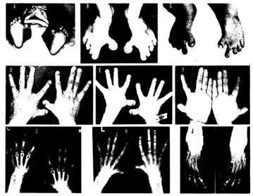
Essa síndrome é caracterizada pela combinação de um dedão do pé virado para dentro com polegares e primeiros dedos dos pés curtos. Nesses casos, os ossos da palma da mão, da planta do pé e as pontas dos dedos são mais curtos, mas as partes do meio e da base dos dedos têm tamanho normal. Além disso, há um afastamento dos dedos afetados.
Introdução
O que você precisa saber de cara
Essa síndrome é caracterizada pela combinação de um dedão do pé virado para dentro com polegares e primeiros dedos dos pés curtos. Nesses casos, os ossos da palma da mão, da planta do pé e as pontas dos dedos são mais curtos, mas as partes do meio e da base dos dedos têm tamanho normal. Além disso, há um afastamento dos dedos afetados.
Escala de raridade
<1/50kMuito rara
1/20kRara
1/10kPouco freq.
1/5kIncomum
1/2k
Encontrou um erro ou informação desatualizada? Sugira uma correção →
Entender a doença
Do básico ao detalhe, leia no seu ritmo
Preparando trilha educativa...
Sinais e sintomas
O que aparece no corpo e com que frequência cada sintoma acontece
Partes do corpo afetadas
+ 9 sintomas em outras categorias
Características mais comuns
Os sintomas variam de pessoa para pessoa. Abaixo estão as 20 características clínicas mais associadas, ordenadas por frequência.
Linha do tempo da pesquisa
Encontrou um erro ou informação desatualizada? Sugira uma correção →
Genética e causas
O que está alterado no DNA e como passa nas famílias
Nenhum gene associado encontrado
Os dados genéticos desta condição ainda estão sendo catalogados.
Diagnóstico
Os sinais que médicos procuram e os exames que confirmam
Tratamento e manejo
Remédios, cuidados de apoio e o que precisa acompanhar
Onde tratar no SUS
Hospitais de referência no Brasil e o protocolo oficial do SUS (PCDT)
🇧🇷 Atendimento SUS — Síndrome de braquidactilia-hálux varo pré-axial
Selecione um estado ou use sua localização para ver resultados.
Dados de DATASUS/CNES, SBGM, ABNeuro e Ministério da Saúde. Sempre confirme a disponibilidade diretamente com o estabelecimento.
Pesquisa ativa
Ensaios clínicos abertos e novidades científicas recentes
Pesquisa e ensaios clínicos
Nenhum ensaio clínico registrado para esta condição.
Publicações mais relevantes
The Incidence of Complications Following Scarf Osteotomy for the Treatment of Hallux Valgus: A Systematic Review With Meta-Analysis.
The Scarf osteotomy is a surgical procedure performed to correct a hallux valgus deformity. Multiple studies have supported use of the procedure with favorable outcomes. In contrast, there have been studies showing a significant complication rate with the procedure. Incidence of complications remains underreported in the literature. We performed a systemic review and meta-analysis examining a wide range of reported complications and associated clinical outcomes from the Scarf osteotomy. One hundred and sixteen publications were identified and 25 (21.6%) met our inclusion criteria. A total of 1583 Scarf procedures were included. Weighted mean follow-up was 26.4 months [range 12-168 months]. We found a 5.1% rate of recurrence, 3.5% rate of troughing, 1.0% rate of avascular necrosis, 1.8% rate of nonunion, 2.7% rate of malunion, 2.4% rate of infection, 5.3% rate of complex regional pain syndrome, and 3.4% rate of hallux varus. An average decrease in intermetatarsal angle of 6.3° was observed. No statistical difference was found in outcomes when comparing Scarf versus Scarf with additional procedure performed at time of surgery. To our knowledge, this systematic review and meta-analysis contains the highest number of Scarf procedures analyzed and presents complication rates on multiple adverse outcomes.
Is the Apert foot an overlooked aspect of this rare genetic disease? Clinical findings and treatment options for foot deformities in Apert syndrome.
Apert syndrome is characterised by the presence of craniosynostosis, midface retrusion and syndactyly of hands and feet, thus, synonymously referred to as acrocephalosyndactyly type I. Considering these multidisciplinary issues, frequently requiring surgical interventions at an early age, deformities of the feet have often been neglected and seem to be underestimated in the management of Apert syndrome. Typical Apert foot features range from complete fusion of the toes and a central nail mass to syndactyly of the second to fifth toe with a medially deviated great toe; however, no clear treatment algorithms were presented so far. This article reviews the current existing literature regarding the treatment approach of foot deformities in Apert syndrome. Overall, the main focus in the literature seems to be on the surgical approach to syndactyly separation of the toes and the management of the great toe deformity (hallux varus). Although the functional benefit of syndactyly separation in the foot has yet to be determined, some authors perform syndactyly separation usually in a staged procedure. Realignment of the great toe and first ray can be performed by multiple means including but not limited to second ray deletion, resection of the proximal phalanx delta bone on one side, corrective open wedge osteotomy, osteotomy of the osseous fusion between metatarsals I and II, and metatarsal I lengthening using gradual osteodistraction. Tarsal fusions and other anatomical variants may be present and have to be corrected on an individual basis. Shoe fitting problems are frequently mentioned as indication for surgery while insole support may be helpful to alleviate abnormal plantar pressures. There is a particular need for multicenter studies to better elaborate surgical indications and treatment plans for this rare entity. Plantar pressure measurements using pedobarography should be enforced in order to document the biomechanical foot development and abnormalities during growth, and to help with indication setting. Treatment options may include conservative means (i.e. insoles, orthopedic shoes) or surgery to improve biomechanics and normalize plantar pressures. Level V.
Further delineation of autosomal recessive intellectual disability syndrome caused by homozygous variant of the NSUN2 gene in a chinese pedigree.
The enzyme NOP2/Sun RNA methyltransferase 2 (NSUN2) catalyzes the methylation of cytosine to 5-methylcytosine (m5C) at position 34 of tRNA(Leu; CAA) precursors containing introns that play a vital role in spindle assembly during mitosis and chromosome segregation. Biallelic variants in the NSUN2 gene cause a rare intellectual disability that has been identified only in a few Middle Eastern patients. Affected individuals usually have other deformities, including developmental delay, short stature, microcephaly, and facial dysmorphism. The aim of this study was to identify the genetic cause of three female patients from a Chinese pedigree, who presented with similar phenotype consisting of the above clinical features. Whole-exome sequencing (WES) was used to screen for causal variants in the genome, and the candidate variants were subsequently verified using Sanger sequencing. WES revealed a previously unreported homozygous nonsense variant (NM_017755.5: c.1004T>A, p.Leu335*) in exon 9 of NSUN2, which was consistent with the clinical phenotype of the patients and co-segregated with the disease in their family. A comparison of this phenotype with that of patients in published reports uncovered several novel clinical features related to NSUN2 variations, including feeding difficulties, slender hands and fingers, severely restricted finger mobility, hallux valgus, varus foot, and elevated α-hydroxybutyrate dehydrogenase (HBDH). These are the first findings of a non-consanguineous Chinese pedigree with a homozygous NSUN2 variant. We expanded the phenotypic spectrum associated with NSUN2 variations.
Minimally Invasive Hallux Interphalangeal Joint Arthrodesis for Hallux Varus in Pfeiffer Syndrome: A Case Report.
Pfeiffer syndrome is a rare hereditary condition with an autosomal dominant transmission caused by a mutation that affects fibroblast growth factor receptors. It is one of the acrocephalosyndactyly diseases causing cranial malformations owing to early suture fusion. In the foot, it is typically associated with hallux varus, first ray hyperplasia, and partial lesser digit syndactyly. We report a clinical case of a 10-year-old patient with Pfeiffer type I syndrome with bilateral severe hallux varus due to a hypoplastic trapezoidal shaped proximal phalanx, a distal, medial-facing articular surface, and interphalangeal instability. This deformity was addressed by minimally invasive hallux interphalangeal joint arthrodesis with internal and external fixation. We report the results at the 2-year follow-up point.
[Arthrodesis of the First Metatarsophalangeal Joint by Locking Plate].
PURPOSE OF THE STUDY The authors in their paper evaluate a group of patients who underwent arthrodesis of the first metatarsophalangeal joint using a locking plate. MATERIAL AND METHODS In the period 2010-2015, we performed surgery in 51 patients (56 forefeet), of which in 5 cases bilaterally and in 46 cases unilaterally, in 38 women and 13 men. The mean age was 57.8 years, the mean follow-up was 3.1 years. The indications for surgery were hallux rigidus in 23 patients, hallux valgus in 15 patients, hallux varus in 3 patients, and hallux erectus in 2 patients. In 4 patients the surgery was performed for valgus deformity associated with rheumatoid arthritis, 9 patients were indicated for a failure of the prior surgical intervention. In all 56 forefeet, the anatomic, low-profile titanium plate Variable Angle LCP 1st MTP Fusion Plate 2.4/2.7 was used. RESULTS According to Gainor s score the surgical outcomes were assessed as excellent in 46 patients who underwent surgery (90%), good in 4 patients (8%), fair in 1 patient (2%), and poor in 0 patient (0%). In 53 forefeet, the control radiographs showed solid bone union. In 2 patients and 3 forefeet, non-union of the arthrodesis occurred. In 2 forefeet, revision arthrodesis was performed, after which solid bone union followed. Malpositioned union was reported in 5 forefeet, of which in 4 cases into valgosity and in 1 case into dorsiflexion. DISCUSSION Numerous fixation materials can be used for arthrodesis of the first metatarsophalangeal joint. The use of the least stable Kirschner wires (cerclage) is being abandoned and substituted with a more stable fixation by screws, memory staples and locking plates. The achievement of excellent results requires proper positioning of the arthrodesis. Impingement syndrome between the big toe and the second toe can result in painful callosities formation, too large dorsiflexion can lead to a hallux hammertoe, with reduced big toe support function, to metatarsalgia. CONCLUSIONS The arthrodesis is indicated in patients with Grade III and IV hallux rigidus, with severe hallux valgus, hallux varus, and in patients in whom the previous surgeries failed. We tend to prefer stable arthrodesis. Fixation by anatomic LCP plate facilitates early rehabilitation, loading and early return to work and sports activities. Key words: arthrodesis, metatarsophalangeal joint, hallux rigidus, hallux valgus.
Publicações recentes
Cerebrovascular involvement in hereditary spherocytosis: observational cohort and case-control MRI study.
Derivation and Validation of the PRECISE-HBR Score to Predict Bleeding After Percutaneous Coronary Intervention.
Demystifying the Contemporary Role of 12-Month Dual Antiplatelet Therapy After Acute Coronary Syndrome.
Brain perfusion changes in beta-thalassemia.
Defining Strategies of Modulation of Antiplatelet Therapy in Patients With Coronary Artery Disease: A Consensus Document from the Academic Research Consortium.
📚 EuropePMCmostrando 9
The Incidence of Complications Following Scarf Osteotomy for the Treatment of Hallux Valgus: A Systematic Review With Meta-Analysis.
The Journal of foot and ankle surgery : official publication of the American College of Foot and Ankle SurgeonsIs the Apert foot an overlooked aspect of this rare genetic disease? Clinical findings and treatment options for foot deformities in Apert syndrome.
BMC musculoskeletal disordersFurther delineation of autosomal recessive intellectual disability syndrome caused by homozygous variant of the NSUN2 gene in a chinese pedigree.
Molecular genetics & genomic medicine[Arthrodesis of the First Metatarsophalangeal Joint by Locking Plate].
Acta chirurgiae orthopaedicae et traumatologiae CechoslovacaMinimally Invasive Hallux Interphalangeal Joint Arthrodesis for Hallux Varus in Pfeiffer Syndrome: A Case Report.
The Journal of foot and ankle surgery : official publication of the American College of Foot and Ankle SurgeonsKaufman oculo-cerebro-facial syndrome in a child with small and absent terminal phalanges and absent nails.
Journal of human genetics[Congenital malformations of the forefoot].
Annales de chirurgie plastique et esthetique[EFFECTIVENESS OF HIGH TIBIAL OSTEOTOMY ASSISTED BY THREE- DIMENSIONAL PRINTING TECHNOLOGY FOR CORRECTION OF VARUS KNEE WITH OSTEOARTHRITIS].
Zhongguo xiu fu chong jian wai ke za zhi = Zhongguo xiufu chongjian waike zazhi = Chinese journal of reparative and reconstructive surgery[Distal soft-tissue procedure in hallux valgus deformity].
Operative Orthopadie und TraumatologieAssociações
Organizações que acompanham esta doença — pra ter apoio e orientação
Ainda não temos associações cadastradas para Síndrome de braquidactilia-hálux varo pré-axial.
É de uma associação que acompanha esta doença? Fale com a gente →
Comunidades
Grupos ativos de quem convive com esta doença aqui no Raras
Ainda não existe comunidade no Raras para Síndrome de braquidactilia-hálux varo pré-axial
Pacientes, familiares e cuidadores se organizam em comunidades pra compartilhar experiências, fazer perguntas e se apoiar. Você pode ser o primeiro.
Tire suas dúvidas
Perguntas, dicas e experiências compartilhadas aqui na página
Participe da discussão
Faça login para postar dúvidas, compartilhar experiências e interagir com especialistas.
Fazer loginDoenças relacionadas
Doenças com sintomas parecidos — ajudam quem ainda está buscando diagnóstico
Referências e fontes
Bases de dados externas citadas neste artigo
Publicações científicas
Artigos indexados no PubMed ligados a esta doença no grafo RarasNet — título, periódico e PMID direto da fonte, sem intermediação de IA.
- The Incidence of Complications Following Scarf Osteotomy for the Treatment of Hallux Valgus: A Systematic Review With Meta-Analysis.The Journal of foot and ankle surgery : official publication of the American College of Foot and Ankle Surgeons· 2023· PMID 37097272mais citado
- Is the Apert foot an overlooked aspect of this rare genetic disease? Clinical findings and treatment options for foot deformities in Apert syndrome.
- Further delineation of autosomal recessive intellectual disability syndrome caused by homozygous variant of the NSUN2 gene in a chinese pedigree.
- Minimally Invasive Hallux Interphalangeal Joint Arthrodesis for Hallux Varus in Pfeiffer Syndrome: A Case Report.The Journal of foot and ankle surgery : official publication of the American College of Foot and Ankle Surgeons· 2018· PMID 29103889mais citado
- [Arthrodesis of the First Metatarsophalangeal Joint by Locking Plate].
- Cerebrovascular involvement in hereditary spherocytosis: observational cohort and case-control MRI study.
- Derivation and Validation of the PRECISE-HBR Score to Predict Bleeding After Percutaneous Coronary Intervention.
- Demystifying the Contemporary Role of 12-Month Dual Antiplatelet Therapy After Acute Coronary Syndrome.
- Brain perfusion changes in beta-thalassemia.
- Defining Strategies of Modulation of Antiplatelet Therapy in Patients With Coronary Artery Disease: A Consensus Document from the Academic Research Consortium.
Bases de dados e fontes oficiais
Identificadores e referências canônicas usadas para montar este verbete.
- ORPHA:1278(Orphanet)
- OMIM OMIM:112450(OMIM)
- MONDO:0007214(MONDO)
- GARD:972(GARD (NIH))
- Busca completa no PubMed(PubMed)
- Q32145314(Wikidata)
Dados compilados pelo RarasNet a partir de fontes abertas (Orphanet, OMIM, MONDO, PubMed/EuropePMC, ClinicalTrials.gov, DATASUS, PCDT/MS). Este conteúdo é informativo e não substitui avaliação médica.
Conteúdo mantido por Agente Raras · Médicos e pesquisadores podem colaborar
Síndrome de braquidactilia-hálux varo pré-axial
📋 Origem dos dados
Esta página agrega dados de fontes públicas e oficiais. Dados sobre cobertura no SUS (PCDT, CEAF) são verificados ativamente por agente proativo (ver badge no infobox). Demais dados têm atribuição de fonte + data da última sincronização — clique para abrir o original.
- Doença rara (ontologia)
- fonte: Orphanet
- Identificador unificado
- fonte: MONDO
- Genética mendeliana
- fonte: OMIM
- Codificação WHO/SUS
- fonte: WHO ICD-10 / DATASUS
- NIH/GARD
- fonte: GARD (NIH)
- Indexação biomédica
- fonte: MeSH (NLM)
- Dado público estruturado
- fonte: Wikidata