As síndromes de Ehlers-Danlos (SED) são um grupo de 13 doenças genéticas do tecido conjuntivo. Os sintomas frequentemente incluem articulações frouxas, dor articular, pele elástica e aveludada, e formação anormal de cicatrizes. Estes podem ser notados ao nascimento ou na primeira infância. As complicações podem incluir dissecção aórtica, luxações articulares, escoliose, dor crônica ou osteoartrite precoce. A classificação existente foi atualizada pela última vez em 2017, quando várias formas mais raras de SED foram adicionadas.
Introdução
O que você precisa saber de cara
Visão geral
A Síndrome de baixa estatura-idade óssea avançada-osteoartrite de início precoce é uma condição genética rara, com prevalência estimada em menos de 1 caso por 1.000.000 de pessoas. Ela se caracteriza por baixa estatura, maturação óssea acelerada (idade óssea avançada) e desenvolvimento precoce de osteoartrite, geralmente já na infância ou adolescência.[1][3]
Sinais e sintomas
Os principais sinais e sintomas incluem baixa estatura (estagnação do desenvolvimento), braquidactilia (dedos das mãos e pés mais curtos que o normal), polegar curto, retrusão médio-facial (face com aparência de “achatada” na região do meio do rosto) e osteoartrite de início precoce, que pode causar dor e rigidez nas articulações já na infância ou adolescência.[1][3]
Causas genéticas
Diagnóstico
O diagnóstico é baseado na avaliação clínica dos sinais característicos (baixa estatura, idade óssea avançada, osteoartrite precoce) e confirmado por teste genético que identifique variantes patogênicas no gene ACAN. Atualmente, há 345 variantes registradas no ClinVar associadas a essa condição. O teste genético está disponível e pode ser solicitado por um médico geneticista.[1][4]
Tratamento e manejo
Não há cura para a síndrome, e o tratamento é focado no manejo dos sintomas, especialmente da osteoartrite. O acompanhamento deve ser multidisciplinar, incluindo ortopedista, reumatologista, fisioterapeuta e geneticista. Medidas como fisioterapia, uso de analgésicos e anti-inflamatórios (sempre sob prescrição médica) podem ajudar a controlar a dor e melhorar a função articular. Não há cobertura específica pelo SUS para esta condição.[1]
Prognóstico e qualidade de vida
O prognóstico varia conforme a gravidade dos sintomas, especialmente o impacto da osteoartrite precoce na mobilidade e qualidade de vida. Com acompanhamento adequado e manejo dos sintomas, muitas pessoas conseguem manter uma vida ativa. O aconselhamento genético é recomendado para famílias afetadas.[1]
Conteúdo informativo gerado e mantido automaticamente a partir de fontes oficiais (Orphanet, HPO, OMIM, SUS). Não substitui avaliação médica.
As síndromes de Ehlers-Danlos (SED) são um grupo de 13 doenças genéticas do tecido conjuntivo. Os sintomas frequentemente incluem articulações frouxas, dor articular, pele elástica e aveludada, e formação anormal de cicatrizes. Estes podem ser notados ao nascimento ou na primeira infância. As complicações podem incluir dissecção aórtica, luxações articulares, escoliose, dor crônica ou osteoartrite precoce. A classificação existente foi atualizada pela última vez em 2017, quando várias formas mais raras de SED foram adicionadas.
Escala de raridade
<1/50kMuito rara
1/20kRara
1/10kPouco freq.
1/5kIncomum
1/2k
Encontrou um erro ou informação desatualizada? Sugira uma correção →
Entender a doença
Do básico ao detalhe, leia no seu ritmo
Preparando trilha educativa...
Sinais e sintomas
O que aparece no corpo e com que frequência cada sintoma acontece
Visão geral
A Síndrome de baixa estatura-idade óssea avançada-osteoartrite de início precoce é uma condição genética rara, com prevalência estimada em menos de 1 caso por 1.000.000 de pessoas. Ela se caracteriza por baixa estatura, maturação óssea acelerada (idade óssea avançada) e desenvolvimento precoce de osteoartrite, geralmente já na infância ou adolescência.[1][3]
Sinais e sintomas
Os principais sinais e sintomas incluem baixa estatura (estagnação do desenvolvimento), braquidactilia (dedos das mãos e pés mais curtos que o normal), polegar curto, retrusão médio-facial (face com aparência de “achatada” na região do meio do rosto) e osteoartrite de início precoce, que pode causar dor e rigidez nas articulações já na infância ou adolescência.[1][3]
Causas genéticas
Diagnóstico
O diagnóstico é baseado na avaliação clínica dos sinais característicos (baixa estatura, idade óssea avançada, osteoartrite precoce) e confirmado por teste genético que identifique variantes patogênicas no gene ACAN. Atualmente, há 345 variantes registradas no ClinVar associadas a essa condição. O teste genético está disponível e pode ser solicitado por um médico geneticista.[1][4]
Tratamento e manejo
Não há cura para a síndrome, e o tratamento é focado no manejo dos sintomas, especialmente da osteoartrite. O acompanhamento deve ser multidisciplinar, incluindo ortopedista, reumatologista, fisioterapeuta e geneticista. Medidas como fisioterapia, uso de analgésicos e anti-inflamatórios (sempre sob prescrição médica) podem ajudar a controlar a dor e melhorar a função articular. Não há cobertura específica pelo SUS para esta condição.[1]
Prognóstico e qualidade de vida
O prognóstico varia conforme a gravidade dos sintomas, especialmente o impacto da osteoartrite precoce na mobilidade e qualidade de vida. Com acompanhamento adequado e manejo dos sintomas, muitas pessoas conseguem manter uma vida ativa. O aconselhamento genético é recomendado para famílias afetadas.[1]
Conteúdo informativo gerado e mantido automaticamente a partir de fontes oficiais (Orphanet, HPO, OMIM, SUS). Não substitui avaliação médica.
Partes do corpo afetadas
Características mais comuns
Os sintomas variam de pessoa para pessoa. Abaixo estão as 5 características clínicas mais associadas, ordenadas por frequência.
Linha do tempo da pesquisa
Encontrou um erro ou informação desatualizada? Sugira uma correção →
Genética e causas
O que está alterado no DNA e como passa nas famílias
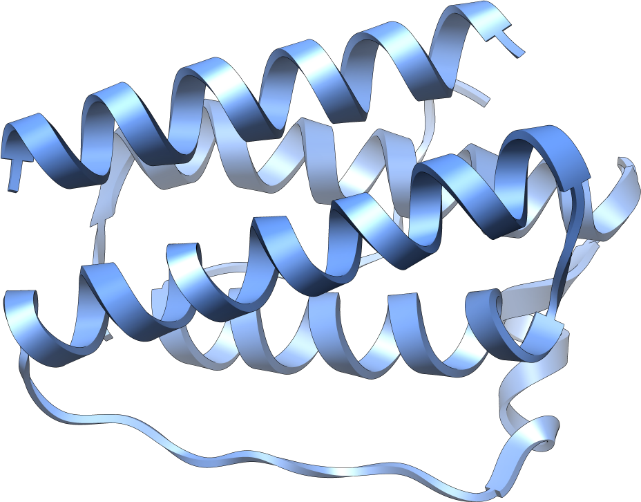
Genes associados
1 gene identificado com associação a esta condição. Padrão de herança: Autosomal dominant.
This proteoglycan is a major component of extracellular matrix of cartilagenous tissues. A major function of this protein is to resist compression in cartilage. It binds avidly to hyaluronic acid via an N-terminal globular region
Secreted, extracellular space, extracellular matrix
Spondyloepiphyseal dysplasia type Kimberley
Spondyloepiphyseal dysplasias are a heterogeneous group of congenital chondrodysplasias that specifically affect epiphyses and vertebrae. The autosomal dominant SEDK is associated with premature degenerative arthropathy.
Variantes genéticas (ClinVar)
345 variantes patogênicas registradas no ClinVar.
Classificação de variantes (ClinVar)
Distribuição de 1 variantes classificadas pelo ClinVar.
Vias biológicas (Reactome)
7 vias biológicas associadas aos genes desta condição.
Diagnóstico
Os sinais que médicos procuram e os exames que confirmam
Tratamento e manejo
Remédios, cuidados de apoio e o que precisa acompanhar
Onde tratar no SUS
Hospitais de referência no Brasil e o protocolo oficial do SUS (PCDT)
🇧🇷 Atendimento SUS — Síndrome de baixa estatura-idade óssea avançada-osteoartrite de início precoce
Selecione um estado ou use sua localização para ver resultados.
Dados de DATASUS/CNES, SBGM, ABNeuro e Ministério da Saúde. Sempre confirme a disponibilidade diretamente com o estabelecimento.
Pesquisa ativa
Ensaios clínicos abertos e novidades científicas recentes
Pesquisa e ensaios clínicos
Nenhum ensaio clínico registrado para esta condição.
Publicações mais relevantes
Engineered GO-Based Hydrogels for Controlled Hyaluronic Acid Release in Knee Osteoarthritis Treatment.
Osteoarthritis (OA) is a prevalent chronic pain syndrome and a leading cause of disability worldwide, characterized by progressive deterioration of articular cartilage. This degradation leads to pain, swelling, inflammation, and eventual stiffness as the cartilage wears down, causing bone-on-bone friction. Current medical treatments primarily aim at pain relief; however, many interventions, especially invasive or surgical ones, carry risks of adverse outcomes. Consequently, intra-articular (IA) therapy, particularly hyaluronic acid (HA) injections, is widely adopted as a conservative treatment option. HA plays a crucial role in maintaining joint homeostasis by supporting proteoglycan synthesis and scaffolding, restoring optimal HA concentrations in synovial fluid, and providing chondroprotective and anti-inflammatory effects. In recent years, hydrogels composed of natural and synthetic materials have emerged as promising candidates for OA treatment. Our research focuses on the biosynthesis and characterization of novel hydrogel composites combining short peptide hydrogelators with aminated graphene oxide (a-GO) nanosheets functionalized with HA (a-GO-HA@Hgel). These a-GO-HA@Hgel nanocomposites are designed to facilitate the controlled release of HA into the extracellular matrix, aiming to promote cartilage regeneration and mitigate inflammation. The strategy is to exploit the oxygen-containing functional groups of GO nanosheets to enable covalent coupling or physical adsorption of HA molecules through various chemical approaches. The resulting a-GO-HA are incorporated within hydrogel matrices to achieve sustained and controlled HA release. We study the influence of a-GO-HA on the native hydrogel structure and its viscoelastic properties, which are critical for mimicking the mechanical environment of native cartilage tissue. Through this multidisciplinary approach combining advanced materials science and cellular biology, this work aims to develop innovative nanocomposite hydrogels capable of delivering HA in a controlled manner, enhancing cartilage repair and providing a potential therapeutic strategy for OA management.
Ultrasound-guided PRP for coracoid syndrome: a novel case of conjoint tendon enthesopathy.
Tendinopathy of the conjoint tendon (CjT), comprising the short head of the biceps brachii and coracobrachialis, represents an uncommon etiology of anterior shoulder pain. To our knowledge, this is the first documented case in which leukocyte-rich platelet-rich plasma (LR-PRP) was employed for its treatment. A 54-year-old male weightlifter presented with right shoulder pain that was refractory to conventional conservative management. Although magnetic resonance imaging revealed mild rotator cuff tendinopathy and acromioclavicular osteoarthritis, point-of-care ultrasound (POCUS) identified a hypoechoic lesion within the CjT accompanied by enthesopathy. An ultrasound-guided injection of LR-PRP resulted in complete symptom resolution within 1 month, with sustained improvement observed at the 12-month follow-up. Adverse effects were limited to mild, transient deep injection-site soreness noted on the first post-procedure day. Furthermore, ultrasound evaluation at 6 months demonstrated notable improvement in tendon morphology. This case underscores the importance of considering CjT tendinopathy in the differential diagnosis of anterior shoulder pain and highlights the critical role of POCUS in enhancing diagnostic accuracy and guiding targeted therapeutic interventions. LR-PRP may serve as a regenerative alternative to corticosteroid therapy, promoting tendon healing while avoiding corticosteroids side-effects. As a single case report, generalizability is limited, and conclusions should be interpreted with caution. Front shoulder pain can happen for many reasons. One rare cause is a problem with the conjoint tendon (CjT) – a tendon that connects two muscles (the short head of the biceps and the coracobrachialis) to the front of the shoulder. Because this condition feels similar to other common shoulder issues, it is often overlooked. We present the case of a 54-year-old man who regularly lifted weights and had pain in his right shoulder for several months. Rest, pain medication, and physiotherapy did not help. An MRI showed common age-related changes, but bedside ultrasound (also called POCUS) clearly showed inflammation in the CjT and where it attaches to the bone. Using ultrasound guidance, we injected a special preparation of platelet-rich plasma (PRP) – a healing substance from his own blood, enriched with white blood cells – directly into the injured tendon. His pain improved quickly and lasted. Within a month, he was pain-free. Six weeks later, he resumed training, and at 1 year, he remained symptom-free. The only side effect was mild soreness the day after the injection. A follow-up ultrasound 6 months later showed a healthier tendon. This case shows how ultrasound can help find and treat unusual causes of shoulder pain. It also suggests that PRP may help tendons heal and could be a safer alternative to steroid injections. However, because this is only one case, more research is needed to confirm these results.
Influence of pelvic incidence-lumbar lordosis mismatch on surgical outcomes of total hip arthroplasty: a retrospective cohort study.
Pelvic incidence-lumbar lordosis (PI-LL) mismatch has been widely studied in spinal disorders; however, its impact on total hip arthroplasty (THA) outcomes remains unclear. This study evaluated the impact of preoperative PI-LL mismatch on postoperative functional recovery and spinopelvic dynamics in patients undergoing THA. This retrospective cohort study included 167 patients who underwent primary unilateral THA. Patients were categorised into two groups based on a PI-LL mismatch threshold of 10°: mismatch group (PI-LL ≥ 10°) and matched group (PI-LL < 10°). Preoperative characteristics, spinopelvic parameters, and clinical outcomes were analysed. The Numerical Rating Scale (NRS) for pain, modified Harris Hip Score (mHHS), and Western Ontario and McMaster Universities Osteoarthritis Index (WOMAC) were used to assess the hip condition preoperatively and 6 months postoperatively. The mismatch group exhibited a significantly higher pelvic tilt (PT) and lower sacral slope (SS) than the matched group. Furthermore, the mismatch group demonstrated reduced spinal flexibility, as indicated by a significantly smaller difference in spinopelvic angle between maximum lateral flexion positions. The spinopelvic parameters (PI, LL, PT, and SS) remained stable from preoperative to postoperative assessments. Preoperative mHHS and WOMAC scores were significantly worse in the mismatch group. However, no significant differences in postoperative outcomes were observed between the groups at 6 months. PI-LL mismatch was associated with altered spinopelvic parameters and poorer preoperative functional scores. However, short-term outcomes after THA remained comparable. These findings underscore the importance of individualised preoperative assessment while supporting the efficacy of THA, irrespective of spinopelvic alignment.
The THA-10 Score for Predicting Conversion to Total Hip Arthroplasty After Contemporary Hip Arthroscopy for Femoroacetabular Impingement Syndrome at a Minimum 10-Year Follow-up.
Previous studies have identified predictors of total hip arthroplasty (THA) conversion after hip arthroscopy (HA) for femoroacetabular impingement syndrome (FAIS) at short- to midterm follow-up, yet no studies to the authors' knowledge have established a scoring system for predicting THA conversion at a minimum 10-year follow-up. To create a scoring system to predict 10-year THA conversion after contemporary HA for FAIS. Case-control study; Level of evidence, 3. Data were prospectively collected from patients undergoing primary contemporary HA for FAIS, including labral repair, osteoplasty of FAIS deformity, and capsular repair, between January 2012 and October 2013, with a minimum 10-year follow-up. Patients who underwent THA conversion were compared with patients who achieved 10-year THA-free survivorship. Significant predictors of THA conversion were identified, and predictor weights were assigned to create the THA-10 score. The score was applied to the cohort, and its clinical utility was evaluated. The threshold score with the greatest sensitivity and specificity for predicting 10-year THA conversion was identified. In total, 280 patients were included; 21 (7.5%) underwent THA conversion by the 10-year follow-up. Patients who underwent THA conversion were of older age (45.4 ± 11.3 vs 33.2 ± 12.1 years; P < .001), had a greater body mass index (28.0 ± 5.2 vs 24.8 ± 4.7 kg/m2; P = .011), and had a greater prevalence of Tönnis grade 1 osteoarthritis (42.9% vs 14.3%; P = .003) and high-grade acetabular (61.9% vs 12.7%; P < .001) and femoral head (33.3% vs 7.3%; P < .001) chondral defects compared with THA-free survivors. After variable weighting, the THA-10 score was established as 1 point for body mass index ≥25 kg/m2, 1 point for Tönnis grade 1, 2 points for age ≥47 years, and 3 points for high-grade defects of the acetabulum or femoral head. The THA-10 score was found to have clinically significant diagnostic value with an area under the receiver operating characteristic curve of 0.823. Patients scoring ≥4 points were 13.2 times more likely to undergo THA conversion (95% CI, 5.0-35.1; P < .001). This study created the THA-10 score and showed it to have clinically significant diagnostic utility in predicting 10-year THA conversion after HA for FAIS. Patients scoring ≥4 points were 13.2 times more likely to undergo THA conversion.
Cementless, Cruciate-Retaining Primary Total Knee Arthroplasty Using Conventional Instrumentation: Technical Pearls and Intraoperative Considerations.
Total knee arthroplasty (TKA) is commonly indicated for patients with severe tibiofemoral osteoarthritis in whom nonoperative treatment has failed. TKA is one of the most commonly performed orthopaedic surgical procedures in the United States and is associated with substantial improvements in pain, function, and quality of life1-3. The procedure may be performed with cemented, cementless, or hybrid cemented and cementless components4,5. Cementless TKA utilizing contemporary implant designs has been demonstrated to have excellent long-term survival and outcomes in patients who are appropriately indicated for this procedure5-8. The preference of the senior author is to perform this procedure with use of a cruciate-retaining implant design when feasible, and according to the principles of mechanical alignment to guide osseous resection. It should be noted that nearly all recent studies on outcomes following cementless TKA utilize traditional mechanical alignment7-9. Alternative alignment strategies, such as gap balancing and kinematic alignment, have not been as well studied in cementless TKA; however, preliminary short-term studies suggest comparable survivorship with restricted kinematic alignment and gap balancing compared with mechanical alignment in patients undergoing cementless TKA10,11. Our preferred surgical technique for cementless TKA begins with the patient in the supine position. A thigh tourniquet is applied, and a valgus post is set at the level of the tourniquet. A flexion pad is also placed at 90°, with a bar at 20°. After sterile skin preparation and draping, a time-out is conducted, and the tourniquet is raised. The surgeon makes a medial parapatellar incision, which begins from 1 cm medial to the medial edge of the patella, extending from the tibial tubercle to 2 fingers above the proximal pole of the patella, using a knife and with the knee at 90° of flexion. Scissors are then used to find the fat above the fascia and dissect distally in the same plane. A knife is used to perform a high vastus-splitting, medial parapatellar arthrotomy. Pickups and scissors are then used to perform a partial medial synovectomy, and electrocautery is used to perform a medial peel. As the procedure progresses further medial, the infrapatellar fat pad is excised, followed by the anterior femoral synovial tissue. The surgeon then cuts through the anterior cruciate ligament footprint and origin with the knee flexed before sawing through the tibial spines to decrease the height of the tibial bone block. To prepare the femur, a step drill is inserted into the femoral canal, and the intramedullary alignment guide is placed with the distal femoral cutting guide set to 5° of valgus. The distal femoral cutting guide is then pressed firmly against the distal femur, making sure that the medial side is touching bone, and threaded pins are inserted in the cutting guide under power. The distal femur is then precisely sectioned with use of an oscillating saw equipped with a 21 mm x 90 mm x 1.27-mm saw blade. The surgeon focuses on initiating the cut at the cortices before proceeding further, to avoid cortical blow-out. The resultant cut is meticulously assessed for uniformity and levelness, employing both the alignment rod and the distal cutting guide for verification. Following this assessment, the pins and guide are removed, and any remaining femoral condylar osteophytes are delicately excised with use of a rongeur. The surgeon uses the femoral sizing guide, measures the size of the femur, and double-checks rotation in preparation for the remaining distal femoral cuts. The holes are then drilled to set the rotation for the 4-in-1 cutting guide. When applying the 4-in-1 cutting guide, care is taken to align the guide with the drilled holes in order to avoid inadvertent malrotation. The secure fixation of the block is ensured through the judicious insertion of 2 threaded pins under power at full speed, followed by a more controlled, slower securing process to avoid stripping the threaded pins. Subsequently, the anterior cut is made with the oscillating saw, again with a focus on initiating the cut at the cortices before proceeding further. The posterior cuts are then made in a controlled manner, employing a gentle bouncing technique to facilitate tactile feedback, and keen attention is given to cutting both the medial and lateral cortices of each of the posterior condyles. The anterior chamfer and posterior chamfer are similarly osteotomized. Subsequently, the 4-in-1 cutting guide is gently removed. To complete this phase of the procedure, a curved osteotome and mallet are employed to delicately extract the resected posterior condyles and remove posterior osteophytes as needed. The concave side of the curved osteotome is used with precision to meticulously trace the contours of the condyles, ensuring a precise result. The surgeon places a bump under the knee and extends it to check the medial collateral ligament, quadriceps tendon, patellar tendon, and posterior cruciate ligament to ensure they are intact. To make the tibial cut, the extramedullary alignment guide is placed, and the height of the slot is set to the level of the subchondral bone, aligning the rotation and coronal axis with the 2nd metatarsal. The tibial slope is also set at this step, with the goal of the resection matching the patient's native tibial slope. Matching is usually achieved by visual inspection of the trajectory of the cutting jig, although the stylus can also be utilized to confirm the appropriate tibial slope. The tibial cut is then completed with use of an oscillating saw. A single-sided reciprocating saw is then used to cut perpendicular to the plateau in the medial compartment while making sure not to extend the cut into the unresected portion of the intact tibial plateau. After removal of the medial plateau fragment, a lamina spreader is placed in the medial compartment; this process is repeated with a second cut in a similar fashion in the lateral compartment to create a triangular bone block that fully preserves the insertion of the posterior cruciate ligament. The medial and lateral menisci are resected, and the gaps are checked with use of a spacer block and alignment rod. The surgeon then sizes the tibia and uses their index fingers to feel both medially and laterally for overhang. An alternative approach is to fully expose the tibia in flexion and to size the tibia under complete visualization of the tibial margins. The tibial trial is then pinned in place after ensuring appropriate external rotation and optimal tibial coverage without overhang. The femoral and tibial trial components are placed, and the surgeon tests 7 things: (1) overall varus-valgus alignment in full extension; (2) degree of extension (specifically noting any amount of recurvatum or flexion contracture); (3) flexion to gravity; (4) anteroposterior stability in flexion (using manual anterior-posterior translation of the tibia); (5) varus-valgus stability in extension, mid-flexion, and full flexion with use of a manual dynamic varus-valgus stress test; (6) patellar tracking; and (7) component rotation. At this point, if any of the above checkpoints are not within acceptable tolerances, additional ligamentous releases or cuts may be performed. After the surgeon is satisfied with the positioning and stability of the trial components, the tibial preparation is completed by seating the feet of the tibial bushing into the tray and drilling the tibia, then punching out the keel. The pins and the tray are removed, the retractors are taken out, and the knee is extended. The surgeon then performs a pulse lavage of the femur and tibia with normal saline solution. The final components are opened, attached to the inserters, and placed in plastic coverings. The final tibial baseplate is inserted and impacted, followed by the femoral component in a similar fashion. We ensure that no soft tissue is incarcerated under the components after impaction. A trial bearing is placed, and the knee is extended. The joint space is then bathed in approximately 500 mL of sterile 0.35% povidone-iodine solution, followed by pulsatile lavage with 1 L of sterile isotonic sodium chloride solution without antibiotics. Stability is then tested again, testing the (7) checkpoints previously discussed. At this point, the only modification that can be made is an increase or decrease in the polyethylene component. Our belief is that any additional changes that require removal or repositioning of the previously implanted cementless femoral and tibial components warrant modification to the cemented TKA. Once satisfied with the stability of the real implants and the trial tibial articular surface, the final polyethylene component is inserted. Finally, the tourniquet is released. The surgeon then irrigates the wound again and closes the arthrotomy and skin. Our preference is to utilize a knotless barbed suture for the arthrotomy closure, followed by 2-0 Vicryl (Ethicon) for subcutaneous closure and 2-0 monofilament knotless barbed suture for skin closure. Some surgeons may choose to utilize a non-barbed suture; however, the use of a barbed suture has been shown to be faster and equally as effective as a non-barbed suture in a large meta-analysis of patients undergoing TKA12. Before final closure, the peri-incisional iodophor-impregnated antimicrobial incise drape is peeled back, and sterile 10% povidone-iodine is applied to the skin surrounding the incision. After subcuticular closure, adhesive skin glue is applied, followed by a waterproof dressing with the knee in flexion. There are numerous nonoperative treatments available for tibiofemoral osteoarthritis. According to the 2021 American Academy of Orthopaedic Surgeons Management of Osteoarthritis of the Knee (Non-Arthroplasty) Clinical Practice Guideline, these include bracing, nonsteroidal anti-inflammatory drugs, acetaminophen, supervised exercise, patient education, weight loss, and intra-articular corticosteroid injection, among others13. When nonoperative treatment has failed, surgical treatment is then indicated for patients who continue to have symptoms that interfere with quality of life. Surgical treatments for tibiofemoral osteoarthritis primarily include unicompartmental knee arthroplasty or TKA, although proximal tibial osteotomy can be performed in some select cases according to disease severity and patient age. Each of these treatments is supported by the recent 2022 American Academy of Orthopaedic Surgeons Management of Osteoarthritis of the Knee (Non-Arthroplasty) Clinical Practice Guideline. Historically, the initial generation of cementless TKA implant designs was associated with relatively high rates of failure and poor clinical outcomes when compared with cemented arthroplasty14,15. However, there has been a renewed interest in cementless TKA with modern implant designs that incorporate newer biomaterials and porous coatings, with several recent studies demonstrating equivalence to cemented components at short-term, mid-term, and in some studies long-term follow-up4,6-8. In a recent study, Kim et al. demonstrated 98% survival free from revision for aseptic loosening at 22 to 25 years postoperatively7. In addition to at least equivalent long-term functional outcomes compared with cemented TKA, across multiple studies4,7, several short-term benefits of cementless fixation have been reported, including decreased costs and the avoidance of complications associated with cement debris8,16,17. Additionally, because there is no need to mix cement, there is a reduced burden of staff training and the elimination of possible variables that may affect cement integrity, in turn leading to improved operative efficiency and shorter operative time8. Bone cement implantation syndrome (BCIS) has been reported in up to 28% of cases of cemented TKA, and has a substantial risk of morbidity and mortality16. Cement debris can also remain in the knee if not retrieved after cement curing and prior to closure17, which is believed to cause discomfort and polyethylene wear. This complication is also avoided when cementless implants are utilized. Additional factors leading to our preference for cementless TKA, when indicated, have not yet been proven in the literature but are intuitive concepts. For example, the lack of cement leads to easier removal of components during revision surgery, and preservation of bone stock is important for performing a successful revision TKA. Cementless TKA using modern implant designs has excellent long-term outcomes at up to 25 years. Kim et al. evaluated 261 patients who underwent bilateral simultaneous TKA with random assignment of cemented and cementless components in contralateral knees. In that study, the mean age was 63 years and the mean follow-up was 24 years. The authors found 98% survival without revision for aseptic loosening at 25 years7. Similar findings have also been shown in older patients. For example, in a 2022 study by Goh et al., 7-year survivorship of modern implant designs was 100%. In that study of patients >75 years old, 120 cementless TKAs were matched in a 1:3 ratio with TKAs using cemented implants of the same modern design. Ultimately, no difference was seen in final postoperative scores or improvement in scores at 2 years. Seven-year survivorship free from aseptic revision was 99.4% for patients with cemented implants and 100% for patients with cementless implants4. When deciding to perform cementless TKA, we consider a variety of preoperative factors, such as a history of osteoporosis, preoperative radiographs showing areas of bone loss, and a history of conditions associated with low bone mineral density.Intraoperative factors can also be considered when deciding between cementless and cemented implants. For example, tactile feedback when sawing can help to determine if bone is hard and sclerotic, which we believe indicates a better candidate for cementless implants.○ Note that during tibial preparation in a varus knee, you will typically have substantial sclerosis of the medial tibial plateau and relative osteopenia in the lateral tibial plateau because of longstanding differences in joint loading. This pattern is reversed in valgus knees.○ In general, we believe that the decision regarding bone integrity should be made primarily on the basis of the non-sclerotic side.With use of the techniques described in the present article, we do not have a preoperative alignment threshold or knee range-of-motion criteria for cementless TKA. More research is needed, however, on the long-term outcomes of cementless TKA when utilizing personalized alignment strategies, which may dictate the placement of components in substantial varus or valgus relative to the anatomic axis.When utilizing keeled tibial implants, we recommend drilling in reverse to pack the walls of the drill hole with bone rather than milling it out, which we believe increases support for bone growth.If there is almost no resistance while drilling in reverse, we believe this to be a poor prognostic sign for cementless TKA, and cementing should be considered.When sizing the tibial baseplate, the goal is to maximize the size of the tibia to fit on top of the rim of cortical bone without overhanging. Undersizing may increase the potential for implant subsidence.Osseous cuts with cementless components need to be perfect. Dome-shaped cuts are at risk for rocking and/or toggling, which could contribute to loosening over time.All 4 quadrants of the tibia should be checked to confirm a flat surface.Soft tissues can get incarcerated under the implant, which is of particular concern for cementless implants as this could impair osseous ingrowth.During trialing, ensure that the trial is completely flush on bone, which is an additional check to guard against toggling and/or loosening.When impacting the femoral component, we recommend applying an extension force so that the weight of the inserter does not pull the component into flexion; however, excessive extension force could also cause a fracture. IV = intravenousAP = anteroposterior.
Publicações recentes
Mast cell mediators in hereditary angioedema.
Prenatal Molecular Diagnosis of COL2A1-Associated Stickler Syndrome: Genotype-Phenotype Correlation in a Resource-Limited Healthcare Setting.
🥉 Relato de casoPlatelet gene signatures detecting pulmonary artery stenosis in patients with pulmonary hypertension.
The global impact of imiglucerase therapy in children with Gaucher disease types 1 and 3: a real-world analysis from the International Collaborative Gaucher Group Gaucher Registry.
Monogenic lupus with SLC7A7 mutations: a retrospective study from a Chinese center.
📚 EuropePMCmostrando 29
Engineered GO-Based Hydrogels for Controlled Hyaluronic Acid Release in Knee Osteoarthritis Treatment.
PolymersUltrasound-guided PRP for coracoid syndrome: a novel case of conjoint tendon enthesopathy.
Pain managementInfluence of pelvic incidence-lumbar lordosis mismatch on surgical outcomes of total hip arthroplasty: a retrospective cohort study.
European journal of orthopaedic surgery & traumatology : orthopedie traumatologieThe THA-10 Score for Predicting Conversion to Total Hip Arthroplasty After Contemporary Hip Arthroscopy for Femoroacetabular Impingement Syndrome at a Minimum 10-Year Follow-up.
The American journal of sports medicineCementless, Cruciate-Retaining Primary Total Knee Arthroplasty Using Conventional Instrumentation: Technical Pearls and Intraoperative Considerations.
JBJS essential surgical techniques[Central band reconstruction in Essex-Lopresti lesions].
Operative Orthopadie und TraumatologieTotal knee arthroplasty in patients with Klippel Trenaunay syndrome and knee osteoarthritis: A case report and a literature review.
MedicineMinced Cartilage Implantation in Acetabular Cartilage Defects: Case Series with 2-Year Results.
Cartilage[Comparative analysis of the use of symptomatic slow acting drugs for osteoarthritis containing chondroitin sulfate or affecting its biosynthesis in patients with non-specific low back pain].
Zhurnal nevrologii i psikhiatrii imeni S.S. KorsakovaOutcomes for Treatment of Capsulolabral Adhesions With a Capsular Spacer During Revision Hip Arthroscopy.
The American journal of sports medicineDeterminants of Quality of Life in Adult Patients with Chronic Non-Bacterial Osteomyelitis (CNO) of the Sternocostoclavicular Region (SCCH): A Dutch Single Center Study.
Journal of clinical medicinePartial arthroscopic trapeziectomy and stabilisation by ligamentoplasty: Outcomes in patients younger than 60years.
Orthopaedics & traumatology, surgery & research : OTSRMidterm Follow-Up and Assessment of Cartilage Thickness by Arthro-Magnetic Resonance Imaging After Arthroscopic Cam Resection, Labral Repair, and Rim Trimming Without Labral Detachment.
Arthroscopy : the journal of arthroscopic & related surgery : official publication of the Arthroscopy Association of North America and the International Arthroscopy Association[Short and medium term follow-up study on the changes of spine pelvic parameters in patients with hip-spine syndrome after total hip arthroplasty].
Zhongguo gu shang = China journal of orthopaedics and traumatologyIsoelastic resurfacing prosthesis for distal radius fractures: Outcomes in 24 cases with at least 2 years' follow-up.
Orthopaedics & traumatology, surgery & research : OTSRTreatment of Labral Calcification in the Setting of Femoroacetabular Impingement Syndrome With Arthroscopic Calcification Excision, Labral Repair, and Osteoplasty Improves Outcomes.
Arthroscopy : the journal of arthroscopic & related surgery : official publication of the Arthroscopy Association of North America and the International Arthroscopy AssociationThe quest for substrates and binding partners: A critical barrier for understanding the role of ADAMTS proteases in musculoskeletal development and disease.
Developmental dynamics : an official publication of the American Association of AnatomistsShort-term improvement of patellofemoral pain in medial unicompartmental knee arthroplasty with patellar denervation: a prospective comparative study.
Musculoskeletal surgeryCombined ACL and Anterolateral Reconstruction Is Not Associated With a Higher Risk of Adverse Outcomes: Preliminary Results From the SANTI Randomized Controlled Trial.
Orthopaedic journal of sports medicineArthroscopic treatment of elbow osteoarthritis.
Orthopaedics & traumatology, surgery & research : OTSRTrapeziometacarpal Joint Arthroplasty of the Thumb without Osseous Tunnels and Carpal Tunnel Release via a Radial Approach; Technique, and Results.
Surgery journal (New York, N.Y.)Development and evaluation of a minimally aggressive method of patellofemoral osteoarthritis surgical treatment.
Journal of orthopaedic surgery (Hong Kong)Pain catastrophizing, anxiety, and depression in hip pathology.
The bone & joint journalRadiotherapy for calcaneodynia, achillodynia, painful gonarthrosis, bursitis trochanterica, and painful shoulder syndrome - Early and late results of a prospective clinical quality assessment.
Radiation oncology (London, England)Multicentre study on capsular closure versus non-capsular closure during hip arthroscopy in Danish patients with femoroacetabular impingement (FAI): protocol for a randomised controlled trial.
BMJ openSpinopelvic parameter changes and low back pain improvement due to femoral neck anteversion in patients with severe unilateral primary hip osteoarthritis undergoing total hip replacement.
European spine journal : official publication of the European Spine Society, the European Spinal Deformity Society, and the European Section of the Cervical Spine Research SocietyFactors associated with functional limitations in the daily living activities of Japanese hip osteoarthritis patients.
International journal of rheumatic diseasesExpressing human SHOX in Shox2SHOX KI/KI mice leads to congenital osteoarthritis‑like disease of the temporomandibular joint in postnatal mice.
Molecular medicine reportsFracture Blisters After Primary Total Knee Arthroplasty.
American journal of orthopedics (Belle Mead, N.J.)Associações
Organizações que acompanham esta doença — pra ter apoio e orientação
Ainda não temos associações cadastradas para Síndrome de baixa estatura-idade óssea avançada-osteoartrite de início precoce.
É de uma associação que acompanha esta doença? Fale com a gente →
Comunidades
Grupos ativos de quem convive com esta doença aqui no Raras
Ainda não existe comunidade no Raras para Síndrome de baixa estatura-idade óssea avançada-osteoartrite de início precoce
Pacientes, familiares e cuidadores se organizam em comunidades pra compartilhar experiências, fazer perguntas e se apoiar. Você pode ser o primeiro.
Tire suas dúvidas
Perguntas, dicas e experiências compartilhadas aqui na página
Participe da discussão
Faça login para postar dúvidas, compartilhar experiências e interagir com especialistas.
Fazer loginDoenças relacionadas
Doenças com sintomas parecidos — ajudam quem ainda está buscando diagnóstico
Referências e fontes
Bases de dados externas citadas neste artigo
Publicações científicas
Artigos indexados no PubMed ligados a esta doença no grafo RarasNet — título, periódico e PMID direto da fonte, sem intermediação de IA.
- Engineered GO-Based Hydrogels for Controlled Hyaluronic Acid Release in Knee Osteoarthritis Treatment.
- Ultrasound-guided PRP for coracoid syndrome: a novel case of conjoint tendon enthesopathy.
- Influence of pelvic incidence-lumbar lordosis mismatch on surgical outcomes of total hip arthroplasty: a retrospective cohort study.European journal of orthopaedic surgery & traumatology : orthopedie traumatologie· 2025· PMID 40526273mais citado
- The THA-10 Score for Predicting Conversion to Total Hip Arthroplasty After Contemporary Hip Arthroscopy for Femoroacetabular Impingement Syndrome at a Minimum 10-Year Follow-up.
- Cementless, Cruciate-Retaining Primary Total Knee Arthroplasty Using Conventional Instrumentation: Technical Pearls and Intraoperative Considerations.
- Mast cell mediators in hereditary angioedema.
- Prenatal Molecular Diagnosis of COL2A1-Associated Stickler Syndrome: Genotype-Phenotype Correlation in a Resource-Limited Healthcare Setting.
- Platelet gene signatures detecting pulmonary artery stenosis in patients with pulmonary hypertension.
- The global impact of imiglucerase therapy in children with Gaucher disease types 1 and 3: a real-world analysis from the International Collaborative Gaucher Group Gaucher Registry.
- Monogenic lupus with SLC7A7 mutations: a retrospective study from a Chinese center.
Bases de dados e fontes oficiais
Identificadores e referências canônicas usadas para montar este verbete.
- ORPHA:435804(Orphanet)
- MONDO:0018566(MONDO)
- GARD:17717(GARD (NIH))
- Variantes catalogadas(ClinVar)
- Busca completa no PubMed(PubMed)
- Q55788183(Wikidata)
Dados compilados pelo RarasNet a partir de fontes abertas (Orphanet, OMIM, MONDO, PubMed/EuropePMC, ClinicalTrials.gov, DATASUS, PCDT/MS). Este conteúdo é informativo e não substitui avaliação médica.
Conteúdo mantido por Agente Raras · Médicos e pesquisadores podem colaborar
Síndrome de baixa estatura-idade óssea avançada-osteoartrite de início precoce
📋 Origem dos dados
Esta página agrega dados de fontes públicas e oficiais. Dados sobre cobertura no SUS (PCDT, CEAF) são verificados ativamente por agente proativo (ver badge no infobox). Demais dados têm atribuição de fonte + data da última sincronização — clique para abrir o original.
- Doença rara (ontologia)
- fonte: Orphanet
- Identificador unificado
- fonte: MONDO
- Codificação WHO/SUS
- fonte: WHO ICD-10 / DATASUS
- NIH/GARD
- fonte: GARD (NIH)
- Dado público estruturado
- fonte: Wikidata
- Reposicionamento
- fonte: Drug Repurposing Hub