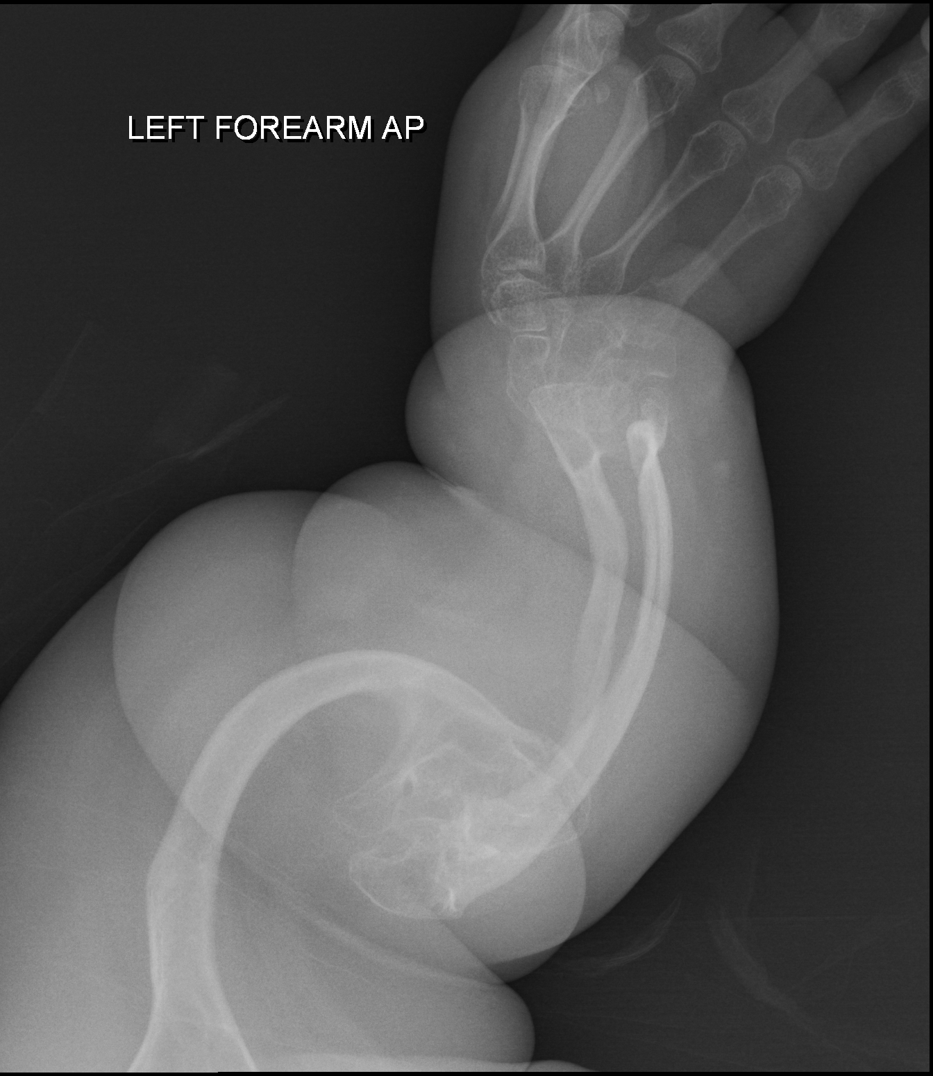
A fibrocondrogênese é uma condrodisplasia rizomélica rara, letal para o neonato. Onze casos foram relatados. O rosto é distinto e caracterizado por olhos protuberantes, rosto médio achatado, nariz pequeno e achatado com narinas antevertidas e boca pequena com lábio superior longo. Podem ocorrer fenda palatina, micrognatia e língua bífida. Os membros apresentam falta acentuada de todos os segmentos com mãos e pés relativamente normais. Nenhuma anomalia interna além da onfalocele foi relatada. A transmissão é provavelmente autossômica recessiva. Foram relatadas recorrências em uma família consanguínea (afetando ambos os sexos) e concordância de gêmeos masculinos afetados.
Introdução
O que você precisa saber de cara
A fibrocondrogênese é uma condrodisplasia rizomélica rara, letal para o neonato. Onze casos foram relatados. O rosto é distinto e caracterizado por olhos protuberantes, rosto médio achatado, nariz pequeno e achatado com narinas antevertidas e boca pequena com lábio superior longo. Podem ocorrer fenda palatina, micrognatia e língua bífida. Os membros apresentam falta acentuada de todos os segmentos com mãos e pés relativamente normais. Nenhuma anomalia interna além da onfalocele foi relatada. A transmissão é provavelmente autossômica recessiva. Foram relatadas recorrências em uma família consanguínea (afetando ambos os sexos) e concordância de gêmeos masculinos afetados.
Escala de raridade
<1/50kMuito rara
1/20kRara
1/10kPouco freq.
1/5kIncomum
1/2k
Encontrou um erro ou informação desatualizada? Sugira uma correção →
Entender a doença
Do básico ao detalhe, leia no seu ritmo
Preparando trilha educativa...
Sinais e sintomas
O que aparece no corpo e com que frequência cada sintoma acontece
Partes do corpo afetadas
+ 34 sintomas em outras categorias
Características mais comuns
Os sintomas variam de pessoa para pessoa. Abaixo estão as 71 características clínicas mais associadas, ordenadas por frequência.
Linha do tempo da pesquisa
Encontrou um erro ou informação desatualizada? Sugira uma correção →
Genética e causas
O que está alterado no DNA e como passa nas famílias
Genes associados
2 genes identificados com associação a esta condição. Padrão de herança: Autosomal dominant, Autosomal recessive.
May play an important role in fibrillogenesis by controlling lateral growth of collagen II fibrils
Secreted, extracellular space, extracellular matrix
Stickler syndrome 2
An autosomal dominant form of Stickler syndrome, an inherited disorder that associates ocular signs with more or less complete forms of Pierre Robin sequence, bone disorders and sensorineural deafness. Ocular disorders may include juvenile cataract, myopia, strabismus, vitreoretinal or chorioretinal degeneration, retinal detachment, and chronic uveitis. Pierre Robin sequence includes an opening in the roof of the mouth (a cleft palate), a large tongue (macroglossia), and a small lower jaw (micrognathia). Bones are affected by slight platyspondylisis and large, often defective epiphyses. Juvenile joint laxity is followed by early signs of arthrosis. The degree of hearing loss varies among affected individuals and may become more severe over time. Syndrome expressivity is variable.
May play an important role in fibrillogenesis by controlling lateral growth of collagen II fibrils
Secreted, extracellular space, extracellular matrix
Otospondylomegaepiphyseal dysplasia, autosomal dominant
An autosomal dominant form of otospondylomegaepiphyseal dysplasia, a disorder characterized by sensorineural deafness, enlarged epiphyses, mild platyspondyly, and disproportionate shortness of the limbs. Total body length is normal. Typical facial features are mid-face hypoplasia, short upturned nose and depressed nasal bridge. Most patients have Pierre Robin sequence including an opening in the roof of the mouth (cleft palate) and a small lower jaw (micrognathia). Ocular symptoms are absent. Some patients have early-onset osteoarthritis.
Variantes genéticas (ClinVar)
1.368 variantes patogênicas registradas no ClinVar.
Classificação de variantes (ClinVar)
Distribuição de 394 variantes classificadas pelo ClinVar.
Vias biológicas (Reactome)
7 vias biológicas associadas aos genes desta condição.
Diagnóstico
Os sinais que médicos procuram e os exames que confirmam
Tratamento e manejo
Remédios, cuidados de apoio e o que precisa acompanhar
Onde tratar no SUS
Hospitais de referência no Brasil e o protocolo oficial do SUS (PCDT)
🇧🇷 Atendimento SUS — Fibrocondrogênese
Selecione um estado ou use sua localização para ver resultados.
Dados de DATASUS/CNES, SBGM, ABNeuro e Ministério da Saúde. Sempre confirme a disponibilidade diretamente com o estabelecimento.
Pesquisa ativa
Ensaios clínicos abertos e novidades científicas recentes
Pesquisa e ensaios clínicos
Nenhum ensaio clínico registrado para esta condição.
Publicações mais relevantes
Distinct mural cells and fibroblasts drive fibrochondrogenesis in retrodiscal tissue following temporomandibular joint disc displacement.
Adaptive remodeling of retrodiscal tissue following anterior disc displacement (ADD) of the temporomandibular joint (TMJ) has been recognized for decades, yet the underlying cellular dynamics and molecular mechanisms remain unclear. Using a porcine ADD model, this study investigated the cellular and molecular basis driving retrodiscal tissue adaptation. Histological staining revealed adaptive remodeling of retrodiscal tissue after ADD induction, with dense connective tissue and cartilaginous masses replacing loose connective tissue. Furthermore, single-cell RNA sequencing (scRNA-seq) captured pronounced fibroblast expansion during tissue remodeling, notably the FB2 subcluster with high developmental potential, and the emergence of a mural cell subcluster MC4 associated with extracellular matrix (ECM) remodeling. CellChat analysis highlighted MC4-FB2 crosstalk via FGF2 and BMP5 signaling. The combination of pathway-aware multi-layered hierarchical network (P-NET) and Seurat with drug database screening identified five promising compounds. Among them, Zaprinast demonstrated the most robust effects by enhancing the remodeling capability of fibroblasts in vitro, and also alleviated TMJ deformation in vivo. Collectively, fibroblast activation is pivotal for early retrodiscal tissue adaptation following ADD, which is driven by MC4-derived FGF2/BMP5 signaling. Zaprinast treatment potentiates this remodeling process. These findings provide new insights into cellular basis of TMJ adaptation and identify potential therapeutic targets for ADD management.
Stem cell warm-up via biomimetic mechanical priming enhances synovial-derived MSCs adaptation for meniscal repair.
Mesenchymal stem cell (MSCs) transplantation shows promise for meniscus repair, but their inadaptability to the in vivo mechanical environment complicates differentiation regulation. This study developed a mechanical priming strategy to enhance MSCs fibrochondrogenic differentiation and promote meniscus repair. Synovial-derived MSCs (SMSCs) were co-cultured with meniscal fibrochondrocytes and exposed to cyclic tensile strain (10%, 0.5 Hz, 4h/day) for 5 days as a "stem cell warm-up system". Differentiation and mechanical adaptation were assessed through gene expression, cytoskeletal and nuclear morphology, and YAP-mediated mechanical transduction in vitro. A 2-mm meniscus defect model in New Zealand white rabbits was used, with histological analysis for repair evaluation. Mechanical priming inhibited SMSCs hypertrophy and promoted fibrochondrogenesis, with effects lasting 72 h post-loading. Meanwhile, priming induced actin cap formation, nuclear flattening, and nuclear pore expansion, facilitating mechanical adaptation. YAP-mediated transduction was essential for the sustained upregulation of differentiation-related genes, alongside increased expression of its downstream targets, Ctgf and Cyr61. siRNA silencing of Ctgf and Cyr61 led to a downregulation of differentiation-related genes, with YAP inhibition further suppressing differentiation, underscoring its pivotal role in regulating this process. Primed SMSCs exhibited faster activation and better phenotype maintenance during secondary loading. In vivo, primed SMSCs demonstrated superior performance in meniscus regeneration, equivalent to the outcomes of growth factor-treated groups. This biomimetic priming system enhances MSC differentiation and mechanical adaptability, offering a clinically translatable strategy for meniscus repair and load-bearing tissue regeneration.
The Characterization of Serum-Free Media on Human Mesenchymal Stem Cell Fibrochondrogenesis.
Developing fibrochondrogenic serum-free media is important for regenerating diseased and injured fibrocartilage but no defined protocols exist. Towards this goal, we characterized the effect of four candidate fibrochondrogenic serum-free media containing transforming growth factor beta-3 (TGF-β3), insulin-like growth factor-1 (IGF-1), and fibroblast growth factor-2 (FGF-2) with high/low glucose and with/without dexamethasone on human mesenchymal stem cells (hMSCs) via proliferation and differentiation assays. In Ki67 proliferation assays, serum-free media containing low glucose and dexamethasone exhibited the highest growth. In gene expression assays, serum-free media containing low glucose and commercially available chondrogenic media (COM) induced high fibrochondrogenic transcription factor expression (scleraxis/SCX and SRY-Box Transcription Factor 9/SOX9) and extracellular matrix (ECM) protein levels (aggrecan/ACAN, collagen type I/COL1A1, and collagen type II/COL2A1), respectively. In immunofluorescence staining, serum-free media containing high glucose and COM induced high fibrochondrogenic transcription factor (SCX and SOX9) and ECM protein (COL1A1, COL2A1, and collagen type X/COL10A1) levels, respectively. In cytochemical staining, COM and serum-free media containing dexamethasone showed a high collagen content whereas serum-free media containing high glucose and dexamethasone exhibited high glycosaminoglycan (GAG) levels. Altogether, defined serum-free media containing high glucose exhibited the highest fibrochondrogenic potential. In summary, this work studied conditions conducive for fibrochondrogenesis, which may be further optimized for potential applications in fibrocartilage tissue engineering.
3D-printed PCL scaffolds combined with injectable sodium alginate/magnesium-doped mesoporous bioactive glass nanosphere hydrogel for meniscus regeneration: In vitro, In vivo, and multiomics-based therapeutic analyses.
Meniscal injury presents a formidable challenge and often leads to functional impairment and osteoarthritic progression. Meniscus tissue engineering (MTE) is a promising solution, as conventional strategies for modulating local immune responses and generating a conducive microenvironment for effective tissue repair are lacking. Recently, magnesium-containing bioactive glass nanospheres (Mg-BGNs) have shown promise in tissue regeneration. However, few studies have explored the ability of Mg-BGNs to promote meniscal regeneration. First, we verified the anti-inflammatory and fibrochondrogenic abilities of Mg-BGNs in vitro. A comprehensive in vivo evaluation of a rabbit critical-size meniscectomy model revealed that Mg-BGNs have multiple effects on meniscal reconstruction and effectively promote fibrochondrogenesis, collagen deposition, and cartilage protection. Multiomics analysis was subsequently performed to further explore the mechanism by which Mg-BGNs regulate the regenerative microenvironment. Mechanistically, Mg-BGNs first activate the TRPM7 ion channel through the PI3K/AKT signaling pathway to promote the cellular function of synovium-derived mesenchymal stem cells and then activate the PPARγ/NF-κB axis to modulate macrophage polarization and inflammatory reactions. We demonstrated that Mg2+ is critical for the crosstalk among biomaterials, immune cells, and effector cells in Mg-BGN-mediated tissue regeneration. This study provides a theoretical basis for the application of Mg-BGNs as nanomedicines to achieve in situ tissue regeneration in complex intrajoint pathological microenvironments.
[Prenatal phenotype and genetic analysis of a fetus with Fibrochondrogenesis 1 due to compound heterozygous variants of COL11A1 gene].
To explore the genetic etiology of a fetus with short limbs identified by prenatal ultrasonography. A fetus detected with short limb malformations at Shengjing Hospital Affiliated to China Medical University on October 25, 2021 was selected as the study subject. Prenatal ultrasound and post-abortion imaging were carried out to determine the phenotypic characteristics of the fetus. Amniotic fluid sample of the fetus and peripheral blood samples of its parents were collected. Following extraction of genomic DNA, whole-exome sequencing was carried out. Candidate variants were verified by Sanger sequencing. Online software was used to predict the structural changes of the mutant proteins. Prenatal ultrasound showed that the fetus had a small bell-shaped thorax, markedly shortened limbs, flat midface, a small nose with anteriorly tilted nostrils, and a small mandible. Post-abortion CT showed typical short and wide fetal ribs, cupped metaphyses at both ends, short long bones with wide metaphyses, resulting in a dumbbell-shaped appearance and curved thoracic vertebrae. Whole-exome sequencing revealed that the fetus had harbored compound heterozygous variants of the COL11A1 gene, namely c.2251G>T and c.3790G>T, both of which were predicted to alter the important Gly-X-Y structure of collagen protein. Sanger sequencing confirmed that the variants were respectively inherited from its parents. A rare fetus with Fibrochondrogenesis type 1 due to compound heterozygous variants of the COL11A1 gene has been diagnosed. Above finding has enabled genetic counseling and reproductive guidance for this family.
Publicações recentes
Distinct mural cells and fibroblasts drive fibrochondrogenesis in retrodiscal tissue following temporomandibular joint disc displacement.
Stem cell warm-up via biomimetic mechanical priming enhances synovial-derived MSCs adaptation for meniscal repair.
The Characterization of Serum-Free Media on Human Mesenchymal Stem Cell Fibrochondrogenesis.
3D-printed PCL scaffolds combined with injectable sodium alginate/magnesium-doped mesoporous bioactive glass nanosphere hydrogel for meniscus regeneration: In vitro, In vivo, and multiomics-based therapeutic analyses.
A novel compound heterozygous variant of the COL11A1 gene in a patient with fibrochondrogenesis type I: the first case in Korea.
📚 EuropePMC35 artigos no totalmostrando 22
Distinct mural cells and fibroblasts drive fibrochondrogenesis in retrodiscal tissue following temporomandibular joint disc displacement.
JCI insightStem cell warm-up via biomimetic mechanical priming enhances synovial-derived MSCs adaptation for meniscal repair.
Osteoarthritis and cartilageThe Characterization of Serum-Free Media on Human Mesenchymal Stem Cell Fibrochondrogenesis.
Bioengineering (Basel, Switzerland)3D-printed PCL scaffolds combined with injectable sodium alginate/magnesium-doped mesoporous bioactive glass nanosphere hydrogel for meniscus regeneration: In vitro, In vivo, and multiomics-based therapeutic analyses.
Bioactive materialsA novel compound heterozygous variant of the COL11A1 gene in a patient with fibrochondrogenesis type I: the first case in Korea.
Annals of pediatric endocrinology & metabolism[Prenatal phenotype and genetic analysis of a fetus with Fibrochondrogenesis 1 due to compound heterozygous variants of COL11A1 gene].
Zhonghua yi xue yi chuan xue za zhi = Zhonghua yixue yichuanxue zazhi = Chinese journal of medical geneticsMeniscal fibrocartilage regeneration inspired by meniscal maturational and regenerative process.
Science advances[Genetic analysis of a child patient with rare fibrochondrogenesis due to COL11A1 gene variant].
Zhonghua yi xue yi chuan xue za zhi = Zhonghua yixue yichuanxue zazhi = Chinese journal of medical geneticsCharacterization of temporomandibular joint articular disc progenitor cell clones.
European cells & materialsClinical whole-exome sequencing analysis reveals a novel missense COL11A1 mutation resulting in an 18-week Iranian male aborted fetus with Fibrochondrogenesis 1: A case report.
Clinical case reportsThe Shape of the Jaw-Zebrafish Col11a1a Regulates Meckel's Cartilage Morphogenesis and Mineralization.
Journal of developmental biologyPulsed Electromagnetic Field Enhances Healing of a Meniscal Tear and Mitigates Posttraumatic Osteoarthritis in a Rat Model.
The American journal of sports medicineLethal and life-limiting skeletal dysplasias: Selected prenatal issues.
Advances in clinical and experimental medicine : official organ Wroclaw Medical University3D cell-printing of biocompatible and functional meniscus constructs using meniscus-derived bioink.
BiomaterialsCol11a1a Expression Is Required for Zebrafish Development.
Journal of developmental biologyInherited and de novo biallelic pathogenic variants in COL11A1 result in type 2 Stickler syndrome with severe hearing loss.
Molecular genetics & genomic medicineNanofiber-based transforming growth factor-β3 release induces fibrochondrogenic differentiation of stem cells.
Acta biomaterialiaLocal Administration of Magnesium Promotes Meniscal Healing Through Homing of Endogenous Stem Cells: A Proof-of-Concept Study.
The American journal of sports medicineThe mechanical impact of col11a2 loss on joints; col11a2 mutant zebrafish show changes to joint development and function, which leads to early-onset osteoarthritis.
Philosophical transactions of the Royal Society of London. Series B, Biological sciencesAnatomical region-dependent enhancement of 3-dimensional chondrogenic differentiation of human mesenchymal stem cells by soluble meniscus extracellular matrix.
Acta biomaterialiaDevelopment of a Micronized Meniscus Extracellular Matrix Scaffold for Potential Augmentation of Meniscal Repair and Regeneration.
Tissue engineering. Part C, MethodsHigh-purity magnesium interference screws promote fibrocartilaginous entheses regeneration in the anterior cruciate ligament reconstruction rabbit model via accumulation of BMP-2 and VEGF.
BiomaterialsAssociações
Organizações que acompanham esta doença — pra ter apoio e orientação
Ainda não temos associações cadastradas para Fibrocondrogênese.
É de uma associação que acompanha esta doença? Fale com a gente →
Comunidades
Grupos ativos de quem convive com esta doença aqui no Raras
Ainda não existe comunidade no Raras para Fibrocondrogênese
Pacientes, familiares e cuidadores se organizam em comunidades pra compartilhar experiências, fazer perguntas e se apoiar. Você pode ser o primeiro.
Tire suas dúvidas
Perguntas, dicas e experiências compartilhadas aqui na página
Participe da discussão
Faça login para postar dúvidas, compartilhar experiências e interagir com especialistas.
Fazer loginDoenças relacionadas
Doenças com sintomas parecidos — ajudam quem ainda está buscando diagnóstico
Referências e fontes
Bases de dados externas citadas neste artigo
Publicações científicas
Artigos indexados no PubMed ligados a esta doença no grafo RarasNet — título, periódico e PMID direto da fonte, sem intermediação de IA.
- Distinct mural cells and fibroblasts drive fibrochondrogenesis in retrodiscal tissue following temporomandibular joint disc displacement.
- Stem cell warm-up via biomimetic mechanical priming enhances synovial-derived MSCs adaptation for meniscal repair.
- The Characterization of Serum-Free Media on Human Mesenchymal Stem Cell Fibrochondrogenesis.
- 3D-printed PCL scaffolds combined with injectable sodium alginate/magnesium-doped mesoporous bioactive glass nanosphere hydrogel for meniscus regeneration: In vitro, In vivo, and multiomics-based therapeutic analyses.
- [Prenatal phenotype and genetic analysis of a fetus with Fibrochondrogenesis 1 due to compound heterozygous variants of COL11A1 gene].Zhonghua yi xue yi chuan xue za zhi = Zhonghua yixue yichuanxue zazhi = Chinese journal of medical genetics· 2024· PMID 38684309mais citado
- A novel compound heterozygous variant of the COL11A1 gene in a patient with fibrochondrogenesis type I: the first case in Korea.
Bases de dados e fontes oficiais
Identificadores e referências canônicas usadas para montar este verbete.
- ORPHA:2021(Orphanet)
- MONDO:0016068(MONDO)
- GARD:2321(GARD (NIH))
- Variantes catalogadas(ClinVar)
- Busca completa no PubMed(PubMed)
- Q3071315(Wikidata)
Dados compilados pelo RarasNet a partir de fontes abertas (Orphanet, OMIM, MONDO, PubMed/EuropePMC, ClinicalTrials.gov, DATASUS, PCDT/MS). Este conteúdo é informativo e não substitui avaliação médica.
Conteúdo mantido por Agente Raras · Médicos e pesquisadores podem colaborar
Fibrocondrogênese
📋 Origem dos dados
Esta página agrega dados de fontes públicas e oficiais. Dados sobre cobertura no SUS (PCDT, CEAF) são verificados ativamente por agente proativo (ver badge no infobox). Demais dados têm atribuição de fonte + data da última sincronização — clique para abrir o original.
- Doença rara (ontologia)
- fonte: Orphanet
- Identificador unificado
- fonte: MONDO
- Codificação WHO/SUS
- fonte: WHO ICD-10 / DATASUS
- CID-11 (futuro)
- fonte: WHO ICD-11
- NIH/GARD
- fonte: GARD (NIH)
- Indexação biomédica
- fonte: MeSH (NLM)
- Dado público estruturado
- fonte: Wikidata