Síndrome rara caracterizada por anomalias faciais, como hipoplasia mandibular/maxilar e trago duplicado, associada a deformidades vertebrais (espinha bífida) e atraso no desenvolvimento neuropsicomotor e da fala. Pode apresentar retardo de crescimento e apneia obstrutiva do sono.
Introdução
O que você precisa saber de cara
Visão geral
A Síndrome Goldenhar é uma condição rara que afeta principalmente o desenvolvimento da face, dos olhos e dos ouvidos. Ela também pode envolver outras partes do corpo, como o coração, os rins e a coluna vertebral. Cada pessoa com a síndrome pode apresentar um conjunto único de características, que variam de leves a mais complexas.[1][3]
Sinais e sintomas
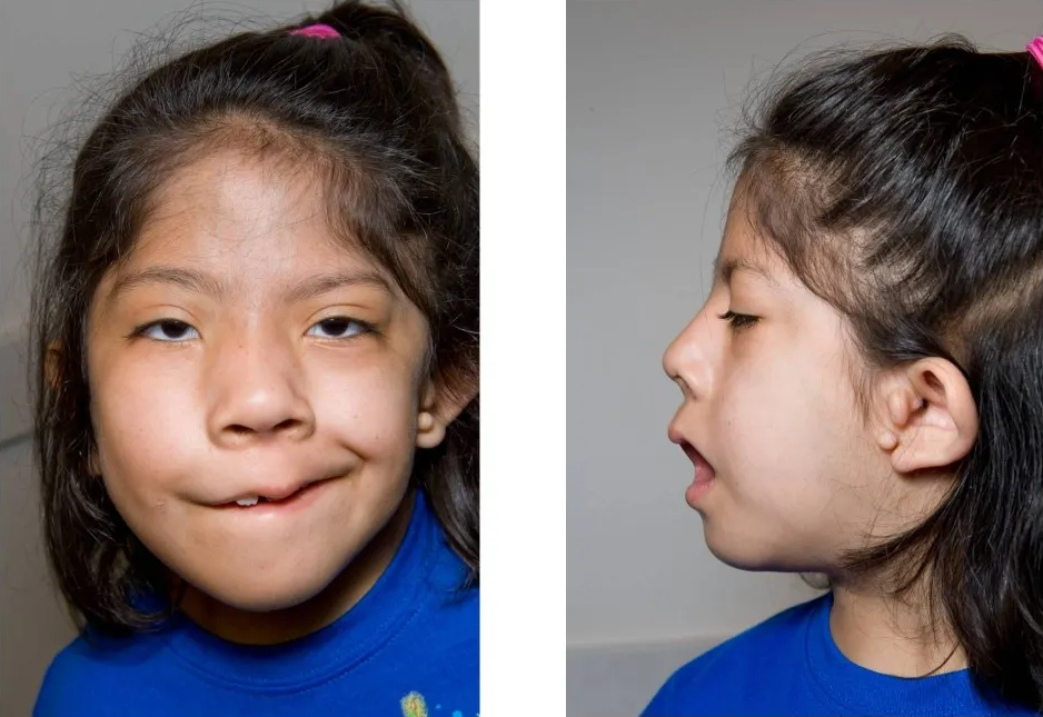
Os sinais e sintomas da Síndrome Goldenhar são variados e podem incluir alterações na face, como assimetria devido à ausência ou subdesenvolvimento da mandíbula (aplasia/hipoplasia da mandíbula) e da maxila (aplasia/hipoplasia da maxila). Na região dos olhos, pode ocorrer coloboma (uma falha na formação de estruturas oculares). As orelhas podem ser pequenas e malformadas (microtia de primeiro, segundo ou terceiro grau), apresentar um trago duplicado (pequena saliência de cartilagem na frente do ouvido) ou acrocórdons (pequenas protuberâncias de pele).[1][3]
Outros sintomas comuns incluem perda auditiva (deficiência auditiva), problemas na boca como úvula bífida (dividida), fenda submucosa do palato (abertura escondida no céu da boca), mordida cruzada e apinhamento dentário. Podem ocorrer também dificuldades respiratórias, como apneia obstrutiva do sono e obstrução das vias aéreas superiores. Em alguns casos, há atraso no neurodesenvolvimento, atraso no desenvolvimento da fala e da linguagem, microcefalia (cabeça menor que o esperado) e alterações na estrutura do cérebro, como aplasia/hipoplasia do corpo caloso.[1][3]
Alterações em outros sistemas do corpo também podem estar presentes, como morfologia anormal do coração, morfologia renal anormal, espinha bífida (malformação da coluna vertebral), trato sinusal dérmico (um pequeno canal na pele) e retardo do crescimento intrauterino. Durante a gestação, pode ser observado polidrâmnio (excesso de líquido amniótico).[1][3]
Causas genéticas
Diagnóstico
O diagnóstico da Síndrome Goldenhar é geralmente clínico, baseado na observação das características físicas típicas. Exames de imagem, como tomografia computadorizada e ressonância magnética, podem ser usados para avaliar a extensão das malformações ósseas e de órgãos internos. O diagnóstico pode ser complementado por testes genéticos. Atualmente, existem 336 testes genéticos disponíveis para a síndrome, e 73 variantes genéticas associadas estão registradas no ClinVar, um banco de dados público.[1][4][5]
Tratamento e manejo
Não existe um tratamento único para a Síndrome Goldenhar, pois o manejo é individualizado e depende dos sintomas apresentados por cada pessoa. O cuidado geralmente envolve uma equipe multidisciplinar, que pode incluir cirurgiões plásticos, otorrinolaringologistas, dentistas, fonoaudiólogos, oftalmologistas e geneticistas. As intervenções podem incluir cirurgias corretivas para as malformações faciais, auditivas e cardíacas, além de terapias de suporte para atrasos no desenvolvimento, como fonoaudiologia e terapia ocupacional.[1][5]
Tratamentos citados na literatura
A literatura científica menciona algumas substâncias em estudos sobre a Síndrome Goldenhar, mas é importante destacar que estas são associações mineradas de publicações e não representam recomendações de tratamento. As substâncias e o número de publicações associadas são: Calcium Sulfate (2), Propofol (2), polyetheretherketone (2), Alcohols (1), Carbon (1), Cholesterol (1), Indoleacetic Acids (1), Oxygen (1), Retinoids (1) e ethylene (1).[5]
Prognóstico e qualidade de vida
O prognóstico para pessoas com Síndrome Goldenhar é variável e depende da gravidade das malformações e do envolvimento de outros órgãos. Com acompanhamento médico adequado e intervenções precoces, muitas pessoas conseguem levar uma vida com boa qualidade. O suporte de uma equipe multidisciplinar e o acesso a terapias de reabilitação são fundamentais para o desenvolvimento e bem-estar.[1][5]
Conteúdo informativo gerado e mantido automaticamente a partir de fontes oficiais (Orphanet, HPO, OMIM, SUS). Não substitui avaliação médica.
Síndrome rara caracterizada por anomalias faciais, como hipoplasia mandibular/maxilar e trago duplicado, associada a deformidades vertebrais (espinha bífida) e atraso no desenvolvimento neuropsicomotor e da fala. Pode apresentar retardo de crescimento e apneia obstrutiva do sono.
Encontrou um erro ou informação desatualizada? Sugira uma correção →
Entender a doença
Do básico ao detalhe, leia no seu ritmo
Preparando trilha educativa...
Sinais e sintomas
O que aparece no corpo e com que frequência cada sintoma acontece
Visão geral
A Síndrome Goldenhar é uma condição rara que afeta principalmente o desenvolvimento da face, dos olhos e dos ouvidos. Ela também pode envolver outras partes do corpo, como o coração, os rins e a coluna vertebral. Cada pessoa com a síndrome pode apresentar um conjunto único de características, que variam de leves a mais complexas.[1][3]
Sinais e sintomas
Os sinais e sintomas da Síndrome Goldenhar são variados e podem incluir alterações na face, como assimetria devido à ausência ou subdesenvolvimento da mandíbula (aplasia/hipoplasia da mandíbula) e da maxila (aplasia/hipoplasia da maxila). Na região dos olhos, pode ocorrer coloboma (uma falha na formação de estruturas oculares). As orelhas podem ser pequenas e malformadas (microtia de primeiro, segundo ou terceiro grau), apresentar um trago duplicado (pequena saliência de cartilagem na frente do ouvido) ou acrocórdons (pequenas protuberâncias de pele).[1][3]
Outros sintomas comuns incluem perda auditiva (deficiência auditiva), problemas na boca como úvula bífida (dividida), fenda submucosa do palato (abertura escondida no céu da boca), mordida cruzada e apinhamento dentário. Podem ocorrer também dificuldades respiratórias, como apneia obstrutiva do sono e obstrução das vias aéreas superiores. Em alguns casos, há atraso no neurodesenvolvimento, atraso no desenvolvimento da fala e da linguagem, microcefalia (cabeça menor que o esperado) e alterações na estrutura do cérebro, como aplasia/hipoplasia do corpo caloso.[1][3]
Alterações em outros sistemas do corpo também podem estar presentes, como morfologia anormal do coração, morfologia renal anormal, espinha bífida (malformação da coluna vertebral), trato sinusal dérmico (um pequeno canal na pele) e retardo do crescimento intrauterino. Durante a gestação, pode ser observado polidrâmnio (excesso de líquido amniótico).[1][3]
Causas genéticas
Diagnóstico
O diagnóstico da Síndrome Goldenhar é geralmente clínico, baseado na observação das características físicas típicas. Exames de imagem, como tomografia computadorizada e ressonância magnética, podem ser usados para avaliar a extensão das malformações ósseas e de órgãos internos. O diagnóstico pode ser complementado por testes genéticos. Atualmente, existem 336 testes genéticos disponíveis para a síndrome, e 73 variantes genéticas associadas estão registradas no ClinVar, um banco de dados público.[1][4][5]
Tratamento e manejo
Não existe um tratamento único para a Síndrome Goldenhar, pois o manejo é individualizado e depende dos sintomas apresentados por cada pessoa. O cuidado geralmente envolve uma equipe multidisciplinar, que pode incluir cirurgiões plásticos, otorrinolaringologistas, dentistas, fonoaudiólogos, oftalmologistas e geneticistas. As intervenções podem incluir cirurgias corretivas para as malformações faciais, auditivas e cardíacas, além de terapias de suporte para atrasos no desenvolvimento, como fonoaudiologia e terapia ocupacional.[1][5]
Tratamentos citados na literatura
A literatura científica menciona algumas substâncias em estudos sobre a Síndrome Goldenhar, mas é importante destacar que estas são associações mineradas de publicações e não representam recomendações de tratamento. As substâncias e o número de publicações associadas são: Calcium Sulfate (2), Propofol (2), polyetheretherketone (2), Alcohols (1), Carbon (1), Cholesterol (1), Indoleacetic Acids (1), Oxygen (1), Retinoids (1) e ethylene (1).[5]
Prognóstico e qualidade de vida
O prognóstico para pessoas com Síndrome Goldenhar é variável e depende da gravidade das malformações e do envolvimento de outros órgãos. Com acompanhamento médico adequado e intervenções precoces, muitas pessoas conseguem levar uma vida com boa qualidade. O suporte de uma equipe multidisciplinar e o acesso a terapias de reabilitação são fundamentais para o desenvolvimento e bem-estar.[1][5]
Conteúdo informativo gerado e mantido automaticamente a partir de fontes oficiais (Orphanet, HPO, OMIM, SUS). Não substitui avaliação médica.
Partes do corpo afetadas
+ 26 sintomas em outras categorias
Características mais comuns
Os sintomas variam de pessoa para pessoa. Abaixo estão as 77 características clínicas mais associadas, ordenadas por frequência.
Linha do tempo da pesquisa
Encontrou um erro ou informação desatualizada? Sugira uma correção →
Genética e causas
O que está alterado no DNA e como passa nas famílias
Genes associados
2 genes identificados com associação a esta condição.
Transcription factor required for pharyngeal arch development, which is involved in hair, ear, jaw and dental development (PubMed:37041148). May act as a pioneer transcription factor during pharyngeal arch development (By similarity). Required for epithelial cell differentiation within the epidermis (By similarity). Acts at multiple stages of otic placode induction: necessary for preplacodal ectoderm to execute an inner ear program (By similarity). Required for hair follicle stem cell specificat
Nucleus
Craniofacial microsomia 2
A form of craniofacial microsomia, a disorder characterized by a spectrum of craniofacial malformations ranging from isolated microtia with or without aural atresia to underdevelopment of the mandible, maxilla, orbit, facial soft tissue, and/or facial nerve. Most CFM2 patients exhibit isolated unilateral or bilateral grade II/III microtia, with or without atresia, although some patients show only minor external ear defects. Mandibular hypoplasia, micrognathia, and dental anomalies have also been observed. CFM2 inheritance can be autosomal dominant or autosomal recessive.
Component of the 17S U2 SnRNP complex of the spliceosome, a large ribonucleoprotein complex that removes introns from transcribed pre-mRNAs (PubMed:12234937, PubMed:32494006, PubMed:34822310). The 17S U2 SnRNP complex (1) directly participates in early spliceosome assembly and (2) mediates recognition of the intron branch site during pre-mRNA splicing by promoting the selection of the pre-mRNA branch-site adenosine, the nucleophile for the first step of splicing (PubMed:12234937, PubMed:32494006
NucleusNucleus speckle
Craniofacial microsomia 1
A form of craniofacial microsomia, a disorder characterized by a spectrum of craniofacial malformations ranging from isolated microtia with or without aural atresia to underdevelopment of the mandible, maxilla, orbit, facial soft tissue, and/or facial nerve. CFM1 is an autosomal dominant form characterized by mandibular hypoplasia, microtia, facial and preauricular skin tags, epibulbar dermoids, and lateral oral clefts. Affected individuals also present skeletal and cardiac abnormalities.
Variantes genéticas (ClinVar)
73 variantes patogênicas registradas no ClinVar.
Vias biológicas (Reactome)
4 vias biológicas associadas aos genes desta condição.
Diagnóstico
Os sinais que médicos procuram e os exames que confirmam
Tratamento e manejo
Remédios, cuidados de apoio e o que precisa acompanhar
Onde tratar no SUS
Hospitais de referência no Brasil e o protocolo oficial do SUS (PCDT)
🇧🇷 Atendimento SUS — Síndrome Goldenhar
Selecione um estado ou use sua localização para ver resultados.
Dados de DATASUS/CNES, SBGM, ABNeuro e Ministério da Saúde. Sempre confirme a disponibilidade diretamente com o estabelecimento.
Pesquisa ativa
Ensaios clínicos abertos e novidades científicas recentes
Ensaios em destaque
Pesquisa e ensaios clínicos
11 ensaios clínicos encontrados.
Publicações mais relevantes
Anaesthetic management of a paediatric patient with Goldenhar syndrome for oral rehabilitation and palatoplasty.
Goldenhar syndrome is a rare congenital condition characterised by craniofacial deformities and vertebral anomalies, which pose significant challenges for anaesthesiologists in airway management and ventilatory support. In this case report, we present a paediatric patient with Goldenhar syndrome for oral rehabilitation and palatoplasty, highlighting the importance of a thorough preoperative evaluation, airway planning and the application of lung protective ventilation strategies. The procedure was successfully done under general anaesthesia without intraoperative or postoperative complications. This case report emphasises the importance of individualised anaesthetic planning in patients with craniofacial and vertebral anomalies. The perioperative risks were significantly reduced by anticipating a potential difficult airway and tailoring ventilatory management. Oculo-auriculo-vertebral spectrum (OAVS) is a congenital disorder of craniofacial morphogenesis, first described by ophthalmologist Maurice Goldenhar in 1952, characterized by an association of ophthalmic, auricular, and facial features. Gorlin et al. subsequently added vertebral anomalies to the classification in 1963. The condition is also known as Goldenhar syndrome, facio-auriculo-vertebral syndrome, or Goldenhar-Gorlin syndrome. The term OAVS, originating from "oculoauriculovertebral dysplasia" as described by Cohen et al in 1989, reflects the phenotypic continuum and significant overlap among malformations of structures derived from the first and second branchial arches. OAVS affects the eyes, mouth (lips, tongue, and palate), ears, maxilla, and mandible. Multisystem involvement frequently includes the central nervous system, heart, kidneys, and skeletal system, distinguishing it from isolated hemifacial microsomia. Recent advances in genetics have identified multiple causative genes, transforming our understanding of this clinically heterogeneous condition. Typical phenotypes include microtia, facial asymmetry, and epibulbar dermoid or lipodermoid. The minimum diagnostic criteria proposed by Tasse et al include either isolated microtia or preauricular tags associated with hemifacial microsomia. The management of patients with OAVS requires an interprofessional approach due to the wide variety of abnormalities and varying severity of presentations. This review highlights current understanding of etiology, pathogenesis, clinical presentations, and evidence-based management options, with particular emphasis on ocular features.
[Application and research progress of mandibular distraction osteogenesis in craniofacial microsomia].
To review the application and research progress of mandibular distraction osteogenesis (MDO) in the treatment of craniofacial microsomia (CFM). Recent domestic and international literature on MDO for CFM was extensively reviewed, with systematic summarization of its indications, device development, key technical points of MDO, complication management, digital surgical applications, biological enhancement strategies, and controversies regarding early intervention. MDO stimulates new bone formation through the "tension-stress" principle, serving as an effective method to elongate the mandible in CFM patients and early improve airway and occlusal functions. Digital technology has enhanced its precision, while biological strategies show potential for optimizing osteogenesis. However, challenges such as postoperative relapse and the long-term efficacy of early intervention remain. MDO is a core component of the sequential treatment for CFM. Future efforts should focus on intelligent technologies and high-quality clinical studies to optimize individualized treatment plans and improve long-term stability. 综述下颌牵引成骨(mandibular distraction osteogenesis,MDO)技术在颅面短小畸形(craniofacial microsomia,CFM)治疗中的应用及研究进展。. 广泛查阅近年国内外关于MDO治疗CFM的相关文献,从其适应证、牵引器发展、MDO技术要点、并发症管理、数字化外科应用、生物学增强策略及早期干预争议等方面进行系统总结。. MDO通过“张力-应力”法则刺激新骨生成,是延长CFM患者下颌骨、早期改善气道与咬合功能的有效方法。数字化技术提升了其精准性,而生物学策略有望优化成骨。然而,该技术仍面临术后复发、早期干预远期疗效不明确等挑战。. MDO是CFM序列治疗的核心手段之一,未来需通过智能化技术与更多高质量临床研究,以优化个体化治疗方案并提升远期稳定性。.
Evaluation of mandibular morphology in untreated growing patients with hemifacial Microsomia: a 3D computed tomography study.
Hemifacial Microsomia (HFM) is the second most common craniofacial malformation after clefts and it is characterized by mandibular hypoplasia and facial asymmetry. This retrospective study aimed to evaluate mandibular morphology in growing HFM patients using three-dimensional (3D) computed tomography (CBCT), focusing on volumetric asymmetries. A total of 26 patients aged 5-13 years with unilateral HFM were analyzed. They were stratified into two age groups (5-8 and 9-13 years) and classified according to Pruzansky-Kaban system in two severity categories (mild/severe deformity). CBCT data were processed to reconstruct 3D mandibular models, segmenting the condylar, coronoid and gonion regions. Volumetric differences between affected and unaffected sides were compared across age and severity subgroups. Results showed that all the three functional units were significantly smaller on the affected side, especially in severe HFM cases. However, no statistically significant differences were found between the two age groups, suggesting that mandibular asymmetry remains substantively unchanged over the two consecutive time points examined. Multivariable regression confirmed that HFM severity was strongly associated with condylar and gonial volume discrepancies. This study highlights the value of CBCT-based 3D reconstruction for precise assessment of mandibular morphology and supports its role in individualized diagnosis and treatment planning for HFM patients.
Surgical treatment of 89 patients with craniofacial microsomia in a craniofacial national reference centre in Finland.
This study aims to evaluate surgical treatments for craniofacial microsomia (CFM) at a national reference centre in Finland, focusing on the age of treatment and the number of surgeries performed. The aim was to describe surgical strategies, explore treatment variations, and assess intervention timing. This retrospective cohort study reviewed the medical records of 191 patients diagnosed with CFM at the Helsinki Cleft Palate and Craniofacial Centre from 2010 to 2022. After excluding patients with insufficient data, 89 patients were included. Patients were classified using the Pruzansky-Kaban system, and their surgeries were categorized by type, indication, and age. Of the 89 patients included, 64 patients (72 %) had undergone surgical treatment. Ear reconstruction surgeries were the most common (41 patients or 46 %), followed by soft tissue surgeries in 29 patients (33 %) and cleft-related surgeries in 21 patients (24 %). Hard tissue surgeries, including orthognathic procedures, were performed on nine patients (10 %), primarily those with severe CFM. The median age for surgeries ranged from 0.8 years for cleft surgery to 21.6 years for lipofilling. No significant gender predominance was found. This study highlights the variety of surgical approaches required for treating CFM, with severity influencing the number and type of surgeries. A shift towards conservative treatments for younger patients and a preference for alloplastic implants over autologous grafts were observed. Further research is needed to standardize surgical protocols and treatment outcomes for CFM. Duane retraction syndrome, previously known as Stilling-Turk-Duane syndrome, is caused by the absence or partial development of the abducens nucleus and nerve. As a result, there is aberrant innervation of the lateral rectus by the oculomotor nerve. Similar developmental anomalies of 1 or more cranial nerves have come to be grouped under congenital cranial dysinnervation disorders. These anomalies may be termed primary due to the absence of normal innervation or secondary following aberrant innervations from other cranial nerves. The evaluation and management of Duane retraction syndrome can be very challenging, and a judicious approach is essential. Duane retraction syndrome is a rare, congenital ocular motility disorder characterized by limitation or absence of horizontal eye movements, globe retraction, and narrowing of the palpebral fissure on attempted adduction. First described by Alexander Duane in 1905, this condition represents approximately 1% to 4% of all cases of strabismus. The pathophysiology is now understood to involve a congenital cranial dysinnervation disorder, in which aberrant innervation of the lateral rectus muscle by branches of the oculomotor nerve leads to paradoxical co-contraction of horizontal recti. Although classically unilateral, Duane retraction syndrome may be bilateral in 10% to 20% of cases and is more commonly observed in females, with a predilection for the left eye. The etiology is linked to developmental anomalies of the abducens nerve (cranial nerve VI) or its nucleus, resulting in absent or hypoplastic innervation and compensatory miswiring from the oculomotor nerve. This neurogenic origin is supported by magnetic resonance imaging (MRI) studies demonstrating absent abducens nerve and by electromyography (EMG) findings of simultaneous lateral and medial rectus contraction during attempted adduction. In some cases, Duane retraction syndrome is associated with systemic anomalies such as Goldenhar syndrome, Klippel-Feil anomaly, or other congenital malformations, reinforcing the notion of a broader embryologic insult affecting cranial nerve development. Clinically, Duane retraction syndrome is classified into 3 primary types according to Huber's classification (1974): Type I: Marked limitation or absence of abduction, with relatively normal adduction and globe retraction on adduction. Type II: Limitation or absence of adduction with relatively normal abduction. Type III: Limitation of both abduction and adduction, often with significant globe retraction. Patients may also exhibit upshoots or downshoots on adduction due to leash effects or mechanical factors within the extraocular muscle pulleys. Abnormal head posture is common as patients adopt compensatory face turns to maintain binocular single vision in primary gaze. The severity of motility restriction and retraction varies widely, influencing both functional and cosmetic concerns. Epidemiologically, Duane retraction syndrome accounts for a small fraction of strabismus cases worldwide but carries substantial clinical importance due to its distinct presentation, surgical challenges, and potential associations with systemic disorders. Population-based studies indicate prevalence rates ranging from 0.1 to 0.7 per 1000 live births, though true incidence may be underestimated due to underdiagnosis in mild cases. Awareness among pediatricians, ophthalmologists, and orthoptists is critical for early recognition and evaluation. From a diagnostic standpoint, Duane retraction syndrome is primarily a clinical diagnosis supported by a detailed ocular motility examination. Key signs include narrowing of the palpebral fissure on adduction, globe retraction, and variable upshoots or downshoots. Forced duction testing may reveal mechanical restrictions, but the hallmark finding is paradoxical co-contraction of horizontal recti, confirmed by EMG. Imaging with high-resolution orbital MRI or diffusion tensor imaging can delineate the absence of the abducens nerve, providing objective confirmation and aiding in surgical planning. Assessment should also include evaluation for associated systemic anomalies, as up to 30% of patients may have other congenital malformations. Management of Duane retraction syndrome is individualized and guided by the severity of motility limitation, presence of abnormal head posture, and patient symptoms. Mild cases with good primary gaze alignment may require only observation and periodic follow-up. Surgical intervention is indicated for significant misalignment in primary gaze, large abnormal head posture, or cosmetically disturbing globe retraction and upshoots or downshoots. Procedures may include recession of the medial rectus, lateral rectus, or a combination, as well as vertical rectus transpositions in selected cases. The surgical approach is often more complex than in other forms of strabismus due to the paradoxical innervation and risk of exacerbating globe retraction. Overcorrection, induced vertical deviations, and persistent limitation are recognized challenges. Duane retraction syndrome is nonprogressive, but long-term follow-up is essential, particularly in children, to monitor ocular alignment, binocular function, and amblyopia risk. Amblyopia occurs in up to 10% of cases, necessitating prompt detection and treatment. Orthoptic therapy plays a supportive role in maintaining binocular vision and managing mild head postures, although it does not correct the underlying innervational anomaly. Recent advances in understanding Duane retraction syndrome pathogenesis have emerged from genetic and neuroimaging studies. Mutations in genes such as CHN1 (encoding alpha2-chimaerin) have been identified in familial cases, implicating axon guidance defects in cranial nerve development. These findings place Duane retraction syndrome within the broader category of congenital cranial dysinnervation disorders, alongside Möbius syndrome and congenital fibrosis of the extraocular muscles. This reclassification has shifted the focus from purely mechanical explanations to neurodevelopmental mechanisms, fostering novel research avenues. The psychosocial impact of Duane retraction syndrome should not be underestimated. Visible eye movement anomalies and abnormal head posture can affect self-esteem, social interactions, and quality of life, particularly in adolescents. Counseling, patient education, and appropriate referral for psychological support may be beneficial in selected cases. In pediatric patients, parental reassurance and guidance are critical to alleviate anxiety and ensure adherence to follow-up schedules. Interprofessional collaboration is central to optimal Duane retraction syndrome management. Pediatric ophthalmologists, orthoptists, neurologists, radiologists, and genetic counselors all contribute to comprehensive evaluation and care. For example, neurologists may assess for associated cranial nerve or central nervous system anomalies, whereas genetic counselors provide insight into inheritance patterns and recurrence risks. Radiologists skilled in high-resolution orbital imaging play an important role in confirming diagnosis and guiding surgical strategy. Orthoptists assist with functional assessment, prism adaptation, and post-operative rehabilitation, enhancing overall outcomes. In conclusion, Duane retraction syndrome represents a distinct, nonprogressive congenital ocular motility disorder with complex neurogenic and mechanical features. Advances in neuroimaging and genetics have enriched our understanding of its pathophysiology, whereas surgical and non-surgical management strategies continue to evolve. Early recognition, thorough evaluation for systemic associations, individualized treatment planning, and long-term multidisciplinary follow-up are key pillars in optimizing functional and cosmetic outcomes for patients. This activity equips learners with an evidence-based, clinically relevant framework for diagnosing and managing Duane retraction syndrome, integrating current guidelines, expert consensus, and interprofessional care principles to improve patient-centered outcomes.
Temporal bone and multisystem phenotypic stratification in oculo-auriculo-vertebral spectrum using high-resolution CT: Correlation with tasse severity score.
To characterize craniofacial, temporal-bone, vertebral, and systemic anomalies in oculo-auriculo-vertebral (OAV) spectrum using high-resolution computed tomography (HRCT) and to examine associations with clinical severity by the Tasse Objective Scoring System. We performed a retrospective study (2015-2024) at a national tertiary center including 223 clinically diagnosed OAV patients; 217 had bilateral temporal-bone HRCT suitable for analysis. HRCT assessed external auditory canal (EAC), ossicular, and intratemporal facial-nerve anatomy; inner-ear/vestibulocochlear-nerve abnormalities were evaluated in a subset. Vertebral anomalies were CT-confirmed when coverage was available; renal and cardiac findings were extracted from clinical records. Statistics included chi-square or Fisher tests with Cramér's V, Cochran-Armitage trend tests across Tasse grades, and Spearman correlation for vertebral anomaly counts (two-sided α = 0.05). Mean age was 7.6 ± 4.2 years; 55.2 % were male. In the HRCT subset, EAC stenosis/atresia and ossicular abnormalities were frequent and increased with Tasse severity (EAC: 48.4 %→59.8 %→82.8 %, p = 0.0078; ossicles: 40.3 %→49.6 %→82.8 %, p < 0.001), as did aberrant intratemporal facial-nerve course (24.2 %/27.4 %/53.3 %, p = 0.010). Inner-ear malformations were identified in 14.3 % and vestibular/vestibulocochlear-nerve anomalies in 42.9 % of those specifically evaluated. CT-confirmed vertebral anomalies occurred in 29.1 % overall; segmentation defects showed a strong grade-wise increase (p < 0.001) and the cumulative vertebral anomaly count correlated with Tasse severity (Spearman ρ = 0.41, p < 0.001). Renal anomalies were present in 16.6 % and rose across grades (p = 0.044; trend p < 0.001), whereas cardiac anomalies occurred in 14.8 % with no significant between-grade difference (p = 0.19). Pairing HRCT phenotyping with Tasse severity stratification provides clinically actionable information for operative planning (canaloplasty/ossiculoplasty/device candidacy) and prioritizes systemic surveillance (spine and renal screening) in OAV spectrum. This integrated approach supports coordinated multidisciplinary care and offers a framework for future standardized screening and outcome-oriented research.
Publicações recentes
Genetic variation near ROBO1 is associated with craniofacial microsomia and related phenotypes in the Finnish population.
Characterization of a Familial Goldenhar Syndrome Case Using Whole-Exome Sequencing.
Anaesthetic management of a paediatric patient with Goldenhar syndrome for oral rehabilitation and palatoplasty.
Oculo-Auriculo-Vertebral Spectrum (Goldenhar Syndrome).
Duane Retraction Syndrome.
📚 EuropePMC357 artigos no totalmostrando 198
Anaesthetic management of a paediatric patient with Goldenhar syndrome for oral rehabilitation and palatoplasty.
BMJ case reports[Application and research progress of mandibular distraction osteogenesis in craniofacial microsomia].
Zhongguo xiu fu chong jian wai ke za zhi = Zhongguo xiufu chongjian waike zazhi = Chinese journal of reparative and reconstructive surgeryEvaluation of mandibular morphology in untreated growing patients with hemifacial Microsomia: a 3D computed tomography study.
Journal of cranio-maxillo-facial surgery : official publication of the European Association for Cranio-Maxillo-Facial SurgerySurgical treatment of 89 patients with craniofacial microsomia in a craniofacial national reference centre in Finland.
Journal of cranio-maxillo-facial surgery : official publication of the European Association for Cranio-Maxillo-Facial SurgeryTemporal bone and multisystem phenotypic stratification in oculo-auriculo-vertebral spectrum using high-resolution CT: Correlation with tasse severity score.
European journal of radiologyPrecision Meets Personalization: The Synergy of Virtual Surgical Planning, 3D Printing, Orthognathic Surgery, and Custom Temporomandibular Joint Prosthesis in Managing Goldenhar Syndrome.
The Journal of craniofacial surgeryAnalysis of Craniofacial Microsomia and Facial Asymmetry: Approaches in Facial Feminization Surgery.
Aesthetic surgery journalCraniofacial Microsomia With Tongue Cleft: Embryological Mechanisms and Clinical Implications From Three Rare Cases.
The Journal of craniofacial surgeryMental Health Needs in Patients With Communication Deficits and Chronic Health Needs: A Case of a Teenager With Goldenhar Syndrome.
CureusBeyond the face: multidimensional care challenges and unmet needs in Hemifacial Microsomia families.
Frontiers in public healthThree-dimensional cephalometric analysis of morphological characteristics in children with bilateral craniofacial microsomia.
Journal of cranio-maxillo-facial surgery : official publication of the European Association for Cranio-Maxillo-Facial SurgeryGenetic correlation and causation of craniofacial microsomia with 33 diseases in Asian populations: insights from large-scale genome-wide cross-trait analysis.
Scientific reportsOccipitocervical surgery rescue: The "Catcher's Mitt" technique.
Journal of craniovertebral junction & spineThe role of adenoid hypertrophy in obstructive sleep apnea outcomes post-mandibular distraction osteogenesis in patients with craniofacial microsomia.
Journal of clinical sleep medicine : JCSM : official publication of the American Academy of Sleep MedicineEmbarking on a Treatment Journey: Experiences of Caregivers of Young Children With Craniofacial Microsomia.
The Journal of craniofacial surgeryHaploinsufficiency of SF3B2 revealed by a craniofacial microsomia with atypical presentation: a case report.
Journal of stomatology, oral and maxillofacial surgeryZSCAN10-Deficiency Mimicking Goldenhar Syndrome.
American journal of medical genetics. Part ALower labial vermilion myomucosal flap for commissuroplasty of Tessier 7 transverse facial cleft.
International journal of pediatric otorhinolaryngologyTreatment Strategies for Craniofacial Microsomia Patients With Obstructive Sleep Apnea: A Systematic Review.
The Journal of craniofacial surgeryPerioperative care in pediatric OTC deficiency and goldenhar syndrome: a case report.
Frontiers in pediatricsOrthognathic Surgery of Goldenhar Syndrome Patient With Absent Bilateral Mental Foramina.
The Journal of craniofacial surgeryA Novel Advantage to Microtia Reconstruction Utilizing Ear Molding: A Case Report of Goldenhar Syndrome's Clinical Presentation and Surgical Reconstruction.
CureusSplicing Defects and Cell Death Cause SF3B2-Linked Craniofacial Microsomia.
Journal of dental researchRetrospective Investigation on Swallowing and Feeding Difficulties in Patients With Oculo-Auriculo-Vertebral-Spectrum.
The Journal of craniofacial surgeryCommon cis-regulatory variation modifies the penetrance of pathogenic SHROOM3 variants in craniofacial microsomia.
Genome researchCephalometric evaluation of the craniofacial complex in hemifacial microsomia treated with an internal distraction osteogenesis device: A case series of 3 patients with a ≥12-year follow-up.
American journal of orthodontics and dentofacial orthopedics : official publication of the American Association of Orthodontists, its constituent societies, and the American Board of OrthodonticsDevelopment of a Novel Large-Animal Model for Craniofacial Microsomia: Proportional Skeletal Defects in Mandibular Ramus and Zygomatic Arch.
The Journal of craniofacial surgeryGoldenhar Syndrome and Surgical Reconstruction: A Case Report of Bilateral Complete Eyelid Colobomas in a 2-Day-Old Patient.
Case reports in ophthalmological medicineRevisiting a Rare Anomaly Described 25 Years Ago in the AJNR: A Journey from Pediatric Hemifacial Microsomia and Middle Cranial Fossa Aplasia to CSF-Lymphatic Fistula and Spontaneous Intracranial Hypotension as an Adult.
AJNR. American journal of neuroradiologyNovel variants in FOXI3 gene confirm its implication in Oculo-Auriculo-Vertebral spectrum.
European journal of human genetics : EJHGComputed tomographic assessment of orbital and maxillary dysmorphology in craniofacial microsomia.
American journal of orthodontics and dentofacial orthopedics : official publication of the American Association of Orthodontists, its constituent societies, and the American Board of OrthodonticsGoldenhar Syndrome and Its Clinical Manifestation With Dentofacial Considerations: A Case Report.
Special care in dentistry : official publication of the American Association of Hospital Dentists, the Academy of Dentistry for the Handicapped, and the American Society for Geriatric DentistrySurgical decision-making regarding hearing and ear reconstruction in craniofacial microsomia: Exploring caregiver narratives.
Journal of cranio-maxillo-facial surgery : official publication of the European Association for Cranio-Maxillo-Facial SurgeryEar malformation in a child with Goldenhar syndrome and its appropriate audiological management.
Anales del sistema sanitario de NavarraOcular Manifestations and Pathological Features in Goldenhar Syndrome: A 10-Year Retrospective Study.
Ophthalmology and therapyPathogenic variants in SHROOM3 associated with hemifacial microsomia.
Journal of human geneticsClinical presentation of hemifacial microsomia in a South African population.
Journal of plastic surgery and hand surgeryOptimal age for mandibular distraction osteogenesis in craniofacial microsomia: A systematic review and meta-analysis.
Journal of stomatology, oral and maxillofacial surgeryFamilial Oculoauriculovertebral Spectrum: A Genomic Investigation of Autosomal Dominant Inheritance.
The Cleft palate-craniofacial journal : official publication of the American Cleft Palate-Craniofacial AssociationThe Biomechanical Properties of A Modified Distraction Device Used in Mandibular Distraction Osteogenesis for Craniofacial Microsomia Patients: A Simulation Finite Element Analysis Study.
The Journal of craniofacial surgeryLongitudinal Study of Epibulbar Dermolipomas Over Five-Year Follow-Up: Growth Analysis, Refractive Errors, and Surgical Outcomes.
Ophthalmic plastic and reconstructive surgeryCervical spine considerations in Goldenhar syndrome: a clinical perspective.
Child's nervous system : ChNS : official journal of the International Society for Pediatric NeurosurgeryUse of Upper Extremity Electromyography Twitch Monitoring in a Patient With Goldenhar Syndrome and Absent Thumbs.
Paediatric anaesthesiaEvaluation of Situs Inversus Combined With Plastic Surgery-Related Malformations in a Chinese Clinic Population.
Annals of plastic surgeryA Curious Case of Multimorbidity in a Patient With Goldenhar Syndrome Presenting With Vomiting.
CureusPrevalence of and Risk Factors for Hearing Impairment in Craniofacial Microsomia.
Journal of oral and maxillofacial surgery : official journal of the American Association of Oral and Maxillofacial SurgeonsMandibular growth following distraction osteogenesis in children with craniofacial microsomia from a skeletal units perspective.
Journal of stomatology, oral and maxillofacial surgeryMost common congenital syndromes with facial asymmetry: A narrative review.
Dental and medical problemsAnalyzing Discrepancies and Correlations in Soft and Hard Tissue Asymmetry: A Focused Study on Hemifacial Microsomia and Isolated Microtia.
Aesthetic plastic surgeryThree-Dimensional Facial Morphology in Patients with Craniofacial Microsomia and Microtia.
Plastic and reconstructive surgeryAnterior segment dysgenesis in Goldenhar syndrome.
Eye (London, England)Managing Limbal Dermoids in Patients with Goldenhar Syndrome: A Case Series.
Romanian journal of ophthalmologyComprehensive treatment approach for hemifacial microsomia: Integrating orthognathic surgery with sequential customized implantation.
Journal of plastic, reconstructive & aesthetic surgery : JPRASA Syndrome Affecting All Five Sense Organs: A Rare Congenital Disorder of Kabuki Makeup Syndrome With Multiple Pre-auricular Skin Tags.
CureusChanges of functional units in type IIA craniofacial microsomia before puberty-a preliminary computed tomography study.
Journal of stomatology, oral and maxillofacial surgeryTwo-step cluster analysis based on three-dimensional CT measurements of craniofacial structures in severe craniofacial microsomia.
Journal of plastic, reconstructive & aesthetic surgery : JPRASChromatin assembly factor subunit CHAF1A as a monogenic cause for oculo-auriculo-vertebral spectrum.
European journal of human genetics : EJHGDistraction Osteotomy Combined With Orthodontics: An Effective Way to Decrease Short-term Recurrence in Treating Child Craniofacial Microsomia.
The Journal of craniofacial surgeryA rare association of monocular elevation deficiency and goldenhar syndrome secondary to vascular insufficiency: A case report.
International journal of surgery case reportsSF3B2 Haploinsufficiency Associated With Hirschprung Disease and Complex Cardiac Defect Without Craniofacial Microsomia.
American journal of medical genetics. Part AGoldenhar Syndrome Complicated by Hemifacial Microsomia and Unilateral Cleft Palate Absence.
The Journal of craniofacial surgeryGoldenhar syndrome associated with increased risk of respiratory failure and reoperations following spinal deformity surgery.
Spine deformityGoldenhar syndrome in a 5-year-old child.
Eye (London, England)Facial asymmetry outcome of orthognathic surgery in mild craniofacial microsomia compared to non-syndromic class II asymmetry.
Clinical oral investigationsHemifacial Microsomia Surgical Approach and Anotia Reconstruction: A Case Report.
In vivo (Athens, Greece)Examination of the masseter muscle in patients with hemifacial microsomia using high-frequency ultrasound and shear wave elastography.
Journal of plastic, reconstructive & aesthetic surgery : JPRASA 3-month-old male infant with Goldenhar syndrome: A clinical case report from Woldia, Northeast Ethiopia.
SAGE open medical case reportsComplex Presentation of Goldenhar Syndrome in a Preterm Neonate: A Case Report.
CureusUsing three-dimensional geometric morphometry for facial analysis in patients with the oculo-auriculo-vertebral spectrum.
Orthodontics & craniofacial researchA case of Abernethy malformation type I combined with hepatoblastoma.
Revista espanola de enfermedades digestivasExploring Progression and Differences in Facial Asymmetry for Hemifacial Microsomia and Isolated Microtia: Insights from Extensive 3D Analysis.
Aesthetic plastic surgeryFBLN2 is associated with Goldenhar syndrome and is essential for cranial neural crest cell development.
Annals of the New York Academy of SciencesHemifacial microsomia: a scoping review on progressive facial asymmetry due to mandibular deformity.
Oral and maxillofacial surgeryThe Chimeric LFC and DCIA Flap in Combined Mandibular and Condylar Head and Neck Reconstruction-A Case Series.
Journal of clinical medicineEvaluation of post-activation mandibular remodelling in children with craniofacial microsomia treated with distraction osteogenesis.
Orthodontics & craniofacial researchAn updated protocol for mandibular reconstruction in nongrowing patients with craniofacial microsomia with temporomandibular joint total prosthesis.
Journal of cranio-maxillo-facial surgery : official publication of the European Association for Cranio-Maxillo-Facial SurgeryManagement of sleep-disordered breathing in patients with syndromic hemifacial macrosomia.
Sleep & breathing = Schlaf & AtmungNavigating Complexity in Mandibular Condyle Aplasia and Temporomandibular Joint Ankylosis in a Five-Year-Old Child: A Case Report.
CureusLacrimal Drainage Anomalies in Goldenhar, Rubinstein-Taybi, and Ectodermal-Ectrodactyly-Clefting Syndromes.
Seminars in ophthalmologyPsychosocial Experiences of Spanish-Speaking Parents of Children With Craniofacial Microsomia.
The Journal of craniofacial surgeryFunctional and Genetic Analyses Unveil the Implication of CDC27 in Hemifacial Microsomia.
International journal of molecular sciences"I can't provide what my child needs": Early feeding experiences of caregivers of children with craniofacial microsomia.
Journal of pediatric nursingDental anomalies in craniofacial microsomia and condylo-mandibular dysplasia: A retrospective study of 103 patients.
Journal of stomatology, oral and maxillofacial surgeryRhabdomyomatous Mesenchymal Hamartoma: Report of 4 Cases With Histochemical and Immunohistochemical Findings and Emphasis on Potential Pitfalls.
The American Journal of dermatopathologyClinical and Genetic Correlation in Neurocristopathies: Bridging a Precision Medicine Gap.
Journal of clinical medicineIdentification of a de novo PUF60 variant associated with craniofacial microsomia.
American journal of medical genetics. Part AVelopharyngeal dysfunction and speech-related characteristics in craniofacial microsomia: a retrospective analysis of 223 patients.
International journal of oral and maxillofacial surgeryAir Embolism-Induced Ischemic Stroke Following Orthognathic Surgery in a Patient With Goldenhar Syndrome.
Clinical medicine & researchA Conceptual Thematic Framework of Psychological Adjustment in Caregivers of Children with Craniofacial Microsomia.
The Cleft palate-craniofacial journal : official publication of the American Cleft Palate-Craniofacial AssociationPrenatal findings in 11 cases with craniofacial microsomia using the Alberta Congenital Anomalies Surveillance System, 1997-2019.
American journal of medical genetics. Part ALoeys-Dietz syndrome and Goldenhar syndrome unveiled together.
BMJ case reportsMorphological and quantitative study of the inferior alveolar nerve canal in hemifacial microsomia.
Scientific reportsA case of 14q terminal deletion syndrome and hemifacial microsomia with review of terminal 14q deletion cases.
Clinical dysmorphologyGoldenhar syndrome with limbal neoformation, microtia and skeletal deformities: a case report and literature review.
BMC ophthalmologyGoldenhar Syndrome: Quality-of-Life Analysis of 43 Consecutive Patients.
The Journal of craniofacial surgery[CT features of abnormally whole-course wide eustachian tubes with microtia and atresia].
Zhonghua er bi yan hou tou jing wai ke za zhi = Chinese journal of otorhinolaryngology head and neck surgerySoft-tissue, non-osteogenic distraction of the mandible and lower face in bilateral hemifacial microsomia-technical report.
Journal of cranio-maxillo-facial surgery : official publication of the European Association for Cranio-Maxillo-Facial SurgeryDiagnostic Imageology of Goldenhar Syndrome: Report of a Rare Case.
Contemporary clinical dentistryEarly Experiences of Parents of Children With Craniofacial Microsomia.
Journal of obstetric, gynecologic, and neonatal nursing : JOGNNBreaking the silence: A qualitative exploration of parental perspectives of children with Goldenhar Syndrome.
HeliyonEfficacy of navigation system-assisted distraction osteogenesis for hemifacial microsomia based on artificial intelligence for 3 to 18 years old: study protocol for a randomized controlled single-blind trial.
Trials3D-CT measurements of facial symmetry in severe CFM patients: A comparative study between mandibular ascending ramus distraction osteogenesis and bone grafting.
Journal of cranio-maxillo-facial surgery : official publication of the European Association for Cranio-Maxillo-Facial SurgeryVascular variation of temporoparietal fascia in microtia associated with hemifacial microsomia.
Journal of cranio-maxillo-facial surgery : official publication of the European Association for Cranio-Maxillo-Facial SurgeryRisk factors and characteristics of the birth of patients with craniofacial microsomia, a case-control study.
Birth defects researchAnalysis of Obstructive Sleep Apnoea in Craniofacial Microsomia Based on Polysomnography.
The Cleft palate-craniofacial journal : official publication of the American Cleft Palate-Craniofacial AssociationVisual Attention toward Patients with Hemifacial Microsomia Reconstruction: A Prospective Eye-Tracking Study.
Plastic and reconstructive surgeryTrans-mastoid anchorage as a novelty in glenoid fossa reconstruction for hemifacial microsomia with agenesis of zygomatic arch and external acoustic meatus-Technical note with a case illustration.
Journal of stomatology, oral and maxillofacial surgeryA clinical epidemiological study on congenital ear malformation (CEM).
Acta oto-laryngologicaVelopharyngeal insufficiency, speech, and language impairment in craniofacial microsomia: a scoping review.
The British journal of oral & maxillofacial surgeryCommentary: Endoscopic Endonasal Odontoidectomy for Upper Cervical Spine and Brainstem Decompression in a Patient With Goldenhar Syndrome: 2-Dimensional Operative Video.
Operative neurosurgery (Hagerstown, Md.)Endoscopic Endonasal Odontoidectomy for Upper Cervical Spine and Brainstem Decompression in a Patient With Goldenhar Syndrome: 2-Dimensional Operative Video.
Operative neurosurgery (Hagerstown, Md.)Interrater Reliability for Classifying Craniofacial Microsomia Severity: A Call for Objective Evaluation.
The Cleft palate-craniofacial journal : official publication of the American Cleft Palate-Craniofacial AssociationManagement of unilateral craniofacial microsomia with orthopaedic functional appliances: A systematic literature review.
Orthodontics & craniofacial researchAn Atypical Presentation of Goldenhar Syndrome With Seizures: A Rare Case Report.
CureusCharacterizing Speech Phenotype in Individuals With Craniofacial Microsomia: A Scoping Review.
American journal of speech-language pathologyCondylar resorption post mandibular distraction osteogenesis in craniofacial microsomia: A retrospective study.
Journal of cranio-maxillo-facial surgery : official publication of the European Association for Cranio-Maxillo-Facial Surgery[Model test study on treatment of Pruzansky type ⅡB and Ⅲ hemifacial microsomia with artificial condyle-mandibular distractor complex].
Zhongguo xiu fu chong jian wai ke za zhi = Zhongguo xiufu chongjian waike zazhi = Chinese journal of reparative and reconstructive surgeryNon-progressive mandibular changes in children with Type I and II craniofacial microsomia.
Orthodontics & craniofacial researchModified horizontal muscle transposition without tenotomy and splitting for a case of inferior rectus and inferior oblique muscles aplasia with hemifacial microsomia.
Journal of AAPOS : the official publication of the American Association for Pediatric Ophthalmology and StrabismusTessier Number 9 Craniofacial Cleft Associated with Goldenhar Syndrome and Its Surgical Management: A Report of a Rare Case.
Journal of current ophthalmologyBioinformatics Analysis of Hub Genes Involved in Smoke-Induced Hemifacial Microsomia Pathogenesis.
The Journal of craniofacial surgeryConcurrent Occurrence of Ear Tag With Posterior Talon Cusp, Fissured Tongue, and Ankyloglossia: A Case Report.
CureusGoldenhar Syndrome Patient with Craniocerebral Lesion.
OphthalmologyUsing a New Deep Learning Method for 3D Cephalometry in Patients With Hemifacial Microsomia.
Annals of plastic surgeryClinical report and genetic analysis of a Chinese neonate with craniofacial microsomia caused by a splicing variant of the splicing factor 3b subunit 2 gene.
Molecular genetics & genomic medicine[Sequential treatment of oral and maxillofacial deformities with hemifacial microsomia].
Zhonghua kou qiang yi xue za zhi = Zhonghua kouqiang yixue zazhi = Chinese journal of stomatologyTotal temporomandibular joint reconstruction prosthesis in hemifacial microsomia: A systematic review.
Orthodontics & craniofacial researchMorphological integration in inferior alveolar canal and mandibular shapes.
Orthodontics & craniofacial researchIdentification of novel mutations in EYA3 and EFTUD2 in a family with craniofacial microsomia: evidence of digenic inheritance.
Frontiers of medicineA missense mutation in the C. elegans src-2 tyrosine-protein kinase reduces brood size and enhances embryonic morphogenesis defects in src-1(RNAi) conditions.
microPublication biologyFocuses, Trends, and Developments in Craniofacial Microsomia From 1992 to 2022: A Bibliometric Analysis.
The Journal of craniofacial surgeryLong-Term Outcomes and Growth Analysis of Costochondral Grafts for Hemifacial Microsomia: 24-Year Experience of a Single Surgeon.
Plastic and reconstructive surgerySingle-stage Repair of Bilateral Cleft Lip and Bilateral Transverse Facial Cleft in Goldenhar Syndrome: A Case Report.
The Cleft palate-craniofacial journal : official publication of the American Cleft Palate-Craniofacial AssociationOrthodontic and Facial Characteristics of Craniofacial Syndromic Children with Obstructive Sleep Apnea.
Diagnostics (Basel, Switzerland)Distribution and Phenotype of Goldenhar Syndrome and Its Association With Other Anomalies.
The Journal of craniofacial surgeryRespiratory outcome of mandibular distraction osteogenesis on obstructive sleep apnea in craniofacial microsomia: A retrospective study.
Journal of cranio-maxillo-facial surgery : official publication of the European Association for Cranio-Maxillo-Facial SurgeryCaruncle dysgeneses - A case series.
American journal of ophthalmology case reportsGoldenhar syndrome complicated with subglottic airway stenosis: a case report.
BMC anesthesiologyOculoauriculofrontonasal syndrome: Refining the phenotype through a new case series and literature review.
American journal of medical genetics. Part AUtilization of extended temporomandibular joint replacements in patients with hemifacial microsomia.
International journal of oral and maxillofacial surgeryCraniofacial Microsomia, Associated Congenital Anomalies, and Risk Factors in 63 Cases from the Alberta Congenital Anomalies Surveillance System.
The Journal of pediatricsEvaluation of Research Diagnostic Criteria in Craniofacial Microsomia.
The Journal of craniofacial surgeryKaban-Pruzansky Grade Predicts Airway Severity in Hemifacial Microsomia.
Plastic and reconstructive surgeryEstablishing an International Interdisciplinary Research Network in Craniofacial Microsomia: The CARE Program.
The Cleft palate-craniofacial journal : official publication of the American Cleft Palate-Craniofacial AssociationModified Nishida's procedure for esotropia in Duane syndrome associated with Goldenhar syndrome.
Journal of AAPOS : the official publication of the American Association for Pediatric Ophthalmology and StrabismusHemifacial microsomia with extensive ipsilateral white matter hyperintensity.
Practical neurologyOverlapping Spectrum of Craniofacial Microsomia Phenotype in Cat-Eye Syndrome.
The Cleft palate-craniofacial journal : official publication of the American Cleft Palate-Craniofacial AssociationSchool participation among young people with craniofacial microsomia and other childhood-onset disabilities.
Developmental medicine and child neurologyCraniofacial Microsomia: New Updates in Spinal Anomalies.
The Journal of craniofacial surgeryQuantitative Analysis on Cartilage Growth Between Ipsilateral and Contralateral Donor Sites in Microtia Patients.
Annals of plastic surgeryGoldenhar Syndrome: An Atypical Presentation With Developmental and Speech Delay.
CureusOculo auriculo vertebral spectrum and CHARGE association.
BMJ case reportsFOXI3 pathogenic variants cause one form of craniofacial microsomia.
Nature communicationsDental manifestations of a paediatric patient with Goldenhar syndrome.
JPMA. The Journal of the Pakistan Medical Association[Treatment of mild hemifacial microsomia in children by autologous nano-fat mixed granule fat transplantation].
Zhongguo xiu fu chong jian wai ke za zhi = Zhongguo xiufu chongjian waike zazhi = Chinese journal of reparative and reconstructive surgeryUseful Genioplasty for Repeated Recurrent Sleep Apnea of Congenital Anomalies and Its Evaluation.
Plastic and reconstructive surgery. Global openBone Density of the Condyle of Children with Craniofacial Microsomia and its Correlation with Condylar Resorption After Mandible Distraction Osteogenesis.
The Cleft palate-craniofacial journal : official publication of the American Cleft Palate-Craniofacial AssociationA 3-Dimensional Evaluation of the Effects of Unilateral Vertical Mandibular Distraction Osteogenesis on Airway Volume Among Patients With Hemifacial Microsomia.
The Cleft palate-craniofacial journal : official publication of the American Cleft Palate-Craniofacial AssociationMorphologic Changes of the Temporomandibular Joint in Pruzansky-Kaban Type IIa Hemifacial Microsomia Postmandibular Distraction Osteogenesis.
The Journal of craniofacial surgeryCharacterising the speech phenotype in individuals with craniofacial microsomia: a scoping review protocol.
BMJ openExploration of Novel Genetic Evidence and Clinical Significance Into Hemifacial Microsomia Pathogenesis.
The Journal of craniofacial surgeryCochlear Implantation in Goldenhar Syndrome.
Indian journal of otolaryngology and head and neck surgery : official publication of the Association of Otolaryngologists of IndiaComputed tomography assessment of hypodontia and crown size in hemifacial microsomia.
Archives of oral biologyDiscrepancy in Mandibular Medullary Cavity on Different Sides: More Hints Towards Understanding Hemifacial Microsomia.
The Journal of craniofacial surgeryUpper and Lower Limb Anomalies in Craniofacial Microsomia and Its Relation to the OMENS+ Classification: A Multicenter Study of 688 Patients.
Plastic and reconstructive surgeryRadiomics and Artificial Intelligence Study of Masseter Muscle Segmentation in Patients With Hemifacial Microsomia.
The Journal of craniofacial surgeryCraniocervical Instability in Oculoauriculovertebral Spectrum.
The Journal of craniofacial surgeryThe etiology, clinical features, and treatment options of hemifacial microsomia.
Oral diseasesThree-dimensional Analysis of the Temporal Bone Morphology in Patients with Craniofacial Microsomia.
The Cleft palate-craniofacial journal : official publication of the American Cleft Palate-Craniofacial AssociationHemifacial microsomia: treatment alternatives-a systematic review of literature.
The Journal of clinical pediatric dentistryExpanding the Etiology of Oculo-Auriculo-Vertebral Spectrum: A Novel Interstitial Microdeletion at 1p36.
International journal of molecular sciencesSpontaneous Closure of Congenital Cranial Defect: Is Early Surgical Intervention Warranted?
The Journal of craniofacial surgeryAstigmatic reduction after dermoid excision in a child with Goldenhar syndrome.
Medical journal, Armed Forces IndiaTreacher Collins syndrome: A case report and review of literature.
Clinical case reportsGoldenhar syndrome associated with lacrimal system agenesis: A case report.
American journal of ophthalmology case reportsDevelopment of the Customized Asymmetric Fixation Plate to Resist Postoperative Relapse of Hemifacial Microsomia Following BSSO: Topology Optimization and Biomechanical Testing.
Annals of biomedical engineeringAirway management in Goldenhar syndrome: See it big, Keep it simple!!
Indian journal of anaesthesiaPreliminary study of the accuracy and safety of robot-assisted mandibular distraction osteogenesis with electromagnetic navigation in hemifacial microsomia using rabbit models.
Scientific reportsOTX2 duplications: a recurrent cause of oculo-auriculo-vertebral spectrum.
Journal of medical genetics[A case report of the first and second branchial arch syndrome with torticollis].
[Zhonghua yan ke za zhi] Chinese journal of ophthalmologyDetermination of Novel, Cranium-Based Relationships for Construct Placement in Microtia Reconstruction for Hemifacial Microsomia Patients.
The Cleft palate-craniofacial journal : official publication of the American Cleft Palate-Craniofacial AssociationAvoidant-restrictive food intake disorder in a male patient with Goldenhar syndrome.
Eating and weight disorders : EWDDamaging variants in FOXI3 cause microtia and craniofacial microsomia.
Genetics in medicine : official journal of the American College of Medical GeneticsAortopulmonary window, aortic arch interruption, and anomalous origin of the right pulmonary artery in a neonate with Goldenhar syndrome.
Cardiology in the youngClinical Outcomes in Orthognathic Surgery for Craniofacial Microsomia Following Mandibular Distraction Using CBCT Analysis: A Retrospective Study.
The Cleft palate-craniofacial journal : official publication of the American Cleft Palate-Craniofacial AssociationFeasibility of a Robot-Assisted Surgical Navigation System for Mandibular Distraction Osteogenesis in Hemifacial Microsomia: A Model Experiment.
The Journal of craniofacial surgeryBilateral cranial nerve involvement with facial asymmetry in a case of goldenhar syndrome.
Medical journal, Armed Forces IndiaVivaSight Single-Lumen Tube Combined With Hyperangulated Videolaryngoscopy to Rescue Failed Tracheal Intubation in a Patient With Goldenhar Syndrome: A Case Report.
A&A practiceHemifacial microsomia is linked to a rare homozygous variant V162I in FRK and validated in zebrafish.
Oral diseasesLong-term efficacy of glycopyrrolate on sialorrhea in Goldenhar syndrome: a case report.
Italian journal of pediatricsBioinformatics Analysis of Hub Genes Involved in Alcohol-Related Hemifacial Microsomia Pathogenesis.
The Journal of craniofacial surgeryOrthognathic Surgery in Goldenhar Syndrome With a Rare Course of the IAN.
The Journal of craniofacial surgeryWormian Bone and Goldenhar Syndrome-Is There Any Association?
Indian journal of pediatricsExtending the PAX1 spectrum: a dominantly inherited variant causes oculo-auriculo-vertebral syndrome.
European journal of human genetics : EJHGThe effect of natural growth on chin point deviation in patients with unilateral craniofacial microsomia: A retrospective study.
Journal of cranio-maxillo-facial surgery : official publication of the European Association for Cranio-Maxillo-Facial SurgeryThree-Dimensional Measurement of the Temporomandibular Joint in Pruzansky-Kaban Type IIa Hemifacial Microsomia.
The Journal of craniofacial surgeryTessier cranio-facial clefts presenting to a tertiary eye care center in Northern India: Ophthalmic features and a review of management.
Indian journal of ophthalmologyVisual and surgical outcomes of limbal dermoid excision at a tertiary care eye hospital.
European journal of ophthalmologyAssociações
Organizações que acompanham esta doença — pra ter apoio e orientação
Ainda não temos associações cadastradas para Síndrome Goldenhar.
É de uma associação que acompanha esta doença? Fale com a gente →
Comunidades
Grupos ativos de quem convive com esta doença aqui no Raras
Ainda não existe comunidade no Raras para Síndrome Goldenhar
Pacientes, familiares e cuidadores se organizam em comunidades pra compartilhar experiências, fazer perguntas e se apoiar. Você pode ser o primeiro.
Tire suas dúvidas
Perguntas, dicas e experiências compartilhadas aqui na página
Participe da discussão
Faça login para postar dúvidas, compartilhar experiências e interagir com especialistas.
Fazer loginDoenças relacionadas
Doenças com sintomas parecidos — ajudam quem ainda está buscando diagnóstico
Referências e fontes
Bases de dados externas citadas neste artigo
Publicações científicas
Artigos indexados no PubMed ligados a esta doença no grafo RarasNet — título, periódico e PMID direto da fonte, sem intermediação de IA.
- Anaesthetic management of a paediatric patient with Goldenhar syndrome for oral rehabilitation and palatoplasty.
- [Application and research progress of mandibular distraction osteogenesis in craniofacial microsomia].Zhongguo xiu fu chong jian wai ke za zhi = Zhongguo xiufu chongjian waike zazhi = Chinese journal of reparative and reconstructive surgery· 2026· PMID 41539712mais citado
- Evaluation of mandibular morphology in untreated growing patients with hemifacial Microsomia: a 3D computed tomography study.Journal of cranio-maxillo-facial surgery : official publication of the European Association for Cranio-Maxillo-Facial Surgery· 2026· PMID 41469317mais citado
- Surgical treatment of 89 patients with craniofacial microsomia in a craniofacial national reference centre in Finland.Journal of cranio-maxillo-facial surgery : official publication of the European Association for Cranio-Maxillo-Facial Surgery· 2026· PMID 41429633mais citado
- Temporal bone and multisystem phenotypic stratification in oculo-auriculo-vertebral spectrum using high-resolution CT: Correlation with tasse severity score.
- Genetic variation near ROBO1 is associated with craniofacial microsomia and related phenotypes in the Finnish population.
- Characterization of a Familial Goldenhar Syndrome Case Using Whole-Exome Sequencing.
- Oculo-Auriculo-Vertebral Spectrum (Goldenhar Syndrome).
- Duane Retraction Syndrome.
Bases de dados e fontes oficiais
Identificadores e referências canônicas usadas para montar este verbete.
- ORPHA:374(Orphanet)
- MONDO:0015397(MONDO)
- GARD:12074(GARD (NIH))
- Variantes catalogadas(ClinVar)
- Busca completa no PubMed(PubMed)
- Q769988(Wikidata)
Dados compilados pelo RarasNet a partir de fontes abertas (Orphanet, OMIM, MONDO, PubMed/EuropePMC, ClinicalTrials.gov, DATASUS, PCDT/MS). Este conteúdo é informativo e não substitui avaliação médica.
Conteúdo mantido por Agente Raras · Médicos e pesquisadores podem colaborar
Síndrome Goldenhar
📋 Origem dos dados
Esta página agrega dados de fontes públicas e oficiais. Dados sobre cobertura no SUS (PCDT, CEAF) são verificados ativamente por agente proativo (ver badge no infobox). Demais dados têm atribuição de fonte + data da última sincronização — clique para abrir o original.
- Doença rara (ontologia)
- fonte: Orphanet
- Identificador unificado
- fonte: MONDO
- NIH/GARD
- fonte: GARD (NIH)
- Indexação biomédica
- fonte: MeSH (NLM)
- Dado público estruturado
- fonte: Wikidata